Materials
Tetraethylorthosilicate (TEOS, 99%) was purchased from Sigma-Aldrich. Cetyltrimethylammonium bromide (CTAB), ascorbic acid (AA),ethanol and hydrochloric acid were acquired from Sinopharm Chemical Reagent Co., Ltd. Sodium borohydride (NaBH4), 4, 6-diamidino-2-phenylindole (DAPI) and sodium hydroxide were purchased from Aladdin (Shanghai, China). Silver nitrate (AgNO3) and Dopamine hydrochloride were purchased from Alfa Aesar. Tetrachloroauric acid (HAuCl4·3H2O) was purchased from Huawei Chemical Reagent Co., Ltd. Doxorubicin hydrochloride (DOX·HCl, 98%) and bortezomib were obtained from Dalian Meloney Biotechnology Co., Ltd (Dalian, China). China). Millipore water with 18.2 MΩ was used in the experiment.
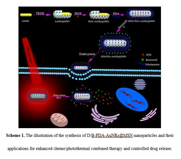
Synthesis of AuNRs
The AuNRs were prepared using a seedless method with slight modifications [24, 25]. Briefly, CTAB solution (30 mL, 0.2 M) was added to 30 mL of 1.0 mM HAuCl4, followed by the addition of 1.8 mL of 4 mM AgNO3 and 72 μL of HCl (37%). Next, 450 μL of 85.8 mM AA was added and gently swirled as the solution became colorless. Finally, 45 μL of 10 mM NaBH4 was rapidly injected. The resulting solution was kept for 6 h at 30 °C.
Preparation of AuNRs with mesoporous silica shell (AuNRs@MSN)
AuNRs@MSN were synthesized according to the reported previously [26]. To remove excess CTAB from AuNRs, 30 mL of the as-synthesized AuNRs was centrifuged at 16 000 rpm for 30 minutes. The precipitate was redispersed in 30 mL of Milli-Q water and 300 μL of 0.1 M NaOH solution was added upon stirring. Then, three injections of 90 μL of 20% TEOS in methanol solution was added into the above solution at 30 minutes intervals. The mixture was stirred for 24 h at 30 °C. The AuNRs@MSN were separated by centrifugation. The precipitate was refluxed with 20 mL of 10 mg/mL NH4NO3-ethanol solution under 60 °C for 12 h to extract the surfactant template CTAB. The final product was collected by centrifugation at 16 000 rpm for 30 min and washed with ethanol three times. The as-synthesized solid was dried in the lyophilizer.
Synthesis of PDA coated DOX-AuNRs@MSN (PDA-DOX-AuNRs@MSN)
The PDA-DOX-AuNRs@MSN were synthesized according to the literatures [27, 28]. AuNRs@MSN (50 mg) was added to the DOX solution ( 1 mg/mL, 5 mL) and stirred in dark at the room temperature for 24 h. The product was acquired by centrifugation and washed with deionized water until the supernatant became colorless. The DOX-AuNRs@MSN (50 mg) nanoparticles were suspended in the 50 mL of Tris-HCl buffer solution (pH 8.5, 10 mM). Then dopamine hydrochloride (25 mg) was added and stirred in dark at room temperature for 24 h. The PDA-DOX-AuNRs@MSN were collected by centrifugation and washed with deionized water several times to remove the unpolymerized dopamine. PDA-AuNRs@MSN were synthesized according to the same procedure without adding the DOX. The loading efficiency (LE%) of DOX on AuNRs@MSN was calculated using the following formula: LE% = (MDOX1-MDOX2)/MDOX1, where MDOX1 is the added DOX content and MDOX2 is the supernatant DOX content.
Synthesis of DOX/Btz-PDA-AuNRs@MSN (D/B-PDA-AuNRs@MSN)
Btz were conjugated to the PDA-DOX-AuNRs@MSN according to the reported literatures [29, 30]. The PDA-DOX-AuNRs@MSN were dispersed in 10 mL of dimethylsulfoxide (DMSO)-deionized water (1:10, v/v) solution containing 5 mg of Btz. The mixture was stirred for 24 h at room temperature. The products were separated by centrifugation and washed with deionized water. The loading efficiency (LE%) of Btz on DOX-PDA-AuNRs@MSN was calculated using the following formula: LE% = (MBtz1-MBtz 2)/MBtz1, where MBtz1 is the added Btz content and MBtz2 is the supernatant Btz content.
In vitro drug release
D/B-PDA-AuNRs@MSN were dispersed in 2 mL of buffer solutions (pH 5.0 and 7.4), respectively. The dispersion solution was then transferred into a dialysis bag (molecular weight cut off =8 000-14 000 kDa) and placed in 100 mL of PBS buffer solution at 37 °C with or without 808 nm light irradiation and shaked at 150 rpm. At timed intervals, 3 mL of solution was withdrawn from the solution. The released DOX and Btz were analyzed by UV-vis spectrum. The volume of the release medium was kept constant by adding 3 mL fresh medium after each sampling.
In vitro cytotoxicity
The cell viability was determined by CCK-8 assay. 4T1 cells were seeded into a 96-well plate at a density of 1 × 10⁴ cells and cultured at 5% CO2 and 37 °C for 24 h. Different concentrations of AuNRs@MSN, PDA-AuNRs@MSN, free DOX+Btz, D/B-PDA-AuNRs@MSN were added to the medium, and the cells were incubated at 5% CO2 and 37 °C for 24 h. In order to evaluate PTT efficacy, PBS, AuNRs@MSN, PDA-AuNRs@MSN, D/B-PDA-AuNRs@MSN were cultured with 12 h before the cells were irradiated with 808 nm laser (1 W/cm2) for 5 min. The cells were further incubated for another 12 h. The cell viability was calculated by measuring the absorbance value at 450 nm.
In vitro cellular uptake
4T1 cells were seeded into confocal dish and cultured for 24 h. The D/B-PDA-AuNRs@MSN solution (DOX concentration = 2 µg/mL, Btz concentration = 0.1 µg/mL) was added and cultured with cells for 4 h. Then, the cells were washed with PBS solution and fixed with 4% formaldehyde for 10 min. After that, the cells were washed with PBS solution several times to remove excess formaldehyde. The cell nuclei was stained by DAPI. The fluorescence images were observed under confocal laser scanning microscopy (CLSM).
Temperature measurement in vitro
The aqueous of AuNRs@MSN and PDA-AuNRs@MSN containing the same Au concentration (20 mg/mL) were added into 0.5 mL centrifuge tube and irradiated by 808 nm laser at a power density of 1 W/cm2 for 600 s. For the control group, 0.5 mL of deionized water was also irradiated under the same condition. To investigate different concentration of PDA-AuNRs@MSN photothermal effect, the as-prepared PDA-AuNRs@MSN was diluted to different concentrations (Au = 10, 20, 40 and 60 mg/mL) and 0.5 mL sample solution was added into the centrifuge tube and was irradiated by 808 nm laser (1 W/cm2) for 600 s. For the control group, 0.5 mL of deionized water was also irradiated under the same condition. A thermal imager was used to measure the temperature changes and obtain the infrared thermal images.
In vivo biosafety analysis
Female BALB/c mice (4 weeks) were purchased from SLAC laboratory animal Co, Ltd. (Shanghai) and the animal procedures were complied with the guidelines of the Institutional Animal Care and Use Committee of Tongji University. Female BALB/c mice (4 weeks) were treated by the PDA-AuNRs@MSN (Au = 32 mmol/L) through tail vein injection. The control group was injected with PBS pH 7.4 solution at the same volume. The main organs (heart, liver, spleen, lung, kidney and intestines) were collected after 15 days and were stained with hematoxylin and eosin (H&E) for histological analysis.
In vivo photothermal treatment
4T1 cells (2×10⁶ cells in PBS pH 7.4 buffer solution) were injected subcutaneously into the flank of the right fore leg of the female BALB/c mice (4 weeks). When the tumor volume reached 80 mm3, the mice were randomly divided into 5 groups (n = 3 each group). The mice were treated via the tail vein with 100 mL solution of PBS+Laser, DOX+Btz, D/B-PDA-AuNRs@MSN, AuNRs@MSN+Laser, D/B-PDA-AuNRs@MSN+Laser, respectively. After 6 h, the mice were irradiated by 808 nm laser (1 W/cm2) for 600 s for laser treating groups. The tumor volume was measured by a digital caliper every 2 days. The tumor volume = length * width2/2.
In vivo photoacoustic (PA) imaging of tumor
4T1 cells (2×10⁶ cells in PBS pH 7.4 buffer solution) were injected subcutaneously into the flank of the right fore leg of the female BALB/c mice (4 weeks). When the tumor volume reached 80 mm3, the PDA-AuNRs@MSN (Au = 32 mmol/L) was intravenously injected into the mice. The PA imaging of the tumor site at different time points (0, 2 h, 4 h and 6.5 h) were scanned using the Vevo LAZR system. The excitation wavelength was 875 nm.
Characterizations
Transmission electron microscopy (TEM) was conducted on JEM-2100 operating at 200 kV. Zeta potentials were measured on a zeta potential analyzer (Zetasizer Nano ZS90, Malvern). The UV-vis spectrum of the sample was measured with a Cary 50 spectrophotometer (Varian). The surface area, pore size, and pore volume were determined by N2 adsorption-desorption isotherms obtained at 77 K on a Quantachrome Autosorb-1 (USA). The sample was outgassed at 10-3 Torr and 60 0C for approximately 6 h prior to the adsorption experiment. The PA imaging was conducted on Vevo LAZR system (FujiFilm VisualSonics Inc., America). Fourier Transform Infrared Spectroscopy (FTIR) was measured on a SHIMADZU IR prestige-21 spectrometer. Cell imaging was conducted on TCS SP5 confocal laser scanning microscope (Leica, Germany).