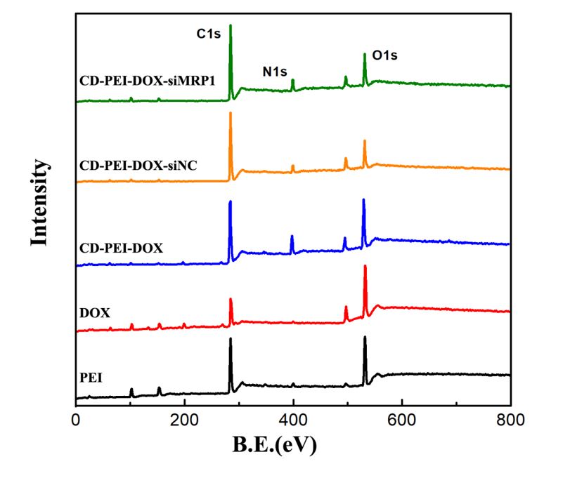
The characteristic of CD-PEI and nanodrug system based on CD-PEI
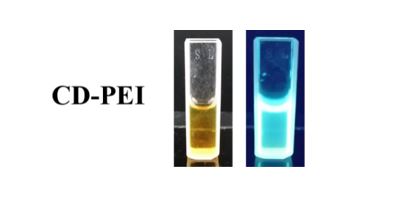
The TEM images showed that the CD-PEI was uniform and spherical, with an average size of 4.25 nm which obtained by measuring the sizes of hundreds CD nanoparticals in TEM images. It is indicated that the average diameter of CD-PEI is 2.58 nm measured by dynamic light scattering (DLS). CD-PEI was dark yellow under white light and well-distributed in aqueous solution. Under the UV light, the CD-PEI solution had obvious green emission indicating that CD-PEI had excellent fluorescence property. The UV-Vis spectrum of CD-PEI had two bands at 304 nm and 346 nm, corresponding to the π-π* transition and n-π* transition[14–16]. The CD-PEI-DOX, CD-PEI-DOX-siNC, and CD-PEI-DOX-siMRP1 had a broad peak band at 480–600 nm among which was the DOX characteristic absorbance peak. These results confirmed that the DOX was loaded on the surface of CD-PEI, and the DLE was 35%. The emission in CD could result from the surface effects and nanometer quantum confinement effect[17, 18]. The photoluminescence (PL) spectra of CD-PEI-DOX-siMRP1 had been characteristic by various excitation wavelengths with 20 nm increments. The PL spectra of CD-PEI and CD-PEI-DOX-siMRP1 were obviously different. Due to the loading of DOX agents, the PL and UV-vis spectrum has changed. The emission intensity of CD-PEI-DOX-siMRP1 is lower than that of CD-PEI in spectra, and the peaks have red-shift. The increase of emission peak confirmed the DOX has been successfully loaded on the surface of CD-PEI[19]. The PL property of CD-PEI has been studied in our previous work, the CD-PEI exhibited excellent and stable PL property that is beneficial for the tracking of drug delivery in vivo. In addition, the CD-PEI possess excitation-dependent emission behavior[20, 21], that when the excitation wavelength increase from 270 nm to 450 nm, the emission peak shift from 400 to 600 nm. When the excitation wavelength is 350 nm, the emission intensity is highest. In this work, we studied the PL behavior of CD-PEI-DOX-siMRP1 through increasing excitation wavelength from 270 to 510 nm. The CD-PEI-DOX-siMRP1 had no obvious emission peak when the excitation wavelength is lower than 330 nm. When excitation wavelengths increase from 350 nm to 510 nm, the emission intensity of CD-PEI-DOX-siMRP1 is increasing, and reaches maximum at 470 nm. The CD-PEI-DOX-siMRP1 has two emission peak centers at 560 and 690 nm.
The drug release behavior of CD-PEI-DOX-siMRP1 was revealed by in vitro release test. The free DOX release totally during 50 hours, while CD-PEI-DOX-siMRP1 prolong the drug release time up to 72 hours when pH was 5.2. CD-PEI-DOX-siMRP1 released fairly minimal DOX under pH 7.4 and illustrated that the nanocarrier would avoid the detrimental release of DOX in normal tissue and cell. When the pH changed from 7.4 to 5.2, the release amount of DOX from CD-PEI-DOX-siMRP1 increased about 5-times indicating that CD-PEI-DOX-siMRP1 was pH-sensitivity and triggered the release of DOX by acid environment because the water solubility of DOX was increased at lower pH. The advantage of selective drug release behavior of nanosystem could enhance the therapeutic effect of tumor and decrease the toxic to normal tissue.
MRP1 was involved in chemoresistance of A549 against CD-PEI-DOX.
Failure of the chemotherapy to malignant tumor was mainly attributable to insensitivity to drugs. Elucidating the mechanisms how tumor cells modulate the chemoresistance was critical for improving the chemotherapeutic effect of various drugs. We previously confirmed that CD-PEI-DOX treatment in hepatocellular carcinoma inhibit tumor growth through actively targeting tumors[7]. This led us to postulate: whether CD-PEI-DOX would elicit side effects on malignant tumors such as increasing chemoresistance in lung cancer. We firstly detected the expression of molecules associated with chemoresistance including p-glycoprotein (P-gp), MRP1, and ABCG2 in adherent, sphere and chemoresistant lung cancer cell line A549. From Fig. 3A, we vividly observed that molecules related with chemoresistance were elevated in spheres formed by A549 cells, which were considered to bear more traits of stemness compared with adherent cells. Consistently, the expression of these molecules elevated most in A549/ADM cells. These results show that A549/ADM cells expressed a panel of molecules involved in chemoresistance. Subsequently, we analyzed the cell viability of A549 and chemoresistant A549/ADM cells after doxorubicin treatment (Fig. 3B). We discovered that A549/ADM cells were resistant to doxorubicin treatment. To investigate the involvement of MRP1 in chemoresistance of A549 against doxorubicin, we tentatively knockdown the expression of MRP1 using siRNA transfection. We determined the best knockdown efficiency of siRNA targeting MRP1 using qPCR methods. From Fig. 3C, we determined siRNA #2 was the most efficient sequence targeting MRP1. CD-PEI-DOX were conjugated with this sequence with best efficiency and CCK8 assays were performed to analyze the cell viability after treatment by various drugs. the IC50 of drugs in both A549 and A549/ADM cells were calculated from three independent cell toxicity experiments. As shown in Fig. 4A-4B and Table 1, free doxorubicin in A549/ADM cells was approximately 5 fold higher compared with A549 cells. Accordingly, the IC50 of CD-PEI-DOX in A549/ADM cells were markedly higher (151.7 µg/mL) than that (6.72 µg/mL) in the A549 cells. When CD-PEI-DOX were loaded with MRP1 siRNA sequences, the cells became more vulnerable to CD-PEI-DOX, IC50 was only 17.4 µg/mL in A549/ADM cells.
Table 1 IC50 of Free DOX, CD-PEI-DOX, CD-PEI-DOX-siMRP1 in A549 and A549/ADM cells
|
IC50(µg/mL)
|
|
A549
|
Free DOX
|
26.993
|
|
CD-PEI-DOX
|
6.72
|
|
CD-PEI-DOX-siMRP1
|
5.692
|
|
A549/ADM
|
Free DOX
|
130.626
|
|
CD-PEI-DOX
|
151.7
|
|
CD-PEI-DOX-siMRP1
|
17.4
|
| Figure capture: |
| Figure S1 The optical images of CD-PEI under white light and UV light |
Based on these findings, we conclude that the free DOX exhibited different toxic behavior to A549 and A549/ADM cells. While CD-PEI-DOX-siMRP1 treatment caused more toxicity to A549/ADM cells compared with free DOX and CD-PEI-DOX groups at all-time points. It indicated that CD-PEI-DOX-siMRP1 possess better antitumor effect than DOX and CD-PEI-DOX. CD-PEI-DOX-siMRP1 possesses high toxic to A549/ADM due to the co-delivery and synergistic effect of siRNA and DOX, indicating that the MRP1 was critically involved in chemoresistance of A549/ADM cells against doxorubicin.
These results led us to postulate: whether MRP1 was induced by CD-PEI-DOX and suppressing MRP1 would reverse this effect? To test our hypothesis, we treated both A549 and A549/ADM cells with the indicated drugs and analyzed the expression of MRP1 after treatment. As demonstrated in Fig. 4C-4D, the expression of MRP1 increased markedly in case of CD-PEI-DOX treatment. In contrast, MRP1 expression decreased after siRNA targeting MRP1 only in A549/ADM cells (Fig. 4D) was loaded on CD-PEI-DOX particles. Combined with the cell viability results, knockdown the expression of MRP1 augment the killing effect of CD-PEI-DOX. The internalization of DOX and nanodrug system based on CD-PEI
The cellular uptake efficiency of free DOX, and CD-PEI-DOX-siMRP1 has been studied by laser scanning confocal microscopy and flow cytometry. The A549 and A549/ADM cells were incubated for 2 hours with PBS, free DOX, CD-PEI-DOX, CD-PEI-DOX-siNC, and CD-PEI-DOX-siMRP1, respectively. The results revealed that more CD-PEI-DOX-siMRP1 is uptaken by the cell and located in the perinuclear regions and the nuclei. The phenomenon revealed that the CD-PEI-DOX-siMRP1 could enhance the permeability of DOX in to the nuclei, which is beneficial to increase the therapeutic efficiency of DOX. In the A549/ADM, the CD-PEI-DOX-siMRP1 may increase the uptake efficiency of the DOX into the cell, and the DOX wrapped by nanocarrier could enter into the A549/ADM more than the free DOX group, for the reason that the nano carrier may increase the permeability of free drug into the cells. The permeability of free DOX, and DOX wrapped by CD-PEI has been investigated using A549 and A549/ADM cell spheres by the Z-stack mode of laser scanning confocal microscopy. The Z-stack mode could be used to observe the penetrate situation of different treatment by gaining the different layers of the cell mammosphere. The results revealed that the group treated by free DOX has the weakest permeability among all the groups. And the CD-PEI-DOX-siMRP1 could penetrate the inner region of mammosphere. This result confirmed that the CD-PEI-DOX-siMRP1 could have the permeability to enter into the tumor tissue, which can maximize the therapeutic effect.
The uptake of DOX and nanodrug system has been measured by flow cytometry. Figure 7 is the uptake in A549 and A549/ADM cells during the different intervals. The group incubated with CD-PEI-DOX-siMRP1 have higher mean fluorescence intensity than other group in both A549 and A549/ADM, indicated that CD-PEI-DOX-siMRP1 could delivery more DOX into cell. The cellular uptake of free DOX is lower than other treated groups at every interval in both A549 and A549/ADM cells. The nanodrug could decrease the drug efflux and gather high intracellular concentration of the drug, because the nanodrugs bypass the efflux pumps. And in the A549/ADM, the uptake amount of CD-PEI-DOX-siMRP1 is higher than other groups, results from the disrupting of siRNA to cellular pathway. This result indicated that the disturbing MRP1 could enhance the uptake of nanodrug effectively. The co-delivery DOX and siRNA would more effective in overcoming resistance of cancer cells.
CD-PEI and CD-PEI-siMRP1 enhanced the inhibitory effect on stemness and metastatic potential elicited by doxorubicin
The ability of metastasis is the critical factor that influence the therapeutic efficiency of tumor. We used the Transwell assay to investigate the migration and invasion in A549 and A549/ADM cells affected by respective drugs. Both A549 and A549/ADM treated with DOX, CD-PEI-DOX and CD-PEI-DOX-siNC and CD-PEI-DOX-siMRP1 exhibited decreased migration and invasion compared to the control, with migration counts shown in Fig. 8, respectively. The results indicated that CD-PEI-DOX and CD-PEI-DOX-siNC and CD-PEI-DOX-siMRP1 generated stronger inhibitory effects than free DOX. We found that CD-PEI-DOX distinctly inhibited the migration and invasion of both A549/ADM and ADM cells compared with the group treated by free DOX. After silencing MRP1, the decline in migration and invasion was reversed elicited by CD-PEI-DOX treatment in chemoresistant A549 cells. These results collectively showed that MRP1 is critical for mediating the chemoresistant effect of A549 against doxorubicin.
To elucidate the regulation of stemness in lung cancer by MRP1, we conducted sphere-forming assays. As revealed in Fig. 9, CD-PEI-DOX-siMRP1 treatment specifically suppressed the size and number of spheres in A549/ADM cells. A conclusion could be drawn from the above findings that suppressing MRP1 expression attenuated the resistance to regulation of stemness, migration and invasion induced by CD-PEI-DOX in DOX-resistant A549 cells.
Targeting and biocompatibility analysis of CD-PEI-DOX-siMRP1