Ultrasound-guided corticosteroid injection for patients with carpal tunnel syndrome: a systematic review and meta-analysis of randomized controlled trials

DOI: https://doi.org/10.21203/rs.3.rs-125732/v1

Abstract

Carpal tunnel syndrome (CTS) refers to the symptoms and signs caused by the compression of the median nerve in the carpal tunnel. It can be treated by corticosteroid injection into the carpal tunnel. Two methods for injection have been employed, namely ultrasound-guided and landmark-guided injection. This systematic review and meta-analysis was conducted to compare these methods in terms of several outcomes. Randomized controlled trials (RCTs) were identified, and data collection was completed on 7 October 7, 2020. Results for continuous variables are expressed as standardized mean differences (SMDs) with 95% confidence intervals (CIs). Analyses were performed using RevMan 5.3 software. The analysis included eight RCTs published between 2013 and 2019 with a total of 448 patients. Ultrasound-guided injection yielded more favorable results for the Boston Carpal Tunnel Syndrome Questionnaire, Symptom Severity Scale [SMD = −0.49, 95% CI (−0.74, −0.25), P < 0.0001], Boston Carpal Tunnel Syndrome Questionnaire, Functional Status Scale [SMD = −0.24, 95% CI (−0.42, −0.06), P = 0.01], distal motor latency [SMD = −0.36, 95% CI (−0.70, −0.02), P = 0.04], and compound muscle action potential [SMD = 0.38, 95% CI (0.16, 0.61), P = 0.0008] . Ultrasound-guided corticosteroid injection is recommended for patients with CTS.

Introduction

Carpal tunnel syndrome (CTS) refers to the symptoms and signs caused by the compression of the median nerve in the carpal tunnel1. This compression leads to nerve ischemia, which thus damages the nerve and affects its function2. The prevalence of CTS in the general population has been estimated between 1% and 5%3-5. Common symptoms of CTS are paresthesia, numbness, tingling, pain, and weakness across the distribution of the median nerve distal to the carpal tunnel6,7. CTS can be diagnosed not only by clinical evaluation but also through electrodiagnostic tests8,9.  Treatment for CTS includes surgical and nonsurgical methods10,11. Among nonsurgical methods, corticosteroid injection into the carpal tunnel is an effective treatment for patients with CTS12,13. Corticosteroid injection into the carpal tunnel is often guided through palpation using anatomical landmarks14,15. However, the injection may be misplaced, resulting in residual symptoms or symptom recurrence16. By contrast, in ultrasound-guided injection, an accurate real-time image of the structure of the wrist enables the physician to inject corticosteroid directly into the carpal tunnel17-19. A systematic review and meta-analysis conducted by Arash et al. indicated that ultrasound-guided injection is more effective than landmark-guided injection in terms of symptom severity but not in terms of functional status and electrodiagnostic outcomes20. However, their meta-analysis featured only three randomized controlled trials (RCTs), and numerous additional studies have recently been conducted. Therefore, we conducted this systematic review and meta-analysis to compare the effects of ultrasound- and landmark-guided corticosteroid injection on symptom severity, functional status, and electrodiagnostic outcomes in patients with CTS. 

Methods

Eligibility criteria

The eligibility criteria for this study were as follows: (1) RCTs; (2) patients with CTS diagnosed through a nerve conduction study; (3) patients with no previous surgical treatment; (4) primary study aim to compare the clinical effectiveness of ultrasound- and landmark-guided (blind) corticosteroid injection in patients with CTS; and (5) outcome measurements including the Boston Carpal Tunnel Syndrome Questionnaire (BCTQ) and electrodiagnostic findings.

Search strategy

The authors independently reviewed the literature, extracted data, and performed crosschecks in accordance with the Preferred Reporting Items for Systematic Reviews and Meta-Analyses guidelines21. We searched electronic databases, such as PubMed, EMBASE, and Cochrane. We defined group A as steroid, synonyms for steroids and several frequently used brands; while group B was formed using CTS and synonyms for it. We intersected groups A and B to prepare our keywords for searching the aforementioned electronic databases (keywords are listed in the appendix). If available, RCTs were identified using the refined search function of the databases. Additional articles were identified through a manual search of the reference lists of the relevant articles. The date of database inception to 7 October, 2020 was the time range for the search. Two reviewers independently reviewed the full texts of all potentially relevant articles to identify articles that met the eligibility criteria. Their decisions were then compared, and disagreements were resolved by a third reviewer.

Data items

The following data were obtained from each RCT: the type of corticosteroid injected; the number, mean age, and mean symptom duration of the participants in the intervention and control groups; the plane of approach in the intervention group; outcome measurements; and follow-up duration.

Outcome measurements

The outcome measurements in this study were the BCTQ [including the Symptom Severity Scale (SSS) and Functional Status Scale (FSS)] and four electrodiagnostic parameters. The BCTQ is a widely applied measurement for CTS in clinical practice; it comprises two parts, namely the SSS (11 questions) and the FSS (eight questions). All questions are answered on a scale from 1 to 5. A higher score indicates more severe symptoms or functional disability22. The BCTQ was used in all of the reviewed studies. The parameters typically reported for electrodiagnosis outcomes were sensory nerve action potential (SNAP), sensory nerve conduction velocity (SNCV), distal motor latency (DML), and compound muscle action potential (CMAP). In summary, the following outcomes were assessed in this study: (1) BCTQ-SSS, (2) BCTQ-FSS, and (3) the SNAP, SNCV, DML, and CMAP electrodiagnostic parameters.

Risk‑of‑bias assessment

The risk of bias was assessed using the RoB 2 tool, a revision of the Cochrane risk-of-bias tool for randomized controlled trials, that is widely applied for assessing the quality of RCTs23. The following domains were considered: (1) randomization process, (2) deviations from intended interventions, (3) missing outcome data, (4) outcome measurement, (5) selection of the reported result, and (6) overall bias23. Following the Cochrane Handbook for Systematic Reviews of Interventions, the risk of bias was assessed by two independent reviewers24. Disagreements between reviewers were resolved through discussion and consultation with a third reviewer.

Statistical analysis

Statistical analyses were performed using RevMan 5.3 software, which was provided by Cochrane Collaboration (https://training.cochrane.org/online-learning/core-sofware-cochrane-reviews/revman/revman-5-download). Results with P < 0.05 were considered statistically significant. We used the I2 test to objectively measure the statistical heterogeneity, with I2 ≥ 50% indicating significant heterogeneity25. A random effects model was used in this meta-analysis. The results for the continuous variables were expressed as standardized mean differences (SMDs) with 95% confidence intervals (CIs). Because various planes of approach may be adopted for ultrasound-guided injection, a subgroup analysis was performed to compare the in-plane and out-plane ulnar approaches.

A funnel plot was not used to test for publication bias because of the limited number of studies included in each analysis (<10).

Results

Search results

From the aforementioned search terms, 631 RCTs were initially retrieved. Of these, 415 duplicates were excluded using EndNote X926. Moreover, 199 citations that were noncompliant with the inclusion criteria were excluded after their titles and abstracts were screened. The full texts of the remaining 17 papers was screened, revealing three articles without a full text available, five that did not compare ultrasound- and landmark-guided corticosteroid injection, and one case of CTS not diagnosed through a nerve conduction test. Finally, eight articles were selected for this systematic review and meta-analysis (Fig. 1)27-34.

Study characteristics

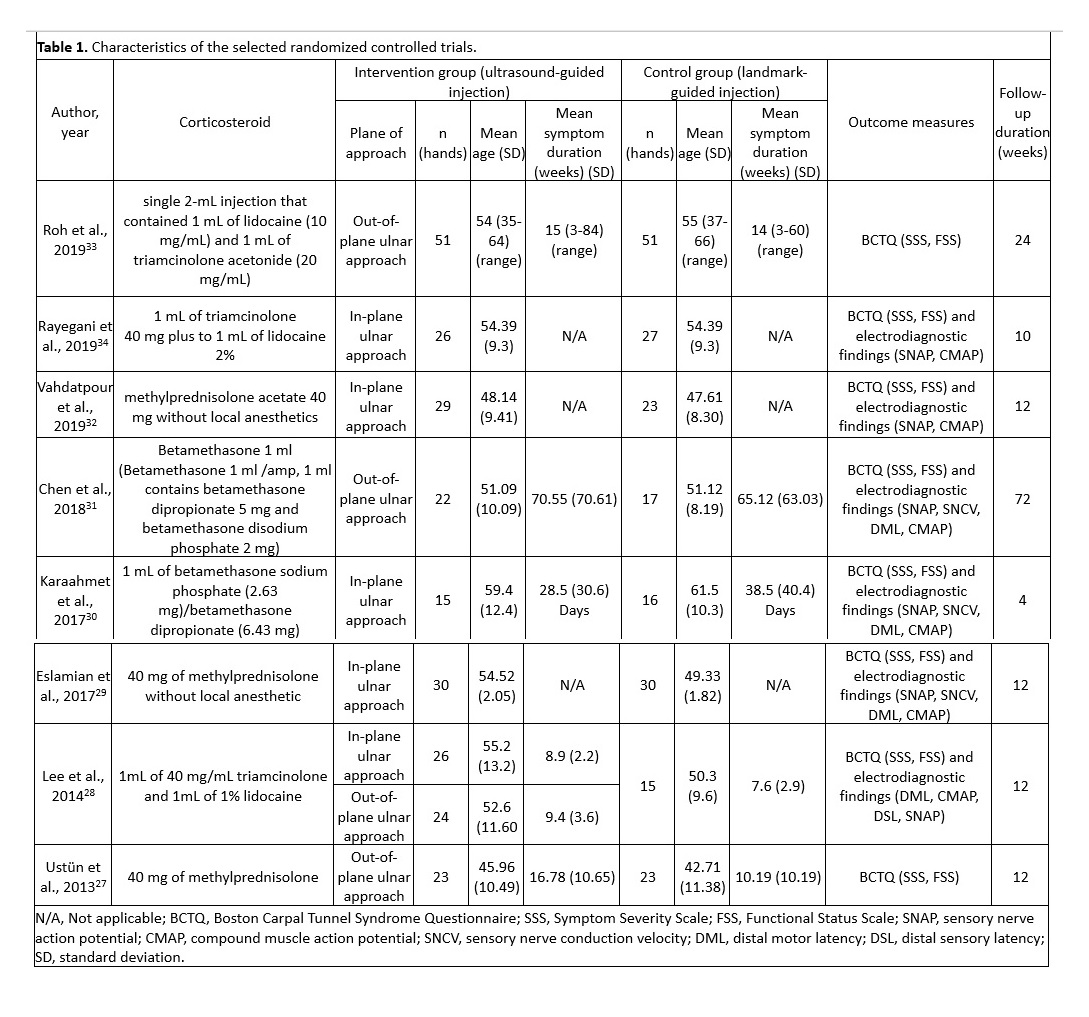
The selected studies were published between 2013 and 2019 and included 448 patients (246 patients in the ultrasound-guided group and 202 in the landmark-guided group). Four studies employed the out-plane ulnar approach27,28,31,33, and five studies adopted the in-plane ulnar approach 28-30,32,34. The main characteristics of the eight RCTs are summarized in Table 1.

Risk‑of‑bias assessment

Two reviewers assessed the quality of the selected RCTs by using the RoB 2 tool, a revision of the Cochrane RoB tool for randomized controlled trials23. Fig. 2 illustrates the risk of bias for each study. Eight studies were identified as having a low risk during randomization27-34. The risk of deviations from intended interventions was low in four studies28,29,31,32, whereas some concerns were noted for the remaining four 27,30,33,34. Eight studies were identified as having a low risk related to missing outcome data27-34. Furthermore, for outcome measures, three studies exhibited uncertain risk 27,33,34, one exhibited high risk30, and four exhibited low risk 28,29,31,32. In terms of the selection of reported results, three studies exhibited low risk29,31,34, but some concerns were noted for the remaining five27,28,30,32,33. The overall risk of bias was low in three studies29,31,32, uncertain in four studies27,28,33,34, and high in one study30.

BCTQ-SSS

BCTQ-SSS scores were reported in all eight studies27-34, including 252 patients in the ultrasound-guided group and 220 in the landmark-guided group. The heterogeneity of the studies was acceptable (I2 = 40%, P = 0.10). The BCTQ-SSS score was significantly lower in the ultrasound-guided group than in the control group [SMD = −0.49, 95% CI (−0.74, −0.25), P < 0.0001]. A subgroup analysis revealed significant differences in BCTQ-SSS score between the ultrasound-guided and control groups for the in-plane ulnar approach [SMD = −0.56, 95% CI (−0.99, −0.14), P = 0.009] and the out-of-plane ulnar approach [SMD = −0.39, 95% CI (−0.65, −0.12), P = 0.004] (Fig. 3).

BCTQ-FSS

BCTQ-FSS scores were reported in all eight studies27-34, which included 252 patients in the ultrasound-guided group and 220 in the landmark-guided group. The homogeneity of the studies was good (I2 = 0%, P = 0.72). BCTQ-FSS scores were significantly lower in the ultrasound-guided group than in the control group [SMD = −0.24, 95% CI (−0.42, −0.06), P = 0.01]. Subgroup analysis revealed a significant difference in BCTQ-FSS score between the ultrasound-guided and control groups for the out-of-plane ulnar approach [SMD = −0.28, 95% CI (−0.55, −0.02), P = 0.04] but not for the in-plane ulnar approach [SMD = −0.20, 95% CI (−0.45, 0.05), P = 0.12] (Fig. 4).

SNAP

SNAP was reported in six studies28-32,34, including 178 patients in the ultrasound-guided group and 146 in the landmark-guided group. The heterogeneity of the studies was moderate (I2 = 62%, P = 0.02). No significant intergroup differences were noted in SNAP [SMD = −0.19, 95% CI (−0.56, 0.17), P = 0.30]. Moreover, subgroup analysis revealed no significant differences in SNAP between the ultrasound-guided and control groups for the in-plane ulnar approach [SMD = −0.27, 95% CI (−0.76, 0.22), P = 0.28] or the out-of-plane ulnar approach [SMD = 0.02, 95% CI (−0.43, 0.47), P = 0.93] (Fig. 5).

SNCV

SNCV was reported in three studies29-31, including 73 patients in the ultrasound-guided group and 66 in the landmark-guided group. The heterogeneity of the studies was moderate (I2 = 53%, P = 0.12). No significant intergroup differences were noted for SNCV [SMD = 0.05, 95% CI (−0.45, 0.55), P = 0.85]. Subgroup analysis revealed no significant differences in SNAP between the ultrasound-guided and control groups for the in-plane ulnar approach [SMD = 0.10, 95% CI (−0.74, 0.94), P = 0.82] or the out-of-plane ulnar approach [SMD = −0.01, 95% CI (−0.64, 0.63), P = 0.98] (Fig. 6).

DML

DML was reported in four studies28-31, including 123 patients in the ultrasound-guided group and 96 in the landmark-guided group. The heterogeneity of the studies was acceptable (I2 = 35%, P = 0.19). DML was significantly lower in the ultrasound-guided group than in the control group [SMD = −0.36, 95% CI (−0.70, −0.02), P = 0.04]. Subgroup analysis revealed a significant difference in DML between the ultrasound-guided and control groups for the in-plane ulnar approach [SMD = −0.51, 95% CI (−0.98, −0.04), P = 0.03] but not for the out-of-plane ulnar approach [SMD = −0.11, 95% CI (−0.57, 0.34), P = 0.63] (Fig. 7).

CMAP

CMAP was reported in six studies28-32,34, including 178 patients in the ultrasound-guided group and 146 in the landmark-guided group. The homogeneity of the studies was high (I2 = 0%, P = 0.82). CMAP was significantly higher in the ultrasound-guided group than in the control group [SMD = 0.38, 95% CI (0.16, 0.61), P = 0.0008]. Subgroup analysis revealed a significant difference in CMAP between the ultrasound-guided and control groups for the in-plane ulnar approach [SMD = 0.42, 95% CI (0.16, 0.67), P = 0.001] but not for the out-of-plane ulnar approach [SMD = 0.27, 95% CI (−0.19, 0.72), P = 0.25] (Fig. 8).

Discussion

In recent years, musculoskeletal physicians have increasingly applied ultrasound-guided injection in their clinical practice35. This enables the dynamic imaging and comparison of the surrounding tissues and ensures the accuracy of injection placement36,37. Therefore, in this systematic review and meta-analysis, we compared the effects of ultrasound-guided and landmark-guided corticosteroid injection on symptomatic severity, functional status, and electrodiagnostic outcomes in patients with CTS. Significant differences in the following outcome measures favored ultrasound-guided injection:

CTS treatment can be assessed using two tools. First, BCTQ is a reliable method comprising two components, namely symptom severity and functional status22,38. In our analysis, both components differed significantly between ultrasound-guided and landmark-guided injection. In the subgroup analysis, the outcomes of the in-plane ulnar approach were more favorable than those of the out-of-plane ulnar approach. Second, electrodiagnostic testing includes SNAP, SNCV, DML, and CMAP8. Overall, significant differences in DML and CMAP favored ultrasound-guided injection; a subgroup analysis also revealed preferable outcomes in the in-plane ulnar approach than in the out-of-plane ulnar approach. In summary, the in-plane ulnar approach is the preferred method of ultrasound-guided injection for patients with CTS not only for symptom improvement but also for electrodiagnostic findings. 

The in-plane ulnar approach has several advantages. First, according to Racasan et al., the flexor carpi radialis tendon proximal to the carpal tunnel is the safest region of the body for injection needle insertion39. However, this method would penetrate the flexor carpi radialis tendon and cause injury. The in-plane ulnar approach enables the visualization of the carpal tunnel structures around the nerve, which facilitates accurate perineural injection and, most importantly, prevents the physician from damaging the surrounding vessels, nerves, and tendons18,19. Second, throughout the procedure, the needle tip and shaft can be visualized in plane relative to the transducer; thus, the physician can adjust the needle to the appropriate site and further hydrodissect the surrounding connective tissues27,28. Third, the method is easy to learn, is not restricted by etiology (i.e., idiopathic or secondary), and can accommodate congenital or postsurgical anatomical variations18,30. Given the combination of these advantages, the in-plane ulnar approach is the recommended method for ultrasound-guided injection. 

The main difference between the present study and that of Arash et al.20 is the sample size. Despite conducting an extensive literature search, Arash et al. included only three RCTs. However, additional studies have been recently published. Thus, we included eight RCTs, with 246 participants in the ultrasound-guided group and 202 participants in the landmark-guided group. Furthermore, Arash et al. identified significant differences in BCTQ-SSS score but not in other outcomes, whereas the present study revealed significant differences in BCTQ-SSS score, BCTQ-FSS score, and electrodiagnostic findings. Finally, the subgroup analysis in the present study revealed that the in-plane ulnar approach for corticosteroid injection is the method of choice for the treatment of CTS.

Our review has several limitations. First, the heterogeneity was moderate for some outcomes. Second, due to the nature of the treatment, blinding the participants and physicians is challenging. Hence, some concerns regarding bias should be expressed. Third, the duration of follow-up in the included studies was not sufficiently long (up to 3 months for the majority of the included studies) to analyze the long-term outcomes. Thus, further reviews of high-quality, large-scale RCTs are required to overcome these limitations.

Conclusion

This study compared the effects of ultrasound-guided and landmark-guided corticosteroid injection on symptomatic severity, functional status, and electrodiagnostic outcomes in patients with CTS. According to our analysis, ultrasound-guided injection yielded the most favorable results for symptom severity, functional status, and electrodiagnostic parameters. Therefore, we recommend ultrasound-guided corticosteroid injection as a treatment for patients with CTS.

Declarations

Acknowledgements

This manuscript was edited by Wallace Academic Editing.

Author contributions

Fu-An Yang and Ya-Chu Shih conceptualized and designed the study and drafted the manuscript. Hung-Chou Chen critically revised the manuscript for intellectual content. Chun-De Liao, Chin-Wen Wu, and Jia-Pei Hong conducted a comprehensive search for articles that met the eligibility criteria. Fu-An Yang and Ya-Chu Shih extracted the relevant data and assessed the quality of the selected trials. Hung-Chou Chen provided statistical expertise, analyzed and interpreted the data, and submitted the manuscript. Fu-An Yang and Ya-Chu Shih contributed equally to this study.

Funding

This research received no external funding.

Conflicts of Interest

The authors have no conflicts of interest to declare.

References

1          Katz, J. N. & Simmons, B. P. Carpal Tunnel Syndrome. New England Journal of Medicine 346, 1807-1812, doi:10.1056/NEJMcp013018 (2002).

2          Aroori, S. & Spence, R. A. Carpal tunnel syndrome. Ulster Med J 77, 6-17 (2008).

3          Atroshi, I. et al. Prevalence of carpal tunnel syndrome in a general population. Jama 282, 153-158, doi:10.1001/jama.282.2.153 (1999).

4          de Krom, M. C., Kester, A. D., Knipschild, P. G. & Spaans, F. Risk factors for carpal tunnel syndrome. Am J Epidemiol 132, 1102-1110, doi:10.1093/oxfordjournals.aje.a115753 (1990).

5          Pourmemari, M. H., Heliövaara, M., Viikari-Juntura, E. & Shiri, R. Carpal tunnel release: Lifetime prevalence, annual incidence, and risk factors. Muscle Nerve 58, 497-502, doi:10.1002/mus.26145 (2018).

6          Hesami, O., Haghighatzadeh, M., Lima, B. S., Emadi, N. & Salehi, S. The effectiveness of gabapentin and exercises in the treatment of carpal tunnel syndrome: a randomized clinical trial. J Exerc Rehabil 14, 1067-1073, doi:10.12965/jer.1836420.210 (2018).

7          Hashempur, M. H. et al. Effect of Linum usitatissimum L. (linseed) oil on mild and moderate carpal tunnel syndrome: a randomized, double-blind, placebo-controlled clinical trial. DARU Journal of Pharmaceutical Sciences 22, 43, doi:10.1186/2008-2231-22-43 (2014).

8          Jablecki, C. K. et al. Practice parameter: Electrodiagnostic studies in carpal tunnel syndrome. Report of the American Association of Electrodiagnostic Medicine, American Academy of Neurology, and the American Academy of Physical Medicine and Rehabilitation. Neurology 58, 1589-1592, doi:10.1212/wnl.58.11.1589 (2002).

9          Bland, J. D. Carpal tunnel syndrome. Bmj 335, 343-346, doi:10.1136/bmj.39282.623553.AD (2007).

10        Andreu, J. L., Ly-Pen, D., Millán, I., de Blas, G. & Sánchez-Olaso, A. Local injection versus surgery in carpal tunnel syndrome: Neurophysiologic outcomes of a randomized clinical trial. Clinical Neurophysiology 125, 1479-1484, doi:https://doi.org/10.1016/j.clinph.2013.11.010 (2014).

11        Graham, B. et al. The American Academy of Orthopaedic Surgeons Evidence-Based Clinical Practice Guideline on: Management of Carpal Tunnel Syndrome. J Bone Joint Surg Am 98, 1750-1754, doi:10.2106/jbjs.16.00719 (2016).

12        Marshall, S., Tardif, G. & Ashworth, N. Local corticosteroid injection for carpal tunnel syndrome. Cochrane Database Syst Rev, Cd001554, doi:10.1002/14651858.CD001554.pub2 (2007).

13        Roh, Y. H., Noh, J. H., Gong, H. S. & Baek, G. H. Comparative study on the effectiveness of a corticosteroid injection for carpal tunnel syndrome in patients with and without Raynaud's phenomenon. Bone Joint J 99-b, 1637-1642, doi:10.1302/0301-620x.99b12.Bjj-2017-0371.R2 (2017).

14        Brooks, R. et al. Anatomic Landmarks to Locate the Median Nerve for Safe Wrist Block or Carpal Tunnel Steroid Injection. Eplasty 19, e19 (2019). <http://europepmc.org/abstract/MED/31501688

https://europepmc.org/articles/PMC6727415

https://europepmc.org/articles/PMC6727415?pdf=render>.

15        Kim, D., Jang, J. E. & Park, B. Anatomical Basis of Ulnar Approach in Carpal Tunnel Injection. Pain physician 16, E191-E198 (2012).

16        Evers, S. et al. Effectiveness of Ultrasound-Guided Compared to Blind Steroid Injections in the Treatment of Carpal Tunnel Syndrome. Arthritis Care Res (Hoboken) 69, 1060-1065, doi:10.1002/acr.23108 (2017).

17        Teh, J. & Vlychou, M. Ultrasound-guided interventional procedures of the wrist and hand. European radiology 19, 1002-1010, doi:10.1007/s00330-008-1209-1 (2008).

18        Smith, J., Wisniewski, S. J., Finnoff, J. T. & Payne, J. M. Sonographically guided carpal tunnel injections: the ulnar approach. J Ultrasound Med 27, 1485-1490, doi:10.7863/jum.2008.27.10.1485 (2008).

19        Grassi, W., Farina, A., Filippucci, E. & Cervini, C. Intralesional therapy in carpal tunnel syndrome: a sonographic-guided approach. Clin Exp Rheumatol 20, 73-76 (2002).

20        Babaei-Ghazani, A. et al. Ultrasound-Guided Versus Landmark-Guided Local Corticosteroid Injection for Carpal Tunnel Syndrome: A Systematic Review and Meta-Analysis of Randomized Controlled Trials. Archives of Physical Medicine and Rehabilitation 99, 766-775, doi:https://doi.org/10.1016/j.apmr.2017.08.484 (2018).

21        Preferred Reporting Items for Systematic Reviews and Meta-Analyses: The PRISMA Statement. Annals of Internal Medicine 151, 264-269, doi:10.7326/0003-4819-151-4-200908180-00135 %m 19622511 (2009).

22        Levine, D. W. et al. A self-administered questionnaire for the assessment of severity of symptoms and functional status in carpal tunnel syndrome. J Bone Joint Surg Am 75, 1585-1592, doi:10.2106/00004623-199311000-00002 (1993).

23        Higgins, J. P. T. et al. The Cochrane Collaboration’s tool for assessing risk of bias in randomised trials. BMJ 343, d5928, doi:10.1136/bmj.d5928 (2011).

24        Higgins JPT, T. J., Chandler J, Cumpston M, Li T, Page MJ, Welch VA (editors). Cochrane Handbook for Systematic Reviews of Interventions version 6.1 (updated September 2020). Cochrane, 2020. Available from www.training.cochrane.org/handbook.

25        Melsen, W. G., Bootsma, M. C., Rovers, M. M. & Bonten, M. J. The effects of clinical and statistical heterogeneity on the predictive values of results from meta-analyses. Clin Microbiol Infect 20, 123-129, doi:10.1111/1469-0691.12494 (2014).

26        EndNote v. EndNote X9 (Clarivate, Philadelphia, PA, 2013).

27        Ustün, N. et al. Ultrasound-guided vs. blind steroid injections in carpal tunnel syndrome: A single-blind randomized prospective study. American journal of physical medicine & rehabilitation / Association of Academic Physiatrists 92, 999-1004, doi:10.1097/PHM.0b013e31829b4d72 (2013).

28        Jin Young Lee, M., Yongbum Park, MD, Ki Deok Park, MD, PhD, Ju Kang Lee, MD, PhD, and Oh Kyung Lim, MD, PhD. Effectiveness of ultrasound-guided carpal tunnel injection using in-plane ulnar approach: A prospective, randomized, single-blinded study. Medicine (United States) 93, e350, doi:10.1097/MD.0000000000000350 (2014).

29        Fariba Eslamian, B. E., Arash Babaei-Ghazani, Fateme Jahanjoo, and Mojgan Zeinali‡. A Randomized Prospective Comparison of Ultrasound-Guided and Landmark-Guided Steroid Injections for Carpal Tunnel Syndrome. Journal of Clinical Neurophysiology 34, 107-113, doi:10.1097/WNP.0000000000000342 (2017).

30        Karaahmet Ö, Z. et al. Comparing the effectiveness of ultrasound-guided versus blind steroid injection in the treatment of severe carpal tunnel syndrome. Turk J Med Sci 47, 1785-1790, doi:10.3906/sag-1704-97 (2017).

31        Po-Cheng CHEN, M., Lin-Yi WANG, MD, Ya-Ping PONG, MD, Yi-Jung HSIN, MD, Mei-Yun LIAW, MD and Chia-Wei CHIANG, MD. EFFECTIVENESS OF ULTRASOUND-GUIDED VS DIRECT APPROACH CORTICOSTEROID INJECTIONS FOR CARPAL TUNNEL SYNDROME: A DOUBLEBLIND RANDOMIZED CONTROLLED TRIAL. J Rehabil Med 50, 200-208, doi:10.2340/16501977-2308 (2018).

32        Babak Vahdatpour, S. H., Zahra Azimi ,  and Hadiseh Ramezanian. Carpal Tunnel Syndrome Treatment Using Ultrasound-Guided Versus Landmark-Guided Corticosteroid Injection: A Randomized Prospective Trial. Shiraz E-Medical Journal 20, e83929, doi:10.5812/semj.83929 (2019).

33        Roh, Y. H., Hwangbo, K., Gong, H. S. & Baek, G. H. Comparison of Ultrasound-Guided Versus Landmark-Based Corticosteroid Injection for Carpal Tunnel Syndrome: A Prospective Randomized Trial. Journal of Hand Surgery 44, 304-310, doi:10.1016/j.jhsa.2019.02.007 (2019).

34        Seyed Mansoor Rayegani, S. A. R. M. A.-D., Nafise Bavaghar, Shahram Rahimi-Dehgolan. Comparing The Efficacy Of Local Triamcinolone Injection In Carpal Tunnel Syndrome Using Three Different Approaches with or without Ultrasound Guidance. Journal of Pain Research 12, 2951–2958, doi:10.2147/JPR.S212948 (2019).

35        Daniels, E. W., Cole, D., Jacobs, B. & Phillips, S. F. Existing Evidence on Ultrasound-Guided Injections in Sports Medicine. Orthop J Sports Med 6, 2325967118756576, doi:10.1177/2325967118756576 (2018).

36        Ozçakar, L., Tok, F., De Muynck, M. & Vanderstraeten, G. Musculoskeletal ultrasonography in physical and rehabilitation medicine. J Rehabil Med 44, 310-318, doi:10.2340/16501977-0959 (2012).

37        Gilliland, C. A., Salazar, L. D. & Borchers, J. R. Ultrasound versus anatomic guidance for intra-articular and periarticular injection: a systematic review. Phys Sportsmed 39, 121-131, doi:10.3810/psm.2011.09.1928 (2011).

38        Vladeva, E. P. The Boston Carpal Tunnel Questionnaire /Bctq/ − a Reliable Method for Diagnosis and Assessment of the Treatment of Carpal Tunnel Syndrome. Web of Scholar, 58-63, doi:10.31435/rsglobal_wos/28022020/6920 (2020).

39        Racasan, O. & Dubert, T. The safest location for steroid injection in the treatment of carpal tunnel syndrome. J Hand Surg Br 30, 412-414, doi:10.1016/j.jhsb.2005.04.009 (2005).