Bleeding from band ligation-induced ulcers following the treatment of oesophageal varices: a retrospective case–control study.

DOI: https://doi.org/10.21203/rs.3.rs-126570/v1

Abstract

Background Band ligation (BL) plays a vital role in the treatment of oesophageal varices; however, the procedure carries a considerable risk of band slippage, variceal site ulcer formation and post-treatment bleeding. Our study aimed to explore the incidence of post-BL ulcer bleeding and to identify possible associated factors.

Methods We retrospectively reviewed the records of patients with oesophageal varices who underwent endoscopic haemostasis by BL at our institution between 2015 and 2020. We statistically compared the patients with post-BL ulcer bleeding and those without (controls). The outcome variable was the development of BL-induced ulcer bleeding. The patients’ demographics, clinical and laboratory parameters, BL procedure outcomes and experts’ opinions were used as the independent variables and possible associated factors.

Results Of the 4579 eligible patients 388 (8.5%) presented with post-BL ulcer bleeding. Proton pump inhibitor (PPI) use was associated with a lower risk of post-BL ulcer bleeding (odds ratio, 0.77; 95% confidence interval [CI]: 0.603–0.983). The presence of high-risk stigmata indicated a 1.276 times higher risk of bleeding (CI: 1.024–1.592), and a greater number of varices was associated with an increased risk of post-BL ulcer bleeding (P = 0.007). The use of fewer bands per variceal site was associated with fewer bleeding incidents (P = 0.008), while lower haemoglobin levels were associated with a higher probability of bleeding (P = 0.007).

Conclusions The overall incidence of post-BL ulcer bleeding was 8.5%. The presence of high-risk stigmata and a higher number of varices and bands per variceal site were associated with an increased risk of bleeding. Adequate haemoglobin levels and the use of adjuvant PPIs and having were protective factors. 

Background

Oesophageal varices result from portal hypertension as a frequent manifestation of liver cirrhosis [1]. About 60%–80% of liver cirrhosis patients develop gastrointestinal varices, with oesophageal varices constituting 17% [2, 3]. The frequency of developing oesophageal varices is firmly attributed to the severity of liver disease. Up to 40% and 85% of Child-Pugh class A and C liver disease patients, respectively, are affected [4]. More prevalent in males (i.e. 60%) than females, the risk of developing oesophageal varices increases by 8% in the second year following the diagnosis of chronic liver disease and 30% in the sixth year post diagnosis [2].

Up to 50% of patients with oesophageal varices will present with bleeding at some point. The incidence of bleeding per year is 15% for large varices and 5% for small varices [1]. Following an episode of bleeding from oesophageal varices, 10%–20% of patients will not survive beyond six weeks [1]. Of the patients who survive the first episode of bleeding, 60%–80% will rebleed in less than a year [5]. Rebleeding episodes are associated with a fatality rate of around 33% [5, 6].

While a small chance of spontaneous bleeding stoppage has been reported in an older study [7], the current guidelines recommend medical interventions [5]. Common interventions include pharmacological (i.e. somatostatin, octreotide, proton pump inhibitors [PPIs] and beta-blockers) and procedural interventions, such as cyanoacrylate injection, balloon tamponade, embolization coils, sclerotherapy and band ligation (BL), among others [3, 5].

BL or rubber-band ligation represents one of the oldest techniques in the treatment of gastrointestinal varices [8]. It is inferior to cyanoacrylate injection and embolization coils in terms of the overall success rate and rebleeding risk [3]. However, BL is cost-effective, less technically demanding than the aforementioned techniques. It is therefore the most commonly used technique globally.

A typical BL procedure involves using an endoscope to suction the variceal site. A rubber band is then wrapped around the base of the sac, thereby strangulating the area from the blood supply. The strangulated variceal sac eventually falls off as a result of ischemia and necrosis, creating a small scar [9]. Seemingly practicable, the procedure carries a considerable risk of band slippage, variceal site ulcer formation and post-treatment bleeding [10, 11]. Our study aimed to explore the incidence of post-BL ulcer bleeding and to identify possible associated factors using a large study sample.

Methods

Study design and setting

This retrospective case–control study was conducted at the Gastroenterology Department of the Third Xiangya Hospital of Central South University, Changsha, Hunan Province, People’s Republic of China. We reviewed all the patient records with oesophageal varices who attended our department between February 2015 and February 2020.

Study population

Our study included participants who met the three main inclusion criteria: (1) older than 18 years, (2) having oesophageal varices from any aetiology and (3) underwent emergent or elective BL as a treatment or prophylaxis. These eligible participants were categorized into either the case group or the control group. The case group comprised participants who met the three inclusion criteria and had endoscopically proven bleeding from a BL-induced ulcer (i.e. post-BL ulcer bleeding) without any other cause of bleeding to explain the symptom. In contrast, the control group did not have endoscopically proven bleeding from a BL-induced ulcer. We excluded patients who (1) underwent BL in combination with other haemostasis procedures such as the use of coils or cyanoacrylate injection, (2) died within the first two days following the BL haemostasis procedure, (3) were lost during the follow-up period, (4) had missing data and (5) did not consent to participate in the study.

Band ligation procedure

The Speedband Superview Super 7TM Multiple Band Ligator (Boston Scientific) was used to tie (i.e. strangulate) high-risk varices or actively bleeding varicose veins. Adjuvant pharmacological treatments (i.e. PPIs and antibiotics) and other treatments, such as blood transfusion, hydration, balloon tamponade, transjugular intrahepatic portosystemic shunt and transplantation, were performed according to the hospital’s guidelines [5] at the time and the gastroenterologist’s discretion. All the BL procedures were performed by consultant gastroenterologists, consultant surgeons or specialist trainees under supervision. All the methods were carried out in accordance with relevant guidelines and regulations.

Data collection process

Before collecting the data, we sought approval from the Institutional Review Board of the Third Xiangya Hospital, Central South University, and were assigned approval number 2019-S475.

We utilized the hospital’s electronic patient database to identify eligible patients. The data collected from the eligible patients included demographics, clinical and laboratory parameters and BL procedure outcomes. We independently collected and recorded the data in Microsoft Excel spreadsheets before cross-checking the data for correctness.

Outcomes and variables

The outcome variable was bleeding from a BL-induced ulcer, and the independent variables were obtained from the patients’ demographics, clinical and laboratory parameters and BL procedure outcomes. Further independent variables were identified by reviewing the literature and obtaining experts’ opinions. One BL procedure was considered per participant.

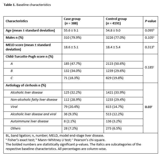
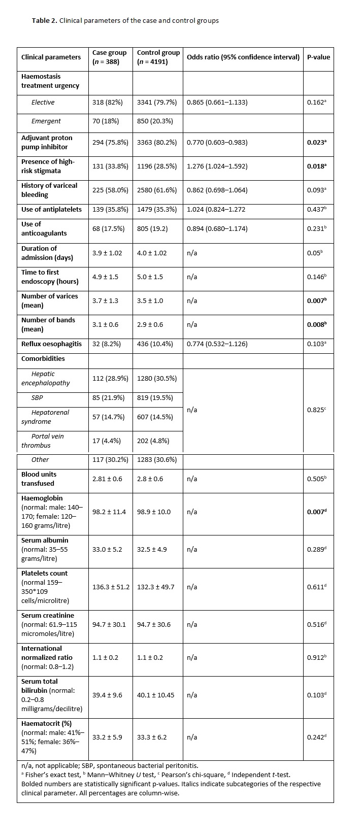
The continuous variables included age, MELD score, duration of admission (in days), time to the first endoscopy (in hours), number of varices, number of bands per variceal site, number of blood units transfused and laboratory investigation results [2, 6]. The categorical variables comprised sex (i.e. male or female), Child-Turcotte-Pugh score (i.e. A, B or C), aetiology of cirrhosis (i.e. alcoholic liver disease, non-alcoholic fatty liver disease, viral, alcoholic liver disease plus viral, autoimmune liver disease or other), haemostasis treatment urgency (i.e. elective or emergent) and adjuvant use of PPIs [12, 13]. The other categorical variables included high-risk stigmata (i.e. yes/no), history of variceal bleeding (i.e. yes/no), use of antiplatelets (i.e. yes/no), use of anticoagulants (i.e. yes/no), reflux oesophagitis (i.e. yes/no) and comorbidities (i.e. hepatic encephalopathy, spontaneous bacterial peritonitis, hepatorenal syndrome, portal vein thrombus and others) [10, 14].

Bias mitigation

Both authors independently performed the data collection followed by data cross-checking to ensure the accuracy of the data. Moreover, we utilized the strengthening the reporting of observational studies in epidemiology (STROBE) tool [15] customized for case–control studies in the write-up of this manuscript to reduce reporting bias.

Data analysis

We initially analyzed the patients’ demographic characteristics using mean (and standard deviation) and proportions. For the categorical variables, we used either Pearson’s, chi-square or Fisher’s exact tests to compare the parameters of the case and control groups depending on the respective tests’ assumptions. We performed post-hoc tests by Bonferroni’s adjustment for the crosstabulation of significant categorical variables and calculated the odds ratios. An odds ratio of <1 or >1 corresponded with reduced or increased odds, respectively, of a post-BL bleeding event, while an odds ratio of 1 suggested no association.

For the continuous variables, we utilized either the independent t-test or Mann–Whitney U-test to compare the case and control groups depending on whether the variables demonstrated normal or non-normal distribution curves, respectively. We used the Shapiro–Wilk’s test to determine the statistical significance of normality. A P-value of <0.05 was considered statistically significant.   

Additional analysis

We explored the number and causes of death in relation to post-BL ulcer bleeding status. We compared the case and control groups in this respect using Fisher’s exact test and calculated the odds ratio.  

Results

A total of 4579 patients were included in our study. The cohort was followed up for six weeks, and 388 (8.5%) patients presented with bleeding from BL-induced ulcers (i.e. case group), while 4198 (91.5%) patients did not (i.e. control group) (Figure 1). The mean time for the occurrence of post-BL ulcer bleeding was 11.4 ± 2.3 days. 

Table 1 summarizes the baseline characteristics of the 4579 patients who participated in our study. There was no statistically significant difference between the case and control groups with respect to age, sex, MELD or Child-Pugh scores. However, a cirrhotic aetiology demonstrated statistical significance (P = 0.03).

Table 2 presents a comparison of the clinical parameters between the case and control groups. The incidence of BL-induced ulcer bleeding was 9.5% (i.e. 318 events) and 8.2% (i.e. 70 events) for elective and emergent BL, respectively. The use of PPIs was associated with a lower risk of BL-induced ulcer bleeding, the odds ratio was 0.77 (95% confidence interval [CI]: 0.603–0.983). Patients with high-risk stigmata observed during endoscopy had a 1.276 times higher risk of bleeding (95% CI: 1.024–1.592).

The case group had a higher mean number of varices compared to the control group (P = 0.007), which means that a higher number of varices was associated with BL-induced ulcer bleeding in our study. Similarly, more bands were used in the case group (mean, 3.1 ± 0.6) compared to the control group (mean, 2.9 ± 0.6), signifying that the use of fewer bands was associated with a lower incidence of BL-induced ulcer bleeding. Moreover, the case group had lower haemoglobin levels compared to the control group (P = 0.007), which indicated that lower haemoglobin levels were associated with a higher probability of bleeding from BL-induced ulcers.

Mortality

Twenty-seven patients died during the follow-up period, 11 of whom were in the case group. The patients in the case group had a higher risk of death compared to those in the control group (odds ratio, 7.6; 95% CI: 3.508–16.524). Figure 2 is a radar chart summarizing the causes and frequency of death in the case and control groups.

Discussion

BL plays a vital role in the treatment of oesophageal varices. However, the procedure carries a small risk of band slippage, variceal site ulcer formation and post-treatment bleeding. Our study aimed to explore the incidence of post-BL ulcer bleeding and to identify possible associated factors.

After a mean follow-up time of 11.4 ± 2.3 days, the incidence of post-BL ulcer bleeding was 8.5%. This finding is higher than that previously reported by Jamwal et al. (3.6%) [14] and Cho et al. (7.7%) [16]. The differences may be attributable to the studies’ methodological differences. Our study included 10 times more participants compared to that of Cho et al. [16], while Jamwal et al. followed up their participants for twice the time we followed up our patients. Older studies reported up to a 15% incidence of post-BL ulcer bleeding [17]; however, this may be due to the lack of advanced technical know-how compared to the present.

In our study, the mean time for the occurrence of post-BL ulcer bleeding was 11.4 ± 2.3 days. This finding is higher than that previously reported by Cho et al. [16], who described a mean of 8.5  ±  5.1 days. As with the incidence of post-BL ulcer bleeding, the difference could be attributed to the use of different methodologies. Our finding, however, was in line with that of Jamwal et al., who reported a range of 10–13 days. While Jamwal et al. used a longer follow-up time compared to that in our study, both studies utilized large sample sizes.    

While there was no association between the bleeding event and BL treatment urgency (P = 0.162) in our study, there was a higher incidence of post-BL bleeding following elective BL (9.5%) than emergent BL (8.2%). This finding is in contrast to those of previous studies [16, 18] where there were higher incidences for emergent BL. While the reason may again be due to methodological differences between the studies, we recommend that robust studies be undertaken to explore this finding.

In our results, the use of PPIs was associated with a reduced risk of post-BL ulcer bleeding (odds ratio, 0.77; 95% CI: 0.603–0.983). This finding is in agreement with those of several other studies [13, 19]. The use of PPIs to reduce the size of an ulcer and lower the risk of reflux oesophagitis and post-prophylactic BL bleeding has been established [12, 20]. However, a study by Wu et al. [21] contradicted this finding. The difference could be ascribed to the patient population. While our study included patients with both elective and emergent BL, Wu et al. included only emergent BL patients in their study.

The presence of high-risk stigmata was associated with a 1.276 times higher risk of post-BL ulcer bleeding (95% CI: 1.024–1.592) in our study. While previous studies have identified numerous risk factors as linked with post-haemostatic procedure rebleeding, we could not locate any studies reporting an association between high-risk stigmata and post-BT ulcer bleeding. Our finding may be explained by the fact that weak mucosa (i.e. high-risk stigmata) provide an unstable site for band placement, which could result in premature band detachment and subsequently ulcer formation and bleeding [22].

The case group in our study had a statistically significantly higher mean number of varices, and more bands were utilized per variceal site compared to the control group. While it might seem logical that the more varices there are, the greater the chance of variceal-related complications, a study by Shaheen et al. [23] demonstrated no relationship between these variables. Notwithstanding, Shaheen et al. used a smaller sample size compared to that in our study. On the other hand, our findings correlated with those of a previous study with regard to the number of bands utilized [24].

The control group in our study had a statistically significantly higher mean haemoglobin level before BL treatment than the case group. This suggests that lower haemoglobin levels are associated with an increased risk of post-BL ulcer bleeding. Singh et al. [25] found that haemoglobin levels decrease spontaneously with the increasing severity of liver disease. This may suggest that our case group had more severe liver cirrhosis and therefore more severe varices than the control group. It could also explain the higher risk of death (i.e. 7.6) observed in the case group.

Study limitations and strengths

The present study was retrospective in design and therefore less robust compared to prospective studies. It also involved only Chinese patients, which could mean less generalizability. On the other hand, our study had a large sample size and thus provides more information, less uncertainty and more reliable results.

Conclusion

The overall incidence of post-BL ulcer bleeding was 8.5% in our study. The presence of high-risk stigmata and an increased number of varices and bands per variceal site was associated with an increased risk of post-BL ulcer bleeding. Nevertheless, the use of adjuvant PPIs and having adequate haemoglobin levels were associated with a lower risk of bleeding from a post-BL ulcer.    

List of Abbreviations

BL – Band ligation

95% CI – 95% confidence interval

PPI - Proton pump inhibitor

MELD - Model for End-Stage Liver Disease

STROBE - Strengthening the reporting of observational studies in epidemiology

Declarations

Ethics approval and consent to participate: Approval to conduct this study was obtained from the Institutional Review Board of the Third Xiangya Hospital, Central South University, (Approval number: 2019-S475). The Institutional Review Board of the Third Xiangya Hospital, Central South University also waived the consent to participate (i.e., informed consent) due to the retrospective nature of this study. All the methods were carried out in accordance with relevant guidelines and regulations.

Consent for publication: Not applicable.

Availability of data and materials: The datasets supporting the conclusions of this article are included within the article (and its additional files).

Competing interests: Authors declared no competing interests

Funding: No funds were given

Author’s contributions: Study conception: ZH and JS; Study design: ZH and JS; Data collection: ZH and JS; Data analysis and interpretation: ZH and JS; Manuscript drafting: JS; All authors read and approved the final version of the manuscript.

Acknowledgements: The authors acknowledge data access support from the department of records and general supervisory input from Shaojun Liu.

References

  1. Meseeha M, Attia M. Esophageal Varices. [Updated 2020 Aug 10]. In: StatPearls [Internet]. Treasure Island (FL): StatPearls Publishing; 2020 Jan-. Available from: https://www.ncbi.nlm.nih.gov/books/NBK448078/.
  2. Sarangapani A, Shanmugam C, Kalyanasundaram M, Rangachari B, Thangavelu P, Subbarayan JK: Noninvasive prediction of large esophageal varices in chronic liver disease patients. Saudi J Gastroenterol 2010, 16(1):38-42.
  3. Hu Z, Zhang D, Swai J, Liu T, Liu S: Risk of rebleeding from gastroesophageal varices after initial treatment with cyanoacrylate; a systematic review and pooled analysis. BMC gastroenterology 2020, 20(1):181.
  4. Mancuso A, D'Amico G, Pasta L, Madonia S, Tarantino I, Fili D, Criscuoli V, Pagliaro L: The incidence esophageal varices in cirrhosis. Journal of Hepatology 2001, 34:71-72.
  5. World Gastroenterology Organisation Global Guidelines [Internet]. Winsconsin (USA): World Gastroenterology Organisation, c2020 [Cited 2020 Oct 21]. Available from: https://www.worldgastroenterology.org/guidelines/global-guidelines/esophageal-varices/esophageal-varices-english. .
  6. Mehmood T, Zia MQ, Latif A, Ansar S: Mortality Related Factors in Patients with Variceal Bleeding with MELD Score ≥ 18. Journal of the College of Physicians and Surgeons--Pakistan : JCPSP 2019, 29(12):1199-1202.
  7. Prandi D, Rueff B, Roche-Sicot J, Sicot C, Maillard JN, Benhamou JP, Fauvert R: Life-threatening hemorrhage of the digestive tract in cirrhotic patients. An assessment of the postoperative mortality after emergency portacaval shunt. American journal of surgery 1976, 131(2):204-209.
  8. Blaisdell PC: Office ligation of internal hemorrhoids. American journal of surgery 1958, 96(3):401-404.
  9. Butt N, Abbasi A, Ali Khan M, Butt S, Ahmad SM: Esophageal Variceal Band Ligation Interval and Number Required for the Obliteration of Varices: A Multi-center Study from Karachi, Pakistan. Cureus 2019, 11(6):e4993-e4993.
  10. Vanbiervliet G, Giudicelli-Bornard S, Piche T, Berthier F, Gelsi E, Filippi J, Anty R, Arab K, Huet PM, Hebuterne X et al: Predictive factors of bleeding related to post-banding ulcer following endoscopic variceal ligation in cirrhotic patients: a case-control study. 2010, 32(2):225-232.
  11. Ozaslan E, Purnak T, Yildiz A, Haznedaroglu IC: Bleeding due to slippage of elastic band during variceal ligation: successful use of Ankaferd blood stopper. Indian Journal of Gastroenterology 2010, 29(4):166-168.
  12. Kang SH, Yim HJ, Kim SY, Suh SJ, Hyun JJ, Jung SW, Jung YK, Koo JS, Lee SW: Proton Pump Inhibitor Therapy Is Associated With Reduction of Early Bleeding Risk After Prophylactic Endoscopic Variceal Band Ligation: A Retrospective Cohort Study. Medicine (Baltimore) 2016, 95(8):e2903-e2903.
  13. Lin L, Cui B, Deng Y, Jiang X, Liu W, Sun C: The Efficacy of Proton Pump Inhibitor in Cirrhotics with Variceal Bleeding: A Systemic Review and Meta-Analysis. Digestion 2020.
  14. Jamwal K, Kumar M, Maiwall R, kumar gC, Sharma BC, Sarin SK: Post EVL (Endoscopic Variceal Ligation) Ulcer Bleeding: A New Classification and Outcomes. Gastroenterology 2017, 152(5):S908.
  15. Cuschieri S: The STROBE guidelines. Saudi J Anaesth 2019, 13(Suppl 1):S31-S34.
  16. Cho E, Jun CH, Cho SB, Park CH, Kim HS, Choi SK, Rew JS: Endoscopic variceal ligation-induced ulcer bleeding: What are the risk factors and treatment strategies? Medicine (Baltimore) 2017, 96(24):e7157-e7157.
  17. D'Amico G, De Franchis R: Upper digestive bleeding in cirrhosis. Post-therapeutic outcome and prognostic indicators. Hepatology 2003, 38(3):599-612.
  18. Vieira da Rocha EC, D'Amico EA, Caldwell SH, Flores da Rocha TR, Soares ESCS, Dos Santos Bomfim V, Felga G, Barbosa WF, Kassab F, Polli DA et al: A prospective study of conventional and expanded coagulation indices in predicting ulcer bleeding after variceal band ligation. Clinical gastroenterology and hepatology : the official clinical practice journal of the American Gastroenterological Association 2009, 7(9):988-993.
  19. Shaheen NJ et al. Pantoprazole reduces the size of postbanding ulcers after variceal band ligation: A randomized, controlled trial. Hepatology 2005 Mar; 41:588-94.
  20. Kang SH, Yim HJ, Kim SY, Suh SJ, Hyun JJ, Jung SW, Jung YK, Koo JS, Lee SW: Proton Pump Inhibitor Therapy Is Associated With Reduction of Early Bleeding Risk After Prophylactic Endoscopic Variceal Band Ligation: A Retrospective Cohort Study. Medicine (Baltimore) 2016, 95(8).
  21. Wu C-K, Liang C-M, Hsu C-N, Hung T-H, Yuan L-T, Nguang S-H, Wang J-W, Tseng K-L, Ku M-K, Yang S-C et al: The Role of Adjuvant Acid Suppression on the Outcomes of Bleeding Esophageal Varices after Endoscopic Variceal Ligation. PLOS ONE 2017, 12(1):e0169884.
  22. Boregowda U, Umapathy C, Halim N, Desai M, Nanjappa A, Arekapudi S, Theethira T, Wong H, Roytman M, Saligram S: Update on the management of gastrointestinal varices. World journal of gastrointestinal pharmacology and therapeutics 2019, 10(1):1-21.
  23. Shaheen NJ, Stuart E, Schmitz SM, Mitchell KL, Fried MW, Zacks S, Russo MW, Galanko J, Shrestha R: Pantoprazole reduces the size of postbanding ulcers after variceal band ligation: a randomized, controlled trial. Hepatology 2005, 41(3):588-594.
  24. Ramirez FC, Colon VJ, Landan D, Grade AJ, Evanich E: The effects of the number of rubber bands placed at each endoscopic session upon variceal outcomes: a prospective, randomized study. The American journal of gastroenterology 2007, 102(7):1372-1376.
  25. Singh S, Manrai M, V S P, Kumar D, Srivastava S, Pathak B: Association of liver cirrhosis severity with anemia: does it matter? Ann Gastroenterol 2020, 33(3):272-276.