The credible evaluation of the effectiveness is of significant importance for the conservative medical treatment of ONFH before collapse. Clinical assessment is currently still relies on radiation scans and symptoms of patients, which includes pain feedback and physical limitations. Kerboul et al. estimated the extent of necrosis radiographically in the early-stages using the sum of the arc of the area of the femoral head involved on anteroposterior and lateral radiographs. They pointed out that clinical outcomes were more favourable if this value was < 200°[25]. Yong-Chan Ha et al. modified Kerboul method using MRI and proposed necrotic index to evaluate the progression of ONFH and pointed that if the index was > 40%, clinical outcomes were worse [15]. The medical images only provide morphologic information while the feedbacks from the patients is highly subjective.
The morphologic change of LDNV is one of the most important indices of the progression of ONFH [22]. However, the quantitative volume of LDNV can hardly be directly assessed by medical scans due to the highly individualized geometric feature of the necrotic region. Moreover, the area and volume ratio of the LDNV are usually calculated to estimate the extent of necrosis [42, 36, 17]. In this study, we firstly compared the morphological change of the LDNV pre- and post-treatment to investigate the effectiveness of the medical treatment. In the morphological comparison, shown in Table 2, the volume ratio of the LDNV decreases in four of the cases (PI, PIV, PV and PVI) in the follow-up models. However, the bone collapse was found in PV and PVI in the one-year follow-up, implicating that the morphological change alone may not sufficient to effectively evaluate the efficacy of the medical treatment and mechanical analysis needed to be incorporated. This result implies that even positive morphologic variations are found (LDNV decreases), the necrotic femoral head may still suffer from collapse risk.
Mechanical evaluations of the bone may assist in better estimation of the effectiveness of the medical treatment. Previously, various biomechanical studies have proposed that biomechanical properties related to treatment results can be provided by finite element analysis[27, 12, 11]. In a large number of relevant studies, necrotic bone tissues were segmented into three regions: necrotic core, cancellous bone and cortical bone region, according to the CT datasets, and the materials for these three bone structures are treated as homogeneous material [58, 27, 50, 47, 3]. Since different levels of stiffness have been assigned in the three components of the bones, the simulation results would be highly dependent on the image segmentation. Thus, the disadvantage of this method is underestimating the load-bearing capability of the overall bone, e.g., the missing of the principal compressive trabecular bone whose major function is loading the principal compressive stress. The morphological characteristics and region of the principal compressive group are estimated by CT images (Fig. 4c).
In the current study, the HU-based Young's modulus was assigned to each discretized model which is able to satisfy the anisotropic feature of bone material, especially for the cancellous bone. The equivalent stress transfer path in the femoral head of the healthy case I is displayed in Fig. 3a, which demonstrates that the region of the equivalent stress transfer path is consistent with the cross-section of CT image. Hence, the finite element results of the present mechanical study appear to reflect the physical phenomenon of the femur.
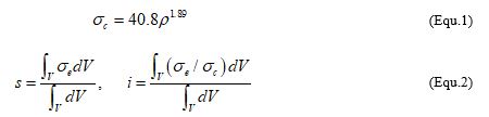
Loading boundary conditions were imposed based on patient-specific weight to take account of the individual condition. As shown in Fig. 4, the difference of the equivalent stress distribution on the coronal plane of the proximal femur between the healthy and ONFH cases is mainly presented in the region of the femoral neck. Ward′s triangle is a space localized at the femoral neck formed by the intersection of the compressive trabeculae groups and the tensile trabeculae groups defining a neutral axis where tensile and compressive forces balance each other. Ward′s triangle rarely bears stress in the normal condition [32]. In healthy cases, in the present FE study, high stress is transferred along with the principal compressive group. Some previous results displayed the similar stress patterns with the finite element results in the present study [58, 21, 31, 2, 20]. While as arrows indicated, in the ONFH cases, the high equivalent stress occurs at the central region of the femoral neck close to Ward triangle, where the distribution in ONFH may induce a potential risk of the bone collapse. After treatment, the non-collapse group (PI-IV) presents an obvious reduction of the stress near the Ward triangle indicating the trend of normalization after medical treatment. However, there is no noticeable change showed in the collapse group (PV and PVI), indicating the stress distribution in the area of Ward triangle may be potential to reveal the efficacy of medical treatment from the mechanical point of view. Furthermore, the mechanical parameters of the necrosis are investigated. High stress occurs in the posterior region of the necrosis that is consistent with the actual fracture region of the ONFH in clinical reports. However, the variation of the LDNV stress pre- and post- medical treatment does not show correspondence to the clinical follow-up results. A previous study investigated the stress and strength distribution on various sizes of necrotic areas by finite element models to predict the fate of the femoral head for early-stage osteonecrosis [54]. An analysis including 28 osteonecrotic femoral head specimens pointed out that the major fracture site appeared at the deep necrotic region of the interface between the necrosis and the healthy proximal femur. The ratio of the equivalent stress and yield strength of the necrosis was calculated. The results exhibited that the site of fracture coincided with the region of the ratio greater than the physiological level (0.10 or less they pointed) in the finite element model study. Thus, the mechanical properties of the necrosis can be represented by the stress and strength of the region. According to published studies, the compressive strength of cancellous femoral bone in the axial load direction is dependent on local density [52, 28, 29]. Thus, the compressive strength of the LDNV is calculated according to the functional relations. In the current study, based on the equivalent stress and compressive strength, averaged equivalent stress, as well as the averaged stress index in the LDNV, were calculated and compared. By calculating the ratio (RSI) between the averaged stress index and equivalent stress, results show that the ratio in most cases of the non-collapse group reduces after treatment; while it increases in the collapse group. In fact, a smaller value of the RSI indicates higher compressive strength, which is directly related to the density of the material; on the other hand, larger RSI indicates smaller compressive strength and thus may be related to the risk of bone collapse.
Morphological results confirm the lack of capability of isolated morphological analysis in evaluating the effectiveness of the medical treatment on ONFH. The finite element analysis seems to be a good non-invasive tool to model the mechanical properties of the femoral and to provide a quantitative evaluation of the load distribution over the proximal femur. Therefore, it has the potential to predict bone collapse risk and to estimate the treatment effects. The currently study preliminarily proposed the relative stress index (RSI) and the distribution of equivalent stress at the Ward triangle as the mechanical parameters that are possible to evaluate medical treatment results.
It should be noted that this study is based on limited patient cases which is due to the rareness of such cases who merely treated by pharmacotherapy without any intervention. To achieve the predictability of such parameter, the proposed RSI, it needs to be validated by larger patient datasets. Further confirmations of the predictive ability of these parameters will be conducted in the near future.