Neuron migration process was perturbed and DG neuron distribution was disordered after HIE, and SPC alleviated these injuries
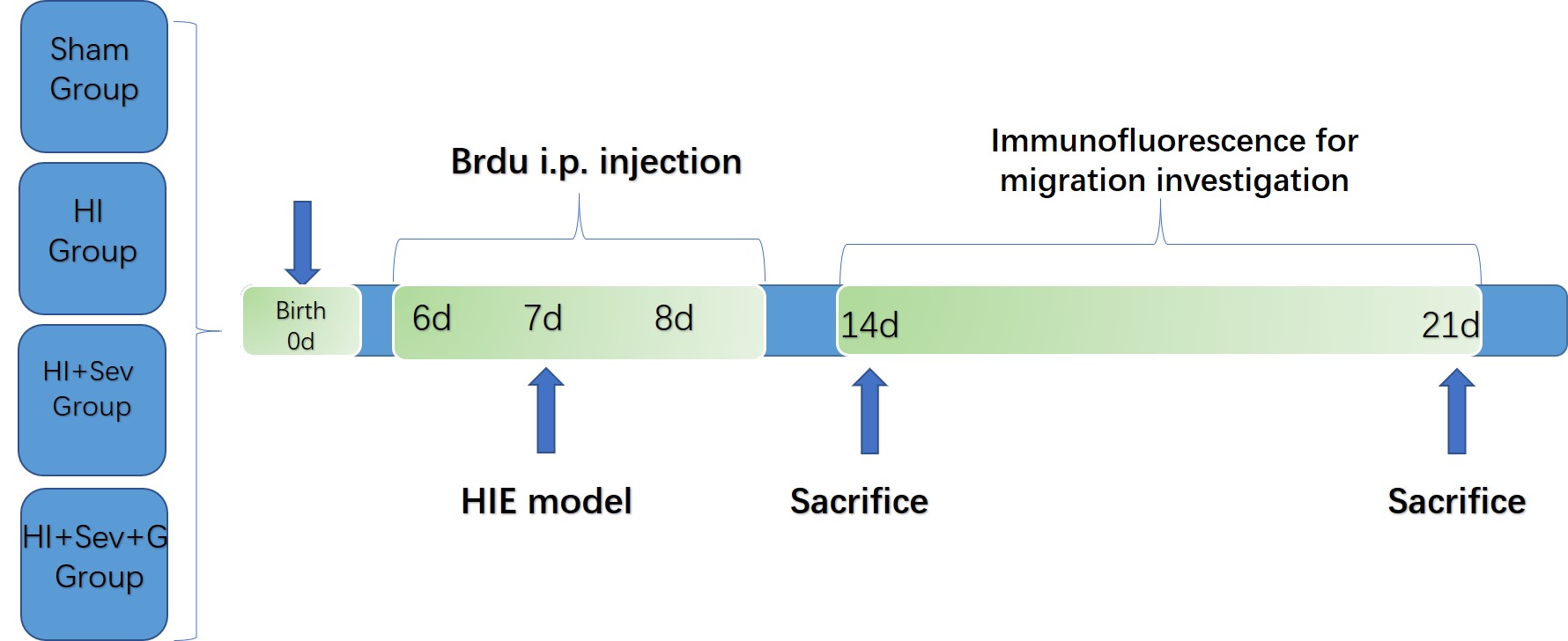
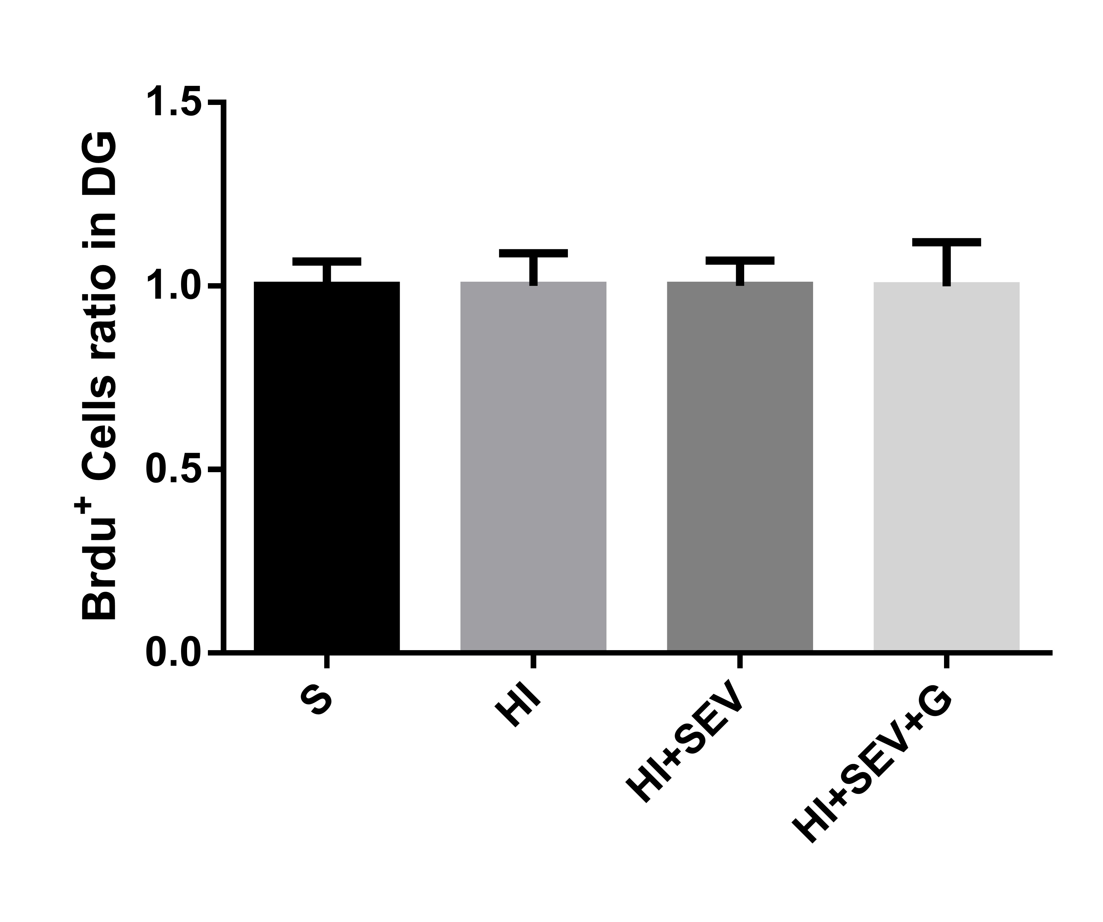
To better visualize neuron migration from the SGZ to the superficial GCL, the BrdU pulse labeling protocol was used (Supplementary Figure S1). Initially, the migration of the pulse-labeled cells to the destination was detected and the ratio of BrdU/neuron cells to the total BrdU-positive cells in each layer was calculated (Fig. 1A, PND14; 1B, PND21). The ratio of BrdU+/neuron + cells in the SGZ layer to BrdU + cells in the whole layer was enhanced in the HI group as compared to that in the Sham group on PND14 and PND21. This change in the ratio indicated that a large number of labeled neurons were restricted to the SGZ rather than migrating outward (Fig. 1C, D, HI versus Sham, p < 0.05 and p < 0.05 on PND14 and PND21, respectively). However, the ratio of BrdU+/neuron + cells in the GCL to the total BrdU + cells in the whole layer was decreased as compared to that in the Sham group (Fig. 1C,D, HI versus Sham, p < 0.01 and p < 0.01 on PND14 and PND21, respectively). To exclude the interference of reactive neurogenesis in BrdU + cells after HIE, the total BrdU + cells were counted, and the data showed no significant difference among all groups (Supplementary Fig. 2). SPC decreased the elevated number of neuronal cells restricted in the SGZ zone and increased the ratio in the GCL (Fig. 1C, D, HI + Sev versus HI, p < 0.05, p < 0.05; Fig. 1C, D, HI + Sev versus HI, p < 0.05, p < 0.01 on PND14 and PND21, respectively).
Nissl staining was conducted on PND14 to study the distribution of hippocampal DG neurons (Fig. 1E). As compared to the Sham group, the HI group showed decreased neuron ratio with rare cytoplasm and few Nissl bodies and disorganized neurons without polarity in DG (Fig. 1F, HI versus Sham, p < 0.001). SPC alleviated neuron loss and promoted reorganization of the neurons with proper orientation (Fig. 1F, HI + Sev versus HI, p < 0.01).
Reelin expression was enhanced after HIE
Here, we investigate the link between Reelin expression and SPC protective effect. Our data showed that rats in the HI group persistently expressed more Reelin-positive cells than those in the Sham group on PND8, PND14, and PND21 (Fig. 2A-F, HI versus Sham, p < 0.05, p < 0.01, and p < 0.01 on PND8, PND14, and PND21, respectively). SPC significantly downregulated Reelin expression (Fig. 2A-F HI + Sev versus HI, p < 0.05, p < 0.01, and p < 0.01 on PND8, PND14, and PND21, respectively).
SPC shows protective effect by repairing the distribution of DG neurons and establishing neuronal migration by suppressing Reelin function.
Piceatannol, which effectively inhibits the activity of the Reelin proteolytic enzyme (ADAMTS-4/5), was used to test the protective effect of SPC on the HIE target Reelin. To verify the Reelin/Dab1 intercellular combined reactions that may activate the downstream intracellular reactions, immunofluorescence staining of co-expression of Reelin and Dab1 was conducted. The co-expression of Reelin and Dab1 was significantly enhanced after HIE, and SPC significantly inhibited the elevated expression (Fig. 3A-F, HI versus Sham, p < 0.01, p < 0.001, and p < 0.001; HI + Sev versus HI, p < 0.01, p < 0.05, and p < 0.01 on PND8, PND14, and PND21, respectively). The beneficial effects of SPC were blocked by Piceatannol (Fig. 3A-F HI + Sev + G versus HI + Sev, p < 0.01, p < 0.01, p < 0.05 on PND8, PND14, and PND21, respectively)
Consistent with the above findings, there were more BrdU/Neun-labeled cells in the SGZ and GCL zones of rats of the HI group than those in rats of the Sham group (Fig. 4C, D, HI versus Sham, p < 0.01, p < 0.05 in SGZ; p < 0.05, p < 0.01 in GCL on PND14 and PND21, respectively). This finding indicated that a large amount of neurons were restricted to the basal zone of DG, and this high ratio was decreased by SPC (Fig. 4C, D, HI versus Sham, p < 0.05, p < 0.05 in SGZ; p < 0.01, p < 0.01 in GCL on PND14 and PND21, respectively). Piceatannol blocked the effect of SPC in the SGZ and GCL (Fig. 4C, D, HI + Sev + G versus HI + Sev, p < 0.05, p < 0.05 in SGZ; p < 0.01, p < 0.001 in GCL on PND14 and PND21, respectively).
Nissl staining was performed on PND14 to study the role of Reelin in the distribution of hippocampal DG neurons (Fig. 5A). The finding was consistent with the previous result that the HI group showed decreased neuron ratio with rare cytoplasm and few Nissl bodies, as well as disorganized neurons without polarity in DG as compared to that in the Sham group (Fig. 5B, HI versus Sham, p < 0.0001). SPC alleviated neuron loss and promoted the reorganization of neurons with proper orientation (Fig. 5B, HI + Sev versus HI, p < 0.01). The beneficial effect of SPC was, however, blocked by Piceatannol (Fig. 5B, HI + Sev + G versus HI + Sev, p < 0.05)
SPC attenuated DG neuron migration disorder in HIE by Reelin/Dab1 Cascade and inhibition of Reelin cleavage blocked this effect
The staining data indicate that HIE may result in disorder of neuron migration and activation of intracellular Dab1 expression by enhanced Reelin expression. Here, we tested the role of the canonical Reelin downstream signaling pathway involving Dab1, GSK-3β, and the microtubule-associated protein Tau in HIE-induced neuron migration disorder. The HI group showed significant upregulation of Reelin expression in 388 kDa fragment, rather than 180 kDa and phosphorylation/activation of Dab1 (Tyr-198), GSK-3β (Ser9), and Tau (Ser396) as compared to that in the Sham group(Fig. 6A-D, HI versus Sham, p < 0.001, p < 0.01, p < 0.01, p < 0.05, respectively). These reductions were significantly attenuated by SPC (Fig. 6A-D, HI + Sev versus HI, p < 0.001, p < 0.05, p < 0.05, p < 0.01, respectively). Piceatannol partially blocked the effect of SPC (Fig. 6A-D, HI + Sev + G versus HI + Sev, p < 0.05, p < 0.01, p < 0.05, p < 0.05, respectively).
SPC promotes hippocampal spatial learning and memory in HIE rats
To exclude anxiety and locomotor ability interference produced by several treatments, we conducted an open field test experiment on PND28. However, there were no difference in the test results among the four groups (Supplementary Figure S3).
In the MWM test, no significant difference in swimming velocities was observed among the four groups (Fig. 7A, p > 0.05 among all groups). Rats in the HI group showed significantly increased escape latency as compared to rats in the Sham group (Fig. 7B, HI versus Sham, p < 0.001, p < 0.001, p < 0.001, and p < 0.01 on the 2nd, 3rd, 4th, and 5th day, respectively). SPC attenuated this phenomenon (Fig. 7B, HI + Sev versus HI, p < 0.01, p < 0.001, and p < 0.01 on the 3rd, 4th, and 5th day, respectively). Rats in the HI + Sev + G group treated with Piceatannol needed more time to find the platform than rats who were administered SPC alone (Fig. 7B, HI + Sev + G versus HI + Sev, p < 0.01 and p < 0.05 on the 4th and 5th day, respectively). In the spatial probe test, the HI group rats showed crossed the platform less number of times than the Sham group rats (Fig. 7C, HI versus Sham, p < 0.01). Rats in the HI + Sev group crossed the platform a larger number of times than rats in the HI group (Fig. 7C, HI + Sev versus HI, p < 0.05). Piceatannol notably blocked the protective effect of SPC on spatial learning ability (Fig. 7C, HI + Sev + G versus HI + Sev, p < 0.05).
SPC facilitates DG-dependent spatial learning and memory in HIE rats
The 8-arm radial maze test was conducted in rats of the four groups from PND28 to PND34, and the path parameters were presented (Fig. 8A). The distance traveled during the reward seeking process was increased in the HI group, but significantly decreased in the HI + Sev group. Rats in the HI + Sev + G group traveled more distance to find the rewards than rats in the HI + Sev group (Fig. 8B, HI versus Sham, p < 0.001, p < 0.0001; HI + Sev versus HI, p < 0.05, p < 0.05; HI + Sev + G versus HI + Sev, p < 0.05, p < 0.05). SPC abolished the increased RME of the HI group as compared to that of the Sham group (Fig. 8C, HI versus Sham, p < 0.001; HI + Sev versus HI, p < 0.05), and Piceatannol blocked this beneficial effect (Fig. 8C, HI + Sev + G versus HI + Sev, p < 0.05). Interestingly, WME, which represents short-term memory, showed no difference among the four groups (Fig. 8D, p > 0.05 compared with all groups). This indicated that the specific hippocampal region that controlled short-term memory production might not be influenced by HIE or SPC.