Necessity for subsequent surgery in women of child-bearing age with positive margins after cold knife conization: A retrospective cohort study

DOI: https://doi.org/10.21203/rs.3.rs-199393/v1

Abstract

Background: Risk factors for positive margins and residual lesions after cold knife conization (CKC) for high-grade cervical intraepithelial neoplasias (CIN) were assessed in women of child-bearing age. A design for postoperative management and avoiding these situations is offered.

Methods: This was a retrospective study on 1,309 premenopausal women with high-grade CIN (including CIN3 and CIN2) based on a cervical biopsy under colposcopy used to diagnose a positive or negative margin. Age, gravidity, parity, HPV species, cytology, transformation zone type, results of endocervical curettage (ECC), quadrant involvement, glandular involvement, and CIN grade were analyzed. Among those with positive margins, 245 underwent surgery within three months, including CKC, a loop electrosurgical excision procedure, and total hysterectomy. Residual lesions were also assessed.

Results: There was no significant difference in age, gravidity, parity, glandular involvement, and CIN grade between the two groups (P>0.3). There was a significant difference in HPV species, cytology, ECC results, and quadrant involvement (P<0.002). Multivariate analysis showed a major cytology abnormality, high-risk HPV infection, type III transformation zone, positive ECC result, and multiple quadrant involvement were independent risk factors for positive margins and residual lesions (P<0.02). Age >35 years was also a risk factor (P<0.03).

Conclusion: High-risk women should be treated appropriately considering fertility. Patients with positive margins should be managed uniquely. Surgery for women without fertility may be appropriate. Close follow-up is necessary for women who have fertility requirements or are unwilling to undergo subsequent surgery if they have no risk factors, especially for women <35 years.

Background

In 2015, the American Society of Colposcopy and Cervical Pathology (ASCCP) recommended that one of the standard treatment options for cervical squamous intraepithelial lesions (SILs), especially high grade SILs [HSILs, including cervical intraepithelial neoplasia 3 (CIN3) and part of CIN2], was cervical conization,[1] including the loop electrosurgical excisional procedure (LEEP) and cold knife conization (CKC). However, these two types of conization have a common limitation; that is, 20–25% of women have residual lesions after an operation.[2] The state of the margin is generally considered to be a risk factor for recurrence or persistent CIN.[3, 4] A comprehensive meta-analysis by Ghaem-Maghami revealed that the rate of positive margins after conization was about 23.0% (8,091/35,109).[5] With improvement of people’s awareness of health care and advances in detection technology, the age of onset for this disease is becoming increasingly younger. Furthermore, due to changes in China’s family planning policy in recent years, an increasing number of women of child-bearing age have fertility requirements. To explore the risk factors of positive margins and residual lesions of high-grade CIN in the women of child-bearing age, the clinicopathological data of 1,309 women of child-bearing age with high-grade CIN (including CIN3 and CIN2) and 245 cases with positive margins who underwent subsequent surgery within three months were retrospectively analyzed. The results of our study can be used to guide the postoperative management of this group of women.

Material And Methods

Clinical data

Case inclusion criteria were women of child-bearing age who had been diagnosed with HSIL by biopsy, including CIN3 and part of CIN2. All women were premenopausal women. Case exclusion criteria were women with complications, such as endometrial carcinomas; cervical cancer including micro-invasion; incomplete information, such as lacking correlations among cytology, biopsy, and colposcopic findings; and no contact information.

A total of 1,309 women of child-bearing age with HSIL (including CIN3 and part of CIN2) in Tianjin Central Hospital of Gynecology and Obstetrics from January 2013 to December 2019, aged from 20 to 49 years old, were diagnosed with a "three-step" method, including cytology, colposcopy, and histology. [6] All women were premenopausal women. Among them, 245 women underwent subsequent surgery within three months. Age, gravidity, parity, HPV species, cytology, transformation zone type, the results of endocervical curettage (ECC), quadrant involvement, glandular involvement, and CIN grade were recorded. The data were collected from the electronic medical records of the institution while preserving patient anonymity. The research ethics committee waived the requirement for ethical approval and informed consent because the study used previously stored data.

Criteria for positive margins and residual lesions

If HSILs were found in the resection margin of about 1 mm or less, including the ectocervical margin, endocervical margin, or both, it was regarded as a positive margin. If HSILs were diagnosed in women who underwent secondary surgery within three months, it was assumed to be a residual lesion. CIN1 was not considered to be a residual lesion in this study.

Statistical methods

SPSS21.0 software was used for statistical analysis (SPSS Inc, Chicago, IL, USA). The statistical methods were examination of exact probabilities in a fourfold table and chi-squared tests. Multivariate analysis was performed using a logistic regression model. All tests were two-sided, and the level of significance was set at P < 0.05.

Results

The average age of women was 38 ± 7.1 (range 20–49) years. According to the state of the margin of specimens, they were divided into two groups: 1) positive group: women with positive margins (321, 24.52%); and 2) negative group: women with negative margins (988,75.48%).

Univariate analysis of risk factors for positive margins

Women in both groups were divided into two levels, depending on age gravidity, parity, HPV species, cytology, transformation zone type, the results of ECC, quadrant involvement, glandular involvement, and CIN grade. There was no significant difference in age, gravidity, and parity between the two groups (P > 0.3). The high-risk HPV infection rate in the positive group was significantly higher than in the negative group (P < 0.001). The preoperative cytology results in the positive group were mainly HSILs and atypical squamous cells. High-grade squamous intraepithelial lesions (ASC-H) were not excluded. These results were significantly different from that in the negative group (P < 0.001). There was a significant difference in the transformation zone type between the two groups (P < 0.001). There was also a significant difference in the results of ECC between the two groups (P = 0.002). There was no significant difference in the percentage of women with glandular involvement and CIN grade between the two groups (P > 0.3). Multiple quadrant involvement was more frequently found in the positive margin group, which was significantly different from that in the negative group (P < 0.001; Table 1).

Logistic analysis of the risk factors for positive margins

The logistic multivariate regression method was used to analyze the risk factors for women with positive margins. Positive margins were the dependent variable of the regression equation, and the severity of cytology, high-risk HPV infection, transformation zone type, ECC result, and quadrant involvement were the independent variables. We found that a major cytology abnormality, high-risk HPV infection, type III transformation zone, positive ECC result, and multiple quadrant involvement were risk factors for the positive margins (P < 0.02; Table 2).

Univariate and multivariate analyses of risk factors for residual lesions in women with positive margins

In women with positive margins, 245 cases underwent subsequent surgery within three months, including secondary CKC, LEEP, and total hysterectomy. Eighty-three cases had residual lesions detected in the postoperative specimens. Univariate analysis showed that age > 35 years, the severity of cytology results, high-risk infection, type of transformation zone, the ECC result, and quadrant involvement were associated with residual lesions of the women with positive margins after CKC (P < 0.05). In a multivariate analysis, a major cytology abnormality (including HSIL and ASC-H), high-risk HPV infection, type III transformation zone, positive ECC result, and multiple quadrant involvement were all risk factors for residual lesions (P < 0.05; Table 3).


Discussion

Cervical conization is the preferred method for the diagnosis and treatment of CIN. CKC is one type of cervical conization. Because CIN lesions are often multipoint in distribution, residual lesions and positive margins are inevitable. Studies have indicated that the incidence of residual lesions in women with positive margins after cervical conization was higher than that in women with negative margins, and the recurrence rate of women with positive margins was also higher in women with positive margins.[7, 8] It has been reported in the literature that the incidence of positive margins after cervical conization was 14% ~ 25% ,[2, 7, 8] which was similar to our study. However, the rate of residual lesions (33.38%) in our study was higher than in a previous study.[9, 10] This may be due to the clinical characteristics of the population who chose subsequent surgeries. In recent years, the onset age of this disease has been increasingly younger. However, there is no unified clinical opinion on the risk factors and further treatment for the women of child-bearing age with positive margins. In women with no fertility requirements, considering the possibility of a poor prognosis, total hysterectomy may be possible. However, for women of child-bearing age who have fertility requirements or want to retain the uterus, it is almost impossible to accept the uterus being removed. Even secondary conization can affect conception and lead to adverse pregnancy outcomes.[11, 12] Therefore, avoiding a positive margin and residual lesions, and reducing unnecessary secondary surgery are particularly important for women of childbearing age.

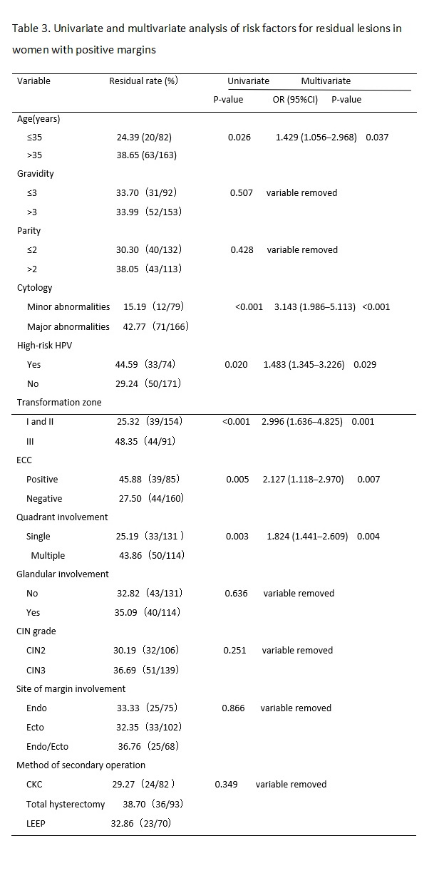
The results of previous studies have suggested that the possible risk factors for residual lesions after cervical conization mainly include positive margins of the specimen, involvement of the lateral margin of the cervical canal, involvement of the lateral margin of the cervical canal and outer orifice, positive specimen from ECC, menopause, persistent infection of high-risk HPV after cervical conization, and decreased or suppressed immune function.[13, 14] Many factors affect the condition of the margin after CKC, including age, menopausal status, glandular involvement, smoking, infection, training level of the operator, and other factors.[15, 16] In this study, we retrospectively analyzed the clinicopathological characteristics of 1,309 women of child-bearing age after initial CKC and 245 cases of women with positive margins who accepted subsequent surgery within three months. We found that a major cytology abnormality (including HSILs and ASC-Hs), high-risk HPV infection type III transformation zone, positive ECC result, and multiple quadrant involvement were the common risk factors for positive margins and residual lesions. Age > 35 years was also a risk factor for residual lesions in women with positive margins after initial CKC.

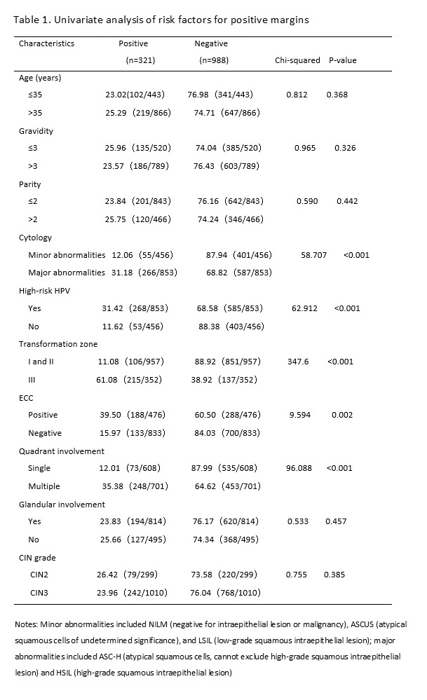
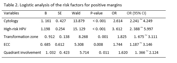
Whether the severity of a cytological abnormality is related to positive margins has been long disputed by researchers. Ryu A reported that the cytologic grade before cervical conization was not a risk factor for residual disease or recurrence. [17] However, Ayhan showed that the result of a smear was an advantageous predictor for a positive ectocervical margin, and it was associated with a decrease in the occurrence rate of positive margins and residual lesions.[18] In this study, the women with major cytology abnormalities (including ASC-Hs and HSILs) were more likely to present with positive margins and residual lesions than women with minor cytology abnormalities (including NILM, ASCUS, and LSIL) before CKC. We found that the severity of cytology before conization was a risk factor for positive margins and residual lesions. With an increase in the cytological grade, CIN levels increased, which meant that the depth of the lesion cells occupying the squamous epithelium increased, and the possibility of positive margins and residual lesions increased.

High-risk HPV infection has been recognized as a necessary condition for the occurrence and development of cervical squamous epithelial lesions and cervical cancer.[19] In this study, the rate of high-risk HPV infection in women with positive margins after CKC was 83.50% (268/321). Multivariate analysis showed that a high-risk HPV infection was the independent risk factor for positive margins and residual lesions, which was consistent with a previous study.[20, 21] However, at present, the pathological mechanism for a high rate of positive margins and residual lesions after cervical conization caused by high-risk HPV infection is not very clear; it may due to the cervical lesion area in women with CIN infected by high-risk HPV was larger and deeper than that of women infected by low-risk HPV.[22] Further study is needed. Based on the above studies, the HPV type can better predict postoperative positive margins and residual lesions.

In addition, we also found that women with a type III transformation zone were more likely to have positive margins and residual lesions after an operation. This may be due to the lesion invading the cervical tube. CKC cannot completely remove diseased tissue. In the same way, the rates for positive margins and residual lesions in women with positive results of ECC were higher than that of negative results. The same results were found in previous studies.[23, 24] Researchers also found that multiple quadrant involvement was a risk factor for positive margins and residual lesions.[25, 26] We came to the same conclusion based on our study results. In this study, the rate of multiple quadrant involvement was 77.26% in the women with positive margins, and that in the women with negative margins was 45.85%. Increases in the range of lesions likely affected observations during the operation, interfered with the judgment of surgical margins, and increased the difficulty of surgery. The above factors should be taken into account in the process of cervical conization to achieve the goal of leaving no residual lesions and preserving cervical function to a greater extent; that is, we should pay more attention to the extent and depth of lesions removed by cervical conization.

There was no significant difference between women of age > 35 years and ≤ 35 years in the rate of positive margins, but there was a significant difference in the rate of residual lesions between the two age groups. The rate of residual lesions in the age > 35 years group was higher than the ≤ 35 years group (P < 0.03). This may be due to the persistent infection of HPV, especially high-risk HPV. Sarian reported that women older than 35 years had a significantly higher risk for persistent infection following LEEP.[27] This could cause multiple quadrant lesions of the cervix.(30) Furthermore, the older the patient, the higher the degree of cervical atrophy. The cervical transformation zone and lesions therefore moved inward to the cervical canal, so cervical conization could not completely remove the diseased tissue. Bilibio also considered that increasing age was the only factor that accurately predicted residual disease.(18) All of these results indicated older age was a predictive factor for residual lesions and it can play a better role in guiding the formulation of a postoperative treatment plan for women of child-bearing age with positive margins. Subsequently, we can preserve normal reproductive function and organ integrity as much as possible in women younger than 35 years old.

Conclusion

A major cytology abnormality (including HSILs and ASC-Hs), high-risk HPV infection, type III transformation zone, positive ECC result, and multiple quadrant involvement were the common risk factors for positive margins and residual lesions. Age > 35 years was also a risk factor for residual lesions in women with positive margins after initial CKC. Experienced doctors should treat these high-risk women appropriately, operate prudently, and expand the scope of the operation while considering the patient’s fertility requirements. Furthermore, the patient with positive margins after CKC should be managed individually. It is reasonable to require further surgical treatment in women without fertility requirement, and close follow-up is necessary for women who have fertility requirements or are unwilling to undergo subsequent surgery if they have no risk factor, especially for women < 35 years.

Abbreviations

HPV: human papillomavirus

CIN: cervical intraepithelial neoplasia

CKC: cold knife conization

ECC: endocervical curettage

TCT: thin prep cytologic test

HSIL: high grade cervical intraepithelial lesions

LEEP: loop electrosurgical excisional procedure

NILM: negative for intraepithelial lesion or malignancy

LSIL: low grade cervical intraepithelial lesions

HSIL: high grade cervical intraepithelial lesions

ASCUS: atypical squamous cells of undetermined significance

ASC-H: atypical squamous cells, cannot exclude high-grade squamous intraepithelial lesion

Declarations

Ethical approval

This retrospective analysis was exempt from ethics committee approval at Tianjin Medical University, Tianjin Central Hospital of Gynecology and Obstetrics, because the committee did not consider approval was necessary for a retrospective chart review.

Informed Consent

The data were collected through the electronic medical records of the institution while preserving patient anonymity, and the research ethics committee waived the requirement for informed consent because the study used previously stored data.

Consent to publish

Not applicable.

 Availability of data and materials

All data generated or analysed during this study are included in this published article [and its supplementary information files].

Competing interests

The authors declare that they have no competing interests.

Conflict of Interest 

All authors declare that: (i) no support, financial or otherwise, has been received from any organization that may have an interest in the submitted work ; and (ii) there are no other relationships or activities that could appear to have influenced the submitted work.

Funding information 

The project name: Establishment and clinical application of early diagnosis system for cervical precancerous lesions caused by HPV E6 and E7

Contract no.: 19YFZCSY00600

Author contribution

Xinmei Wang:Project development, Data Collection, Manuscript writing.

Juan Xu:Data collection and Data analysis.

Yang Gao:Data collection

Pengpeng Qu: Project development.

All authors have read and approved the final manuscript.Acknowledgement

We thank all participants in the study for their invaluable contribution.

References

  1. Darragh TM, Colgan TJ, Cox JT et al (2012) The lower anogenital squamous terminology standardization project for HPV-associated lesions: background and consensus recommendations from the College of American Pathologists and the American Society for Colposcopy and Cervical Pathology. J Low Genit Tract Dis 16:205–242.
  2. Arbyn M, Redman CWE, Verdoodt F et al (2017) Incomplete excision of cervical precancer as a predictor of treatment failure: a systematic review and meta-analysis. Lancet Oncol 18:1665–1679.
  3. Kietpeerakool C, Khunamornpong S, Srisomboon J, Siriaunkgul S, Suprasert P (2007) Cervical intraepithelial neoplasia II–III with endocervical cone margin involvement after cervical loop conization: is there any predictor for residual disease? J Obstet Gynaecol Res 33:660–664.
  4. Park JY, Lee SM, Yoo CW, Kang S, Park SY, Seo SS (2007) Risk factors predicting residual disease in subsequent hysterectomy following conization for cervical intraepithelial neoplasia (CIN) III and microinvasive cervical cancer. Gynecol Oncol 107:39–44.
  5. Ghaem-Maghami S, Sagi S, Majeed G, Soutter WP (2007) Incomplete excision of cervical intraepithelial neoplasia and risk of treatment failure: a meta-analysis. Lancet Oncol 8:985–993.
  6. Coccia PF, Pappo AS, Beaupin L et al (2018) Adolescent and Young Adult Oncology, Version 2.2018, NCCN Clinical Practice Guidelines in Oncology. J Natl Compr Canc Netw 16:66–97.
  7. Fu Y, Chen C, Feng S et al (2015) Residual disease and risk factors in women with high-grade cervical intraepithelial neoplasia and positive margins after initial conization. Ther Clin Risk Manag 11:851–856.
  8. Chen JY, Wang ZL, Wang ZY, Yang XS (2018) The risk factors of residual lesions and recurrence of the high-grade cervical intraepithelial lesions (HSIL) women with positive-margin after conization. Medicine (Baltimore) 97:e12792.
  9. Kocken M, Helmerhorst TJ, Berkhof J et al (2011) Risk of recurrent high-grade cervical intraepithelial neoplasia after successful treatment: a long-term multi-cohort study. Lancet Oncol 12:441–450.
  10. Serati M, Siesta G, Carollo S et al (2012) Risk factors for cervical intraepithelial neoplasia recurrence after conization: a 10-year study. Eur J Obstet Gynecol Reprod Biol 165:86–90.
  11. Pils S, Eppel W, Seemann R, Natter C, Ott J (2014) Sequential cervical length screening in pregnancies after loop excision of the transformation zone conization: a retrospective analysis. Brit J Obstet Gynaecol 121:457–462.
  12. Sozen H, Namazov A, Cakir S et al (2014) Pregnancy outcomes after cold knife conization related to excised cone dimensions. A retrospective cohort study. J Reprod Med 59:81–86.
  13. Ruano Y, Torrents M, Ferrer FJ (2015) Human papillomavirus combined with cytology and margin status identifies women at risk for recurrence after conization for high-grade cervical intraepithelial neoplasia. Eur J Gynaecol Oncol 36:245–251.
  14. 20. Dos SMPP, Duarte G, Quintana SM (2016) Multivariate analysis of risk factors for the persistence of high-grade squamous intraepithelial lesions following loop electrosurgical excision procedure. Int J Gynaecol Obstet 133:234-237.
  15. Bilibio JP, Monego HI, Binda MLA, Dos Reis R (2019) Menopausal status is associated with a high risk for residual disease after cervical conization with positive margins. PLoS One 14:e0217562.
  16. Montanari E, Grimm C, Schwameis R et al (2018) Influence of training level on cervical cone size and resection margin status at conization: a retrospective study. Arch Gynecol Obstet 297:1517–1523.
  17. Ryu A, Nam K, Kwak J, Kim J, Jeon S (2012) Early human papillomavirus testing predicts residual/recurrent disease after LEEP. J Gynecol Oncol 23:217–225.
  18. Ayhan A, Boynukalin FK, Guven S, Dogan NU, Esinler I, Usubutun A (2009) Repeat LEEP conization in women with cervical intraepithelial neoplasia grade3 and positive ectocervical margins. Int J Gynaecol Obstet 105:14–17.
  19. Wentzensen N (2016) Epidemiology, prevention and early detection of cervical cancer. Der Onkologe 22:725–736.
  20. Tasci T, Turan T, Ureyen I et al (2015) Is there any predictor for residual disease after cervical conization with positive surgical margins for hsil or microinvasive cervical cancer. J Low Genit Tract Dis 19:115–118.
  21. Dae KW, Chul JU, Mo KS (2016) A human papillomavirus (HPV)-16 or HPV-18 genotype is a reliable predictor of residual disease in a subsequent hysterectomy following a loop electrosurgical excision procedure for cervical intraepithelial Neoplasia 3. J Gynec Oncol 27:e2.
  22. Kyehyun N, Jeongja K, Kim J (2013) Human papillomavirus type 16 causes larger colposcopic lesions than other HPV types in women with grade 3 cervical intraepithelial neoplasia. American Society for Colposcopy and Cervical Pathology. J Low Genit Tract Dis 17:1–5.
  23. Diaz E, Aoyama C, Baquing M et al (2014) Predictors of residual carcinoma or carcinoma-in-situ at hysterectomy following cervical conization with positive margins. Gynecol Oncol 132:76–80.
  24. Kim H, Kim K, Mok J et al (2007) Pathologic risk factors for predicting residual disease in subsequent hysterectomy following LEEP conization. Gynecol Oncol 105:434–438.
  25. Tasci T, Turan T, Ureyen I et al (2015) Is there any predictor for residual disease after cervical conization with positive surgical margins for HSIL or microinvasive cervical cancer? J Low Genit Tract Dis 19:115–118.
  26. Hamontri S, Israngura N, Rochanawutanon M, Bullangpoti S, Tangtrakul S (2010) Predictive factors for residual disease in the uterine cervix after large loop excision of the transformation zone in women with cervical intraepithelial neoplasia III. J Med Assoc Thai 93Suppl 2:S74–80.
  27. Sarian LO, Derchain SF, Andrade LA, Tambascia J, Morais SS, Syrjanen KJ (2004) HPV DNA test and Pap smear in detection of residual and recurrent disease following loop electrosurgical excision procedure of high-grade cervical intraepithelial neoplasia. Gynecol Oncol 94:181–186.