Chemicals and reagents
The research was approved by the ethical committee of Kerman University of Medical Sciences with the Ethics Approval Code: IrKMU REC: 1397-135.
Dulbecco’s modified Eagle’s medium (DMEM) was purchased from Gibco (Grand Island, USA). Fetal bovine serum (FBS) and Penicillin streptomycin were purchased from Hangzhou Sijiqing Biological Engineering Materials Co. Ltd (Hangzhou, China). Mouse Leydig (TM3) cell line was purchased from Pasteur Institute (Tehran, Iran). Mouse Testosterone ELISA Kit was purchased from ZellBio GmbH/Germany. RNX plus solution (Cinnagen, Iran). cDNA synthesis kit and all reverse transcription (RT) substances were purchased from Yektatajhiz Company (Tehran, Iran). SYBR Green I Master was purchased from Genaxxon bioscience (Ulm, Germany). hCG was purchased from Merck/Germany. Thrombin was provided as a generous gift by Dr M. Farsinejad from department of Hematology, Faculty of Paramedicine, Kerman University of Medical Science, Kerman, Iran. Other chemicals used in this study were purchased from Sigma-Aldrich Chemical Company (St. Louis. MO. USA).
Cell culture and experimental groups
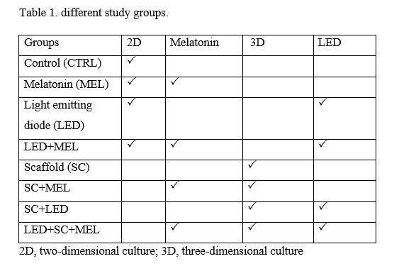
TM3 cells were cultured in DMEM-F12 supplemented with 10% FBS, 1% penicillin & streptomycin and 1% amphotericin (complete culture medium) at 37ºC with 5% CO2 in the atmosphere. After reaching 80% confluence, the cells were harvested by trypsinization and different treatments were enrolled according to the following groups (table 1).
2D culture
The harvested cells were cultured at 1×10⁴ in 96 well culture plates for 24 h in DMEM-F12 supplemented with 10% FBS and 1% antibiotics at 37ºC with 5% CO2 in the humidified atmosphere as 2D control group. Some wells received 10-7 M melatonin as MEL group.
Fibrin 3D culture
Ten μl of fibrinogen extracted from human plasma, 10 μl cell suspension (1×10⁴) in PBS and 10 μl thrombin were transferred into a 1.5 ml microtube and mixed thoroughly. The samples were then incubated in a 37ºC incubator with 5% CO2 in the air for 10 min. After fibrin clot formation, it was pushed out of microtube and transferred into 96 well culture plate (3D, SC group). 10-7 M melatonin was added to some wells 24 h later as SC+MEL group.
LED irradiation
A handmade LED device with green light emission was used in the LED exposure groups (LED, MEL+LED, SC+LED and LED+SC+MEL). Twenty-four hr later the medium was refreshed and the samples were once irradiated for 10 minutes at radiation energy of 1.5 J/cm2, and 530 nm wavelength 31. All the exposure procedures were carried out inside a 37ºC CO2 incubator. In the MEL+LED group the cells/scaffolds were treated with 10-7 M melatonin and were then irradiated as explained above.
TM3 proliferation assessment
MTT assay was used to estimate the proliferation of TM3 cells. Briefly, cell density was adjusted to 7500 cells per well/ fibrin scaffold in 96-well plates. After different treatments, the cells/scaffolds were incubated for 72 hr at 37 ºC under 5% CO2 in the air. Twenty µl MTT solution (5 mg/ml) was added into each well and incubation was continued for 4 hr. The supernatant was discarded afterward and 150 µl DMSO was added into each well. The absorbance of each well was measured at 490 nm against a reference wavelength of 630 nm. All the experiments were done in triplicates 32.
Cell viability assessment
3×10⁴ TM3 cells were cultured in the 24 well plates with 300 µl complete culture medium. After 72 hr of the different treatments, the supernatants containing floating cells were transferred into 1.5 ml microtubes. The attached cells were washed with PBS, detached by trypsin EDTA solution, and added to the microtubes with gently pipetting. Ten µl of the cell suspension was mixed with 10 µl of trypan blue, loaded onto a Neubauer chamber and the number of viable and dead cells were counted in 4 large square under an inverted microscope. This assessment was not applicable for the scaffold-containing groups.
The cell viability rate was assessed using the following formula: cell viability rate = number of viable cells/total cell number
Testosterone assessment
The level of testosterone in the supernatant was assessed using an ELISA kit was purchased from ZellBio GmbH (Germany). In brief, 3×10⁴ viable TM3 cells were cultured in 24 well plates with complete culture medium for 24 hr, and different treatments were applied according to the study groups. 48 hr later, 10 IU/ml hCG was added to each well, and 24 h later the supernatant was removed and stored at -20 ºC for testosterone measurement. The testosterone level was evaluated according to the protocol provided by the manufacturer 33.
RNA extraction and cDNA synthesis
Total RNA was isolated using RNX plus (1 ml) and chloroform (200 μl) solutions at room temperature. After centrifugation at 12000 rpm at 4°C for 15 min, the aqueous phase was replaced into a new microtube, cold isopropanol solution was added and was then gently inverted 10 times. The tubes were transferred to a -20 °C freezer overnight. The next day, after centrifugation at 12000 rpm (4°C) for 10 min, one ml 75% ethanol was rinsed on the pellet, the tubes were centrifuged at 7500 rpm (4°C) for 5 min, the ethanol was removed and the pellet was dissolved in diethyl pyrocarbonate (DEPC) water and stored at -20 °C. The concentration and purity of the isolated RNA were assessed by absorbance readings on a UV spectrophotometer (Thermo Scientific™ NanoDrop 2000) at the wavelengths of 260 and 280 nm to obtain a ratio of 1.81 ± 0.06. The RNA integrity was then determined by 1% agarose gel electrophoresis. The cDNA synthesis was performed using cDNA synthesis kit (Yektatajhiz azma, Tehran, Iran) according to the manufacturer’s protocol: 70 °C for 5 min (RNA denaturing), 42°C for 60 min (cDNA synthesis) and then 70 °C for 10 min (enzyme inactivation).
Quantitative real time PCR
Quantitative real time PCR was carried out to evaluate the mRNA expression level of PCNA, CYCLIND1, CDC2, CDKN1B, GATA4 and RORα genes in the TM3 cells by using magnetic induction cycler (mic) Real-time PCR system (Australia). For the amplification reactions, the synthetized cDNA solution (1 μl) was mixed with specific primers (1 μl) and SYBR Green I Master (10 μl) at a total volume of 20 μl based on the following PCR program (table 2). Primer sequences are described in Table 3. GAPDH was used as a housekeeping gene to normalize the qRT-PCR reactions. The 2−ΔΔCT method was used to measure gene expression levels.