Neoadjuvant chemoradiotherapy followed by minimally invasive esophagectomy for borderline resectable esophageal squamous cell carcinoma; A retrospective cohort study with short- and mid-term oncological outcomes

DOI: https://doi.org/10.21203/rs.3.rs-2950806/v1

Abstract

Background

The best treatment strategy for borderline resectable locally advanced esophageal squamous cell carcinoma (ESCC) is unknown. Although neoadjuvant chemoradiotherapy (NACRT) provides good local control, the subsequent surgery is complicated by edema or fibrosis, particularly in minimally invasive esophagectomy (MIE). This retrospective cohort study sought to assess the feasibility of NACRT followed by MIE for borderline resectable ESCC.

Methods

Consecutive patients who underwent NACRT (40Gy radiotherapy with chemotherapy of 5-FU and Cisplatin) followed by MIE with gastric tube reconstruction for locally advanced ESCC were included. Following a review of pretreatment CT by 2 independent physicians, clinically borderline resectable T3 (cT3br) tumors were chosen, with resectable (cT3r) and unresectable (cT4b) tumors excluded. The short- and mid-term oncological outcomes were assessed.

Results

Patients with 33 cT3br tumors were included. There was no postoperative mortality, and 11 and 7 patients had postoperative Clavien-Dindo Grade ≥ II and Grade ≥ III complications, respectively. R0 resection was achieved in 32 patients, with 4 patients showing pathological complete response. With a median follow-up of 675 days, 15 patients had relapsed, the majority of which included distant metastasis, and only one patient had a pure locoregional recurrence. Patients who experienced postoperative complications had significantly lower relapse-free survival (RFS) than those who did not (p = 0.049) (2-year RFS: 0% and 65%, respectively).

Conclusions

NACRT followed by MIE for cT3br tumors was feasible with adequate R0 resection rates. Although locoregional control was adequate, distant metastasis was poorly controlled. MIE with fewer postoperative complications would provide better oncological results.

Introduction

Esophageal cancers tend to invade adjacent organs such as the trachea, bronchus, and aorta, due to the lack of serosa in the esophagus and its location in a very narrow mediastinal space. Definitive chemoradiotherapy is the major treatment strategy for T4b esophageal squamous cell carcinoma (ESCC) with unresectable invasion to other vital organs [1], whereas neoadjuvant chemotherapy (NAC) followed by surgery is the standard treatment for resectable locally advanced ESCC in Japan [2, 3]. In between them, borderline resectable cases are clinically difficult to diagnose as either T3 or T4b, which makes deciding on a therapeutic strategy difficult. Neoadjuvant chemoradiotherapy (NACRT) is another promising treatment strategy for locally advanced esophageal cancer, with higher R0 resection rates, better local control, and a better prognosis than surgery alone [4, 5], and several studies have also demonstrated the feasibility of NACRT followed by esophagectomy for borderline resectable ESCC [68]. In the following surgery, however, the tissue edema or fibrosis where tumor invasion was suspected before radiotherapy would make adequate dissection technically challenging, so open esophagectomy has been chosen so far. Minimally invasive esophagectomy (MIE) has the advantage of better short-term and equivalent long-term oncological outcomes when compared to open esophagectomy [9, 10], but it is unclear whether MIE after NACRT is technically and oncologically feasible.

This study sought to examine the feasibility of NACRT followed by MIE for clinically borderline resectable locally advanced ESCC, by assessing the short- and mid-term oncological outcomes.

Patients and methods

Study design and patient selection (Fig. 1)

The cohort of this retrospective single-center study included patients who received NACRT for locally advanced ESCC between April 2011 and September 2022 at the Japanese Red Cross Osaka Hospital. Pretreatment clinical examinations included endoscopy, Computed Tomography (CT), upper gastrointestinal series, positron emission tomography, and/or magnetic resonance imaging. Patients who had a two-staged operation (n = 3), palliative resection for distant metastasis (n = 3), total pharyngolaryngoesophagectomy (n = 1), cervical esophageal resection (n = 1), and reconstruction with jejunal (n = 2) or colonic (n = 1) conduit, were exempted. A critical review of pretreatment CT was performed independently and blindly by two physicians, and clinically borderline resectable T3 (cT3br) tumors were included (n = 34), with resectable (cT3r, n = 8) and unresectable (cT4b, n = 3) tumors excluded. A patient whose tumor became unresectable due to disease progression during NACRT was exempted. Those patients who underwent NACRT followed by MIE with gastric tube reconstruction (n = 33) were included in the study.

Review process with pretreatment CT images

Pretreatment CT images were retrospectively reviewed by 2 physicians (HH and KS) independently to characterize the locally advanced ESCC into cT3r, cT3br, and cT4b tumors based on the circumferential location of the primary tumor by endoscopic examination. All clinical information except CT and endoscopy was blinded for this review process. A discussion was held to reach an agreement on the discordant diagnosis. The trachea, bronchus, aorta, pulmonary vein, and spine were among the organs with suspicious invasion. Diagnosis criteria of cT4b by CT images included the following; the presence of flattening and indentation of the membranous part of the trachea/bronchus and tracheal deformation, changes in the venous lumen with severe protrusion into the pulmonary vein, and contact of > 90° or loss of triangular fat space between the esophagus, aorta, and spine adjacent to the tumor [1113]. cT3br tumors were classified as those with a suspected invasion of adjacent organs but could not be definitively diagnosed as cT4b.

Neoadjuvant chemoradiotherapy and the following surgery

The Intensity Modulated Radiation Therapy technique was used for preoperative radiotherapy, and each patient received conventional fractionation of 2Gy/d 5 days per week for 4 weeks, for a total dose of 40 Gy. The gross tumor volume included the primary tumor as well as metastatic lymph nodes (LN) with possible invasion into other organs. Preoperative chemotherapy included cisplatin + 5-FU (CF) for 4 weeks. Each patient received 70mg/m2 cisplatin on day 1 and 700mg/m2 5-FU on days 1–4 intravenously. The chemotherapy was administered over 2 cycles in some cases. MIE was performed 4–8 weeks after radiotherapy was completed.

Surgical procedure

For the MIE with a thoracoscopic/robotic approach, the procedure has previously been described [1416]. All thoracoscopic/robotic esophagectomies (TE/RE) were performed using the same surgical principle in a semi-prone position. Briefly, using the surgical theory of mesenteric excision in upper mediastinal lymphadenectomy [1416], complete lymphadenectomy along the bilateral recurrent laryngeal nerves was conducted. The thoracic duct and its surrounding tissues were preserved in cases where there was no obvious tumor invasion. Cervical, total mediastinal, and upper abdominal lymphadenectomy were performed following the Japanese guidelines [17]. After mediastinal lymphadenectomy, the patient was placed in the supine position, and laparoscopic/robotic gastric mobilization, upper abdominal lymphadenectomy, conduit creation, cervical lymphadenectomy, and cervical anastomosis were performed. The da Vinci Xi surgical robot (Intuitive Surgical, Inc, Sunnyvale, CA) was used for RE.

Short- and Mid-term analysis

The hospital records were used to retrospectively retrieve the clinical characteristics of the patients including their age, sex, body mass index, Eastern Cooperative Oncology Group performance status (ECOG-PS) score, serum SCC antigen before treatment, tumor location, and clinical/pathological cancer stage. LNs with major diameters ≥ 5 mm were diagnosed as clinically positive LNs. The cancer stage was determined using the TNM classification of the American Joint Committee on Cancer (AJCC), 8th Edition [18]. Surgical complications were classified using the Clavien-Dindo system [19]. The treatment’s efficacy was assessed using the Response Evaluation Criteria in Solid Tumors guidelines [20]. The histological efficacy of chemoradiotherapy was graded as follows: Viable cancer cells account for ≥ 2/3 of the tumor tissue in Grade 1a; 1/3 to 2/3 of the tumor tissue in Grade 1b; ≤ 1/3 of the tumor tissue in Grade 2; and no viable cancer cells are visible in Grade 3 [21]. Residual tumor status was classified as follows: R0 represents no residual tumor, R1 represents microscopic residual tumor, and R2 represents macroscopic residual tumor [21].

Statistical analysis

The Mann–Whitney U test was used to evaluate continuous variables and to compare categorical variables, either Fisher’s exact test or the chi-squared test was used. All statistical analyses were carried out using STATA ver. 16.1 (STATA Corp, College Station, TX, USA). A p-value of less than 0.05 was deemed statistically significant. OS and RFS curves were created by the Kaplan–Meier method, which was then compared using the log-rank test.

Results

During the review process of pretreatment CT, one discordant case was discovered among 45 cases and was classified as cT3r after discussion. Following this review, 34 patients with cT3br tumors were chosen. Because one tumor became unresectable due to progressive disease with newly developed lung metastasis during NACRT, 33 patients who completed NACRT followed by MIE were included in the analysis. Figure 2 shows typical cT3br CT images in this study. Table 1 displays the clinical characteristics of the patients. The adjacent organs with suspected invasion included, the trachea/bronchus in 20 cases, followed by the aorta in 12, the spine in 2, and the pulmonary vein and subclavian artery in one each. Table 2 shows the short-term outcomes of NACRT followed by MIE. NACRT was completed in all cases without major complications. Seventy percent (23 out of 33) of patients received one cycle of chemotherapy. In terms of the MIE approach, TE was chosen in 20 cases, while RE was chosen in 13 cases. There was no conversion to open thoracotomy or postoperative mortality. Grade II or higher (Grade ≥ II) postoperative complications were observed in 11 (33%) cases and those of Grade III or higher (Grade ≥ III) in 7 (21%) cases. Table 2 shows the details of complications of Grade ≥ III, and the other Grade II complications included ileus (n = 1), cholecystitis (n = 1), pneumonia (n = 1), and surgical site infection (n = 1). Table 2also included pathological results. R0 resection was obtained in 32 (97%) of the patients. Regarding the histological pretreatment efficacy, 8 (24%) patients achieved Grade 3, and 4 (12%) patients achieved pathological complete response (pCR). 


Variables

n = 33

Table 1

Clinical features of the patients of clinically borderline resectable ESCC a

Age, median (range)

66 (47–80)

Sex, n, Male/Female

22/11

SCC antigen before treatment (ng/ml), median (range)

2.4 (0.5–21.3)

Body Mass Index, median (range)

19.5 (13.9–25.9)

ECOG Performance Status, n (0/1/2)

22/10/1

Tumor location (Ceb/Utb/Mtb/Ltb)

1/10/20/2

Clinical Nc, n (N0/1/2)

7/15/11

Clinical Stagec (II/III)

7/26

Adjacent organ with suspected invasiond


Trachea/Bronchus (invasion by MLNe)

20 (7)

Aorta (invasion by MLNe)

12 (1)

Spine

2

Pulmonary vein

1

Subclavian artery

1

a esophageal squamous cell carcinoma


b Ce; Cervical esophagus, Ut; Upper thoracic, Mt; Middle thoracic, Lt; Lower thoracic

c Determined by the 8th edition of the AJCC TNM classification


d contains duplicate organs, e metastatic lymph node


Variables

n = 33

Table 2

Short-term results of neoadjuvant chemoradiotherapy followed by minimally invasive surgery

Number of chemotherapy cycles, n, (1/2)

23/10

Response of neoadjuvant chemoradiotherapy


Non-target lesions (CRa/IR,SDa)

2/31

Target lesions (PRa/SDa)

4/3

Approach for esophagectomy, n, (Thoracoscopic/Robotic)

20/13

LN dissection, n, (2 field/3 field)

14/19

Reconstruction route, n, (Posterior mediastinal/Retrosternal/Intrathoracic)

20/12/1

Total operative time, min, median (range)

549 (412–846)

Thoracoscopic time, min, median (range)

232 (156–399)

Blood loss, g, median (range)

50 (0–680)

Hospital Stay, days, median (range)

23 (11–137)

Postoperative complicationb, ≥ Grade II, n (%)

11 (33)

Postoperative complicationb, ≥ Grade III, n (%)

7f (21)

Recurrent laryngeal nerve palsy

4

Tracheoesophageal fistula

2

Lymphorrea

1

Anastomotic leakage

1

Pneumonia

1

Pathological Tc, n (T0d/T1a/T1b/T2/T3)

8/1/1/3/20

Pathological Nc, n (N0/1/2/3)

8/17/6/2

Pathological Stagec (0e/IIA/IIB/IIIA/IIIB/IVa)

4/1/9/1/16/2

Residual tumor, n, (R0/1/2)

32/1/0

Pretreatment efficacy, Grade, n, (1a/1b/2/3)

9/5/11/8

Adjuvant chemotherapy, n, (None/S-1/5-FU + Cisplatin/Nivolumab)

15/7/6/5

a CR; Complete Response, PR; Partial Response, IR; Incomplete Response, SD; Stable Disease,

b by Clavien-Dindo classification, c Determined by the 8th edition of AJCC TNM classification

d No residual tumor cells, e pathological complete response, f contains duplicates,


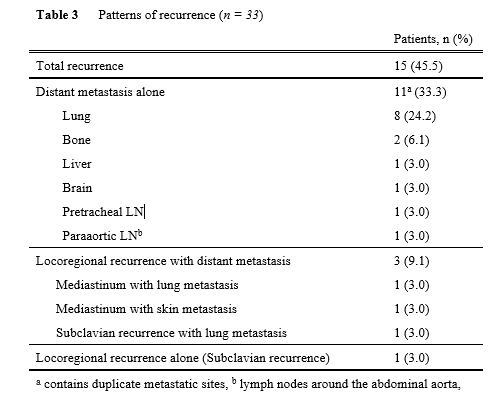
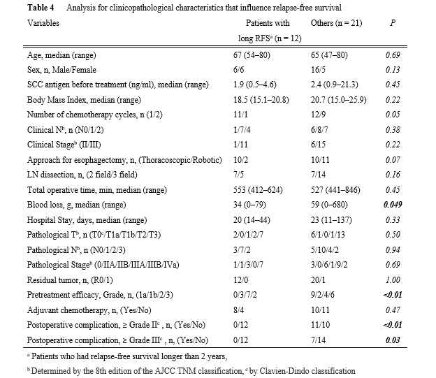
Next, mid-term oncological outcomes were assessed (Table 3 and Fig. 3). Figures 3a and b show the RFS and OS curves. The 2-year RFS/OS in all populations was 54%/73%, respectively. With a median follow-up of 675 days, recurrence was found in 15 cases (45.5%), with 14 (93%) recurring within 2 years (Table 3). Regarding the recurrence pattern, distant metastases alone were found in 11 cases (33.3%). Three cases (9.1%) had locoregional recurrences with distant metastasis, while only one case (3.0%) had locoregional recurrence alone. To determine which factors influenced relapse-free survival, the clinicopathological characteristics of 12 patients with RFS longer than 2 years (long RFS, n = 12) were compared to those of the other patients (n = 21) (Table 4). Long RFS patients had considerably better histological pretreatment efficacy than the others (p < 0.01). Interestingly, postoperative complications were significantly lower in patients with long RFS compared to other patients. Postoperative complications of Grade ≥ II or ≥ III were never observed in patients with long RFS, whereas those of Grade ≥ II or ≥ III were observed in 11 (52%), and 7 (33%), respectively, of the other patients. The significant difference was confirmed regardless of whether the complication was of Grade ≥ II (p < 0.01) or Grade ≥ III (p = 0.03) was selected for the analysis. Finally, the clinicopathological characteristics of patients with and without postoperative complications (Grade ≥ II) were examined (Supplementary Table 1). Male patients (p = 0.04), longer operation time (p = 0.03), and longer hospital stays (p < 0.01) were associated with postoperative complications. Figures 3c and d depict RFS and OS curves with and without postoperative complications (Grade ≥ II). RFS in patients with complications was significantly lower than in patients without complications (p = 0.049) (2-year RFS: 0% and 65%, respectively). There was no significant difference in OS (p = 0.35) (2-year OS: 65% and 74%, respectively).


Discussion

The current study demonstrated the feasibility of NACRT followed by MIE with an adequate R0 resection rate and acceptable local control as mid-term oncological outcomes for cT3br ESCC. Furthermore, patients who experienced postoperative complications had significantly lower RFS than those who did not. To the best of our knowledge, this is the first study to show that MIE after NACRT is feasible and that postoperative complications have a negative prognostic impact in patients with cT3br ESCC.

Surgery alone cannot control advanced esophageal cancer, and preoperative CF therapy was the standard treatment in Japan based on the findings of the JCOG9907 study [2]. Recently, the JCOG1109 study revealed a significant increase in survival in the preoperative docetaxel + cisplatin + 5-FU (DCF) therapy group compared to the preoperative CF group [3], and preoperative DCF therapy is now considered the new standard treatment for resectable ESCC in Japan [17].

However, when the tumor invades adjacent vital organs, definitive chemoradiotherapy is the main treatment option for T4b tumors [1], because combined resection of neighboring organs does not improve survival and has a high incidence of mortality and morbidity [24]. Some locally advanced tumors are difficult to diagnose as cT3 or cT4b and are commonly referred to as ‘borderline resectable ESCC’ [68]. The treatment strategy for these tumors has not been standardized, possibly due to the difficulty in determining whether it has adjacent organ invasion. Recently, attention has been drawn to the diagnosis of T classification in locally advanced ESCC [25], which revealed inter-observer variations in clinical diagnosis of the T category. The recently published Japanese Classification of Esophageal Cancer, 12th edition, categorized the clinical T category of locally advanced ESCC into cT3r, cT3br, and cT4b. The CT criteria for cT4b diagnosis were defined as, “flattening,” “deformity,” and “indentation” of the lumen of the trachea/bronchus, and “contact ≥ 110° for longer than 10 mm,” “contact of 90°–110° for longer than 20 mm,” or “encasement” for aortic invasion. We critically reviewed the pretreatment CT images using the diagnostic criteria described above [1113], following the Japanese classification. The review was conducted independently and blindly by the 2 reviewers to avoid the study’s ambiguity.

Although the standard treatment for cT3br tumors has not been established, the feasibility of NACRT followed by esophagectomy with the advantage of better local control for such tumors has been investigated [68]. The preferred chemotherapeutic regimen and the dose of radiotherapy were CF with 40Gy in general. R0 resection rates ranged from 44–85%, probably owing to variations in cT3br diagnosis. These studies used open esophagectomy, and there have been no studies using the MIE approach. After NACRT, the normal tissue gap disappears and is replaced by hard and fibrous tissue, particularly where the tumor invades the border of the preserving organs before treatment. Furthermore, strong adhesion to vital organs may result in lethal intraoperative complications. These factors may have prevented the use of MIE for this type of tumor. A magnified view in MIE has enabled more precise dissection, and in our study, there were no conversions, and we achieved a high R0 resection rate with similar postoperative complication rates as previous studies [7, 8]. Furthermore, locoregional recurrence alone was observed in only one case (3.0%), and our treatment strategy maintained acceptable locoregional control, even in this type of severely advanced tumor.

Despite good local control, 14 of the 15 recurrences had distant metastases. Good histological treatment efficacy could not predict survival, because 4 of 8 patients with Grade 3 pretreatment efficacy had distant metastases. Better pretreatment efficacy helped prevent relapse, as shown in Table 4, but sufficient survival could not be achieved even with complete local control. We had the possible explanations; this chemotherapeutic regimen lacked the power to control distant metastasis. To better control distant metastasis, other treatment strategies may be required. Recently, chemoselection of DCF and subsequent conversion surgery for clinical T4 tumors [26] provided favorable long-term outcomes with fewer distant metastases than ours. Strong chemotherapeutic regimens, such as DCF, may show promise for systemic control and better oncological outcomes.

Our research found that postoperative complications harmed survival. Similar to ours, previous research has suggested that postoperative complications worsened prognosis in patients with esophageal cancer [2729]. Some studies found that pneumonia/infectious complications [27], anastomotic leakage [28], and all complications [29] had a negative prognostic impact. Changes in cytokines caused by postoperative complications may accelerate the development of microscopic residual disease into a clinically manifest recurrence. Postoperative complications would cause adjuvant chemotherapy to be delayed, and compliance would suffer as a result. In our study, the presence of whole postoperative complications (Grade ≥ II) worsened RFS. It will be interesting to see if the advantage of robotic surgery with refined visualization of surgical anatomy and precise movements of the stable articulated instruments, can be demonstrated in this patient cohort. In our study, recurrent laryngeal nerve palsy of Grade I was less common in RE than in TE (23% vs 55%, data not shown, p = 0.07). More research is needed to determine which clinicopathological factors are associated with postoperative complications, and it is critical to pursue reliable perioperative management to avoid complications.

There are several limitations to this study. First, this is a retrospective single-center study with a small number of patients, which may have resulted in selection bias. The number of preoperative chemotherapy cycles, MIE approach (thoracoscopic or robotic), and adjuvant chemotherapy indication and regimen have not been standardized. To reduce the selection bias in the inclusion of cT3br, the review process was conducted strictly. Second, the follow-up period was too short for a long-term analysis. Because the majority of relapses occurred within the first 2 years, patients with RFS ≥ 2 years were considered an “effectively treated population,” and were compared to other patients who may have had an insufficient follow-up period with the possibility of recurrence. We believe that our follow-up period was long enough to evaluate RFS. More research is needed to reveal solid long-term outcomes.

Finally, NACRT followed by MIE for cT3br tumors was feasible with sufficient R0 resection rates. In terms of mid-term oncological outcomes, it also provided adequate locoregional control but poor control of distant metastasis. Furthermore, postoperative complications harmed survival. NACRT followed by MIE for cT3br tumors would provide more acceptable oncological outcomes if we can successfully avoid postoperative complications.

Declarations

Acknowledgments

The authors sincerely thank Daisuke Yagi and Masato Sakamoto (Japanese Red Cross Osaka Hospital) for the fruitful discussions and insights that they provided.

Author Contribution 

HH: Acquisition, analysis, and interpretation of data. Drafting and revising. SY: Analysis and interpretation of data. MS: Acquisition of data. Revising. KS: Interpretation of data. Drafting and revising. All authors have approved the final version of the manuscript and agreed to be accountable for all aspects of the work.

Compliance with Ethical Standards 

All the studies were conducted following the ethics of the Declaration of Helsinki, and approved by the ethics committee of the Japanese Red Cross Osaka Hospital (IRB J-0480). The requirements to obtain individual patient consent was waived given the retrospective nature of the study.

Conflict of interest

All authors have no conflicts of interest or financial ties to disclose. 

References

  1. Ishida K, Ando N, Yamamoto S et al (2004) Phase II study of cisplatin and 5-fluorouracil with concurrent radiotherapy in advanced squamous cell carcinoma of the esophagus: a Japan esophageal oncology group (JEOG)/Japan clinical oncology group trial (JCOG9516). Jpn J Clin Oncol 34:615-619
  2. Ando N, Kato H, Igaki H et al (2012) A randomized trial comparing postoperative adjuvant chemotherapy with cisplatin and 5‐fluorouracil versus preoperative chemotherapy for localized advanced squamous cell carcinoma of the thoracic esophagus (JCOG9907). Ann Surg Oncol 19:68-74
  3. Kato K, Ito Y, Daiko H et al (2022) A randomized controlled phase III trial comparing two chemotherapy regimen and chemoradiotherapy regimen as neoadjuvant treatment for locally advanced esophageal cancer, JCOG1109 NExT study. J Clin Oncol 40:238
  4. van Hagen P, Hulshof MC, van Lanschot JJ et al (2012) Preoperative chemoradiotherapy for esophageal or junctional cancer. N Engl J Med 366:2074-2084
  5. Yang H, Liu H, Chen Y et al (2018) Neoadjuvant chemoradiotherapy followed by surgery versus surgery alone for locally advanced squamous cell carcinoma of the esophagus (NEOCRTEC5010): a phase III multicenter, randomized, open-label clinical trial. J Clin Oncol 36:2796-2803
  6. Hirohata R, Hamai Y, Hihara J et al (2022) Evaluation of Neoadjuvant Chemoradiotherapy Followed by Surgery for Borderline Resectable Esophageal Squamous Cell Carcinoma. World J Surg 46:1934-1943
  7. Shiraishi O, Yasuda T, Kato H et al (2021) Comparison of Aggressive Planned Salvage Surgery Versus Neoadjuvant Chemoradiotherapy Plus Surgery for Borderline Resectable T4 Squamous Cell Carcinoma. Ann Surg Oncol 28:6366-6375
  8. Suzuki T, Okamura A, Watanabe M et al (2020) Neoadjuvant Chemoradiotherapy with Cisplatin Plus Fluorouracil for BorderlineResectable Esophageal Squamous Cell Carcinoma. Ann Surg Oncol 27:1510-1517
  9. Biere SS, van Berge Henegouwen MI, Maas KW et al (2012) Minimally invasive versus open oesophagectomy for patients with oesophageal cancer: A multicentre, open-label, randomized controlled trial. Lancet 379:1887-1892
  10. Straatman J, van der Wielen N, Cuesta MA et al (2017) Minimally invasive versus open esophageal resection: Three-year follow-up of the previously reported randomized controlled trial: The Time trial. Ann Surg 266:232-236
  11. Thompson WM, Halvorsen RA, Foster WL Jr et al (1983) Computed tomography for staging esophageal and gastroesophageal cancer: reevaluation. AJR Am J Roentgenol 141:951-958
  12. Picus D, Balfe DM, Koehler RE et al (1983) CT in the staging of esophageal carcinoma. Radiology 146:433-438
  13. Kumbasar B. (2002) Carcinoma of esophagus: radiologic diagnosis and staging. Eur J Radiol 42:170-180
  14. Akagawa S, Hosogi H, Yoshimura F et al (2018) Mesenteric excision for esophageal cancer surgery: based on the concept of mesotracheoesophagus. Int Cancer Conf J 7:117-120
  15. Yagi D, Hosogi H, Akagawa S et al (2019) Is complete right cervical paraesophageal lymph node dissection possible in the prone position during thoracoscopic esophagectomy? Esophagus 16:324-329
  16. Hosogi H, Yagi D, Sakaguchi M et al (2021) Upper mediastinal lymph node dissection based on mesenteric excision in esophageal cancer surgery; confirmation by near-infrared image-guided lymphatic mapping and the impact on locoregional control. Esophagus 18:219-227
  17. Kitagawa Y, Ishihara R, Ishikawa H et al (2023) Esophageal cancer practice guidelines 2022 edited by the Japan Esophageal Society: part 2. Esophagus doi.org/10.1007/s10388-023-00994-1
  18. Rice TW, Patil DT, Blackstone EH (2017) 8th edition AJCC/UICC staging of cancers of the esophagus and esophagogastric junction: application to clinical practice. Ann Cardiothorac Surg 6:119-130
  19. Clavien PA, Barkun J, de Oliveira ML et al (2009) The Clavien-Dindo classification of surgical complications: five-year experience. Ann Surg 250:187-196
  20. Eisenhauer EA, Therasse P, Bogaerts J et al (2009) New response evaluation criteria in solid tumours: revised RECIST guideline (version 1.1). Eur J Cancer 45:228-247
  21. Japanese Classification of Esophageal Cancer. 11th Edition: part I. (2016) Esophagus 14:1-36
  22. Wang H, Tang H, Fang Y et al (2021) Morbidity and Mortality of Patients Who Underwent Minimally InvasiveEsophagectomy After Neoadjuvant Chemoradiotherapy vs Neoadjuvant Chemotherapy for Locally Advanced Esophageal Squamous Cell Carcinoma: A Randomized Clinical Trial. JAMA Surg 156:444-451
  23. Tang H, Wang H, Fang Y et al (2023) Neoadjuvant chemoradiotherapy versus neoadjuvant chemotherapy followed by minimally invasive esophagectomy for locally advanced esophageal squamous cell carcinoma: a prospective multicenter randomized clinical trial. Ann Oncol 34:163-172
  24. Makino T, Yamasaki M, Tanaka K et al (2018) Treatment and clinical outcome of clinical T4 esophageal cancer: A systematic review. Ann Gastroenterol Surg 3:169-180
  25. Yokota T, Yasuda T, Kato H et al (2018) Concordance of clinical diagnosis of T classification among physicians for locally advanced unresectable thoracic esophageal cancer. Int J Clin Oncol 23:73-80
  26. Yokota T, Kato K, Hamamoto Y et al (2020) A 3-Year Overall Survival Update From a Phase 2 Study of Chemoselection With DCF and Subsequent Conversion Surgery for Locally Advanced Unresectable Esophageal Cancer. Ann Surg Oncol 27:460-467
  27. Kataoka K, Takeuchi H, Mizusawa J et al (2017) Prognostic Impact of postoperative morbidity after esophagectomy for esophageal cancer: Exploratory analysis of JCOG9907. Ann Surg 265:1152-1157
  28. Markar S, Gronnier C, Duhamel A et al (2015) The impact of severe anastomotic leak on long-term survival and cancer recurrence after surgical resection for esophageal malignancy. Ann Surg 262:972-980
  29. Rutegard M, Lagergren P, Rouvelas I et al (2012) Surgical complications and long-term survival after esophagectomy for cancer in a nationwide Swedish cohort study. Eur J Surg Oncol 38:555-561