Ethic statement. The Declaration of Helsinki Principles was followed and patients gave written informed consent to collect samples of human material for research. Further, the Institutional Research Ethics Committee (Istituti Regina Elena e San Gallicano) approved all research activities involving human subjects.
Preparation of hyaluronan-paclitaxel bioconjugate. The preparation of HA-paclitaxel bioconjugate (Oncofid-P20) with ~20% w/w of paclitaxel (taxol) loading has been previously described [38]. In brief, Oncofid-P20 (Onco-P20) is a chemical conjugate between hyaluronic acid (Mw ranging between 100 and 220 kDa) and paclitaxel covalently bound by an ester linkage through a molecular spacer. Onco-P20 and paclitaxel (donated by Fidia Farmaceutici, Abano Terme, Italy) were diluted with DMSO. Solutions were further diluted at each experimental day in order to achieve a 0.05% final DMSO concentration. Hyaluronic acid (HA) used as reference to compare receptor stimulation to Onco-P20 treatment have a molecular weight of 403 kDa (BioWORLD Inc., Dublin, HO, USA). Equivalent amount of hyaluronan was calculated considering ~80% w/w. For competition experiments a rabbit polyclonal anti-CD44 blocking antibody (Santa Cruz Biotechnologies Inc., Santa Cruz, CA, USA) was incubated (10µg/mL) in addition to Onco-P20 (or alone as control).
Cell cultures. Primary cultures of normal human keratinocytes (NHK) and fibroblasts (NHF) were isolated from human skin fragments obtained from surgery. Briefly, skin was catted into approximately 4 mm2 sized pieces and digested overnight at 4°C with dispase (2.5 mg/mL) to separate epidermis from dermis. Then, keratinocytes were dissociated from the epidermis by trypsin and propagated in serum-free M154 medium with Human Keratinocytes Growth Supplement (HKGS) supplements (Cascade Biologics Inc., Portland, OR USA), Ca2+ (0.07 mM) and antibiotics. Dermis was digested with collagenase for 2 hours at 37°C and obtained NHF were maintained in culture with DMEM (EuroClone S.p.A., Milan Italy) supplemented with 10% FBS and antibiotics (Hyclone Laboratories, South Logan, UT, USA). Cancer‐associated fibroblasts (perilesional CAFs) and paired normal associated fibroblasts (NAFs) were isolated from 6 tumor samples of patients, 6 SCC (SCC63-TG; SCC376-DMG; SCC439-PG, SCC1300-UC, SCC233-FG, SCC138-CI and SCC316-BA and 2 BBC (BCC263-DP and BCC233-FG) using the same method. After having surgically separated peritumoral tissue from the tumor lesion, the above described 6 carcinoma samples were washed three times with PBS, then dissected into approximately 1‐2 mm2 sized pieces and digested using 5 mL of a 0.1% trypsin solution (Gibco, CA, USA). After variable digestion time (2-6 hours), the homogenate was collected and passed through a 70µm strainer and cultured in M154 plus HKGS. A small portion of the carcinoma (~10%) was digested with collagenase to obtain intratumoral fibroblasts (lesional CAFs). A431 and SCC1300-UC cell lines were cultured in DMEM containing 10% FBS or in M154 plus supplements where indicated. Images were recorded using an Axiovert 25 inverted microscope (Carl Zeiss, Oberkochen, Germany) and a Power Shot G5 digital camera (Canon, Inc., Tokyo, Japan).
Proliferation assays. Briefly, 2.5 x 10⁴ keratinocyte or 0.8 x 10⁴ fibroblasts were seeded into the 24-well plates for 24 hours to adhere. Then, growth medium was changed with fresh medium containing treatments (or not for control cells) at the appropriate concentrations. Culture medium and drugs were refreshed twice a week. At the experimental end point cells were incubated with 3-(4,5 dimethylthiazol)-2,5-diphenyl tetrazolium bromide (MTT) for 2 hours. After this time, the medium was removed and the resulting crystals were solubilized in DMSO. The absorbance was measured at 570 nm with a reference wavelength of 690 nm. Absorbance readings were subtracted from the value of blank wells, and results were calculated as a percentage of absorbance respect to control samples. Experiments were performed in duplicates.
Coomassie staining. Cell monolayers were fixed with 4% paraformaldehyde for 20 minutes at room temperature followed by an incubation period of 30 minutes at room temperature with Comassie Brillant Blue Staining Solution (BioRad). Then for the destaining step cells were rinsed with PBS for 1 hour with gentle agitation. Images were recorded using an Axiovert 25 inverted microscope (Carl Zeiss) and a Power Shot G5 digital camera (Canon, Inc.).
Preparation of CAF conditioned medium (CM). CAFs were plated into a 10 cm2 dish and a conventional culture was carried out for 24 hours before Onco-P20 treatment. Subsequently, fresh DMEM + 10% FBS containing treatment (or not for control cells) was replaced twice a week. After 2 weeks and an additional 48 h period without treatment, medium was replaced with DMEM without FBS before CM harvesting. The supernatant was collected, centrifuged at 1,000 rpm, filtered with a 0.22-μm membrane for sterilization. CM of untreated proliferating fibroblasts was used as control medium. Experiments were performed in duplicates.
Trans-well co-culture. Briefly, 12 mm trans-well with 3.0 µm pore membrane insert were used with CAFs and cancers cells alternatively in the lower or upper compartment. 2.0 x 10⁴ fibroblasts per well were seeded in the lower compartment or alternatively 0.8 x 10⁴ fibroblasts were seeded in upper compartment and incubated at 37°C 5% CO2 overnight. Then, fresh DMEM + 10% FBS containing treatment (or not) was replaced twice a week. After 2 weeks DMEM was replaced with M154 without treatment. After 48 hours, trans-well inserts containing 2.0 x 10⁴ carcinoma cells were added. Otherwise, 2.0 x 10⁴ carcinoma cells were seeded in lower compartment before insert addition. After 96 hours MTT assay was developed. Experiments were performed in duplicates.
Western blot analysis: Cell extracts were prepared with RIPA buffer containing proteases and phosphatases inhibitors. Proteins were separated on SDS-polyacrylamide gels, transferred to nitrocellulose membranes and then treated with the following primary antibodies: mouse monoclonal CD44, anti-β-catenin, anti-cyclinE (1:1000) (Santa Cruz Biotechnology Inc.), anti-bFGF (1:500) (Upstate Biotechnology, Inc., USA), anti-p53, anti-cyclinD1 (1:1000) (Dako, Agilent Technology Italia, Milan, Italy), rabbit polyclonal anti-p21, anti-p27 (1:500), anti-cyclinB1 (1:1000) (Cell Signaling Biotechnology), and goat anti-KGF (1:200), (Santa Cruz Biotechnology) antibodies. Anti-β-actin mouse monoclonal antibody (1:5000) (Sigma Aldrich, Merck KGaA, Darmstadt, Germany) was used to normalize protein content. Horseradish peroxide-conjugated goat anti-mouse, goat anti-rabbit or bovine anti-goat secondary antibody complexes were detected by chemiluminescence (Cell Signalling Technology, MA, USA). Imaging and densitometric analysis were performed with UVITEC Mini HD9 acquisition system (Alliance UVItec Ldt, Cambridge, UK).
Elisa assay. Growth factors and bioactive molecules concentration released in culture medium were measured after 2 weeks treatment using commercially available enzyme-linked immunosorbent assay (ELISA) kits according with manufacturer’s instructions: HGF (Cusabio Technology LLC, Baltimore, MD, USA), IGFBP4 and 6 (Aviva Systems, Biology, CA, USA) and VEGF-A (eBioscience, Inc. San Diego, CA, USA). Proliferating fibroblasts were used as control. Cells were incubated for 48 hours with DMEM without FBS before medium collection. Results were normalized against protein concentration.
Immunofluorescence analysis. Cells on coverslips were fixed with 4% paraformaldehyde for 20 minutes at room temperature followed by 0.1% Triton X-100 to allow cell permeabilization. Cells were then incubated with anti-PML rabbit polyclonal (1:500) (Santa Cruz Biotechnology), for 1 hour. Primary antibodies were visualized using anti-rabbit IgG Alexa Fluor 488 (BD Bioscences, Milan, Italy). Incubation with secondary antibody alone was used as negative control. Nuclei were visualized with 4’,6’-diamino-2-phenylindole (DAPI). Fluorescence signals were recorded using a CCD camera (Zeiss, Oberkochen, Germany). To determine the amount of functional CD44 exposed on the membrane, cells were fixed as described above and then incubated with a mouse monoclonal CD44-FITC antibody (1:500) (BD Bioscences) without a cell permeabilization step. Unstained cells were used as negative control.
Flow cytometry analysis. Cell death and apoptosis were analyzed by annexin-V FITC/propidium iodide (PI) double staining method after 48 hours of treatment. Cells were harvested by trypsinization, suspended in the staining buffer (10 mm HEPES ⁄ NaOH, pH 7.4, 140 mm NaCl, 2.5 mm CaCl2), stained with FITC-labeled annexin V and PI for 15 min at RT in the dark and then kept on ice until be analyzed. For cell cycle distribution, after cells harvesting by trypsinization, cells were fixed in cold 70% ethanol for 10 minutes and then stained with propidium iodide (PI) solution (1 μg/μL PI and 0.125% RNaseA; Sigma Aldrich, St. Louis, MO) at room temperature for 15 minutes. 20,000/sample cells were analyzed using a FACS Calibur instrument (BD) equipped with a 488nm argon ion laser. The percentage of cells in each phase of the cell cycle was determined using FlowJo software v8.0. The amount of membrane bound CD44 was measured incubating unfixed cells with a CD44-PE antibody (BD Biosciences) before cells wash and cytofluorimetric analysis. Unstained cells were used as negative control.
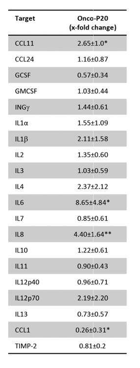
Cytokines protein array. The expression of 20 human cytokines was analyzed using a commercially available antibody array system (RayBio® C-Series Human Inflammation Array C1 Map, RayBiotech, Inc) that uses membrane-bound cytokine-specific antibodies to capture cytokines in biological fluids. The procedure was performed as per the manufacturer’s instructions. Cells were seeded in 10-cm culture dishes and treated (or not) with Onco-P20 0.15 µg/mL for 2 weeks. After removing drug cells (and untreated proliferating fibroblasts) were maintained in serum-free medium for 48 hours. The cytokine array membranes were blocked in 2 ml 1 × blocking buffer for 30 min at room temperature (RT) and then were incubated with 1 ml of conditioned medium at 4°C overnight. The medium was then decanted from each container, and the membranes were washed three times with 2 ml 1 × wash buffer I, followed by two washes with 2 ml 1 × wash buffer II at room temperature. Next, the membranes were incubated in biotin-conjugated primary antibodies for 2 hours at RT and then washed as described above before incubation in 1:1000-diluted horseradish peroxidase-conjugated streptavidin for 2 hours. The membranes were then washed thoroughly and incubated with a chemiluminescent ECL substrate at RT for 5 min. Imaging and densitometric analysis were performed with UVITEC Mini HD9 acquisition system (Alliance UVItec Ldt, Cambridge, UK).
Statistical Analysis: Results in the figures are representative of several experiments we performed with at least five cell lines from different donors. Quantitative data were reported as mean ± standard deviation (SD). Student t-test was used to assess statistical significance with thresholds of *p ≤0.05 and **p ≤0.01.