The expression and significance of AKR1B10 in laryngeal squamous cell carcinoma

DOI: https://doi.org/10.21203/rs.3.rs-403673/v1

Abstract

Aldosterone reductase family 1 member B10 (AKR1B10) is a nicotinamide adenine dinucleotide phosphate (NADPH) (reduced coenzyme II)-dependent oxidoreductase, and its biological functions include carbonyl detoxification, hormone metabolism, osmotic adjustment, and lipid synthesis. Studies suggested that AKR1B10 is a new biomarker for cancer based on its overexpression in epithelial tumors, such as breast cancer, cervical cancer, and lung cancer. At present, studies on the expression of AKR1B10 in laryngeal cancer have not been reported. However, we found that AKR1B10 is upregulated in laryngeal carcinoma, and its expression was negatively correlated with the degree of differentiation. In addition, AKR1B10 expression was positively correlated with tumor size; lymph node metastasis; long-term drinking; and Ki-67, mutant p53, and matrix metalloproteinase 2 expression. AKR1B10 was overexpressed in Hep-2 laryngeal carcinoma cells. Oleanolic acid inhibited AKR1B10 activity and expression in Hep-2 cells and suppressed Hep-2 cell proliferation, migration, and invasion. Therefore, AKR1B10 may be related to the development of laryngeal carcinoma, suggesting its use as a prognostic indicator for laryngeal cancer.

Introduction

Laryngeal squamous cell carcinoma (LSCC) is a malignant tumor originating from the epithelial tissue of the laryngeal mucosa. Although significant progress has been made in the development of surgery and radiotherapy in recent decades, the mortality rate of LSCC remains high [1]. Therefore, exploring the molecular mechanism of the occurrence and development of LSCC and developing new treatment options are current research hotspots. Aldosterone reductase family 1 member B10 (AKR1B10), which belongs to the AKR1B subfamily, is also known as aldose reductase-like enzyme, and it is present in limited tissues in humans, primarily the small intestine and colon. AKR1B10 is also expressed in the liver, thymus, prostate, and testis. Conversely, AKR1B10 is absent in other tissues (e.g., brain, spleen, heart, ovaries, lungs, placenta, pancreas, skeletal muscle, kidneys) [2]. AKR1B10 protein is a nicotinamide adenine dinucleotide phosphate (NADPH) (reduced coenzyme II)-dependent oxidoreductase that efficiently catalyzes the reduction of various cytotoxic carbonyl compounds in cells, such as oxidative stress products and various drugs. Moreover, AKR1B10 can reduce the highly reactive carbonyl group to a less toxic hydroxyl group, thereby protecting cells from carbonyl damage [3-6]. AKR1B10 also catalyzes the reduction of 13-cis retinal, 9-cis-retinal, and all-trans retinal to their corresponding retinols [7].

Studies revealed the involvement of AKR1B10 in the development and carcinogenesis of some tumors through effects on tumor cell growth and survival. The enzyme is reported to be overexpressed in lung cancer (including lung squamous cell carcinoma and smoking-related lung adenocarcinoma), cholangiocarcinoma, oral squamous cell carcinoma, liver cancer, breast cancer, and pancreatic cancer [8-13]. Interestingly, AKR1B10 is downregulated in gastrointestinal cancer [14]. Abnormal AKR1B10 expression, such as its overexpression in lung cancer [15], is currently considered a useful biomarker for the diagnosis and prognosis of some cancers.

At present, there is no report on the expression of AKR1B10 in LSCC. This study analyzed the expression of AKR1B10 in LSCC and the correlations of its expression with laryngeal carcinoma differentiation, tumor size, lymph node metastasis, and prognostic indices. This may provide new ideas for further exploration of the occurrence, development, and prevention of LSCC.

Results

AKR1B10 is upregulated in laryngeal carcinoma and correlated with tumor size, lymph node metastasis, long-term drinking, and differentiation

AKR1B10 expression in laryngeal carcinoma cells was mainly localized in the cytoplasm, and the individual nuclei also displayed positive staining (Figure 1A). Positive staining in the adjacent squamous epithelial tissues was mainly localized in the nuclei of cells near the basal layer (Figure 1B). The expression pattern differed from that of laryngeal carcinoma. AKR1B10 expression was higher in LSCC tissues than in adjacent tissues (P < 0.001, Table 1). In addition, AKR1B10 is diffusely expressed in laryngeal carcinoma cells (Fig. 1A). However, in Figure 1B, AKR1B10 was only expressed in the area near the basal layer of the advanced squamous epithelium. AKR1B10 protein expression in laryngeal carcinoma was found higher than that in the adjacent squamous epithelium.

AKR1B10 expression in laryngeal carcinoma was correlated with tumor size, lymph node metastasis, tumor differentiation, and long-term drinking (all P < 0.05), but not with gender, age, and smoking status (Table 2).

Expression of Ki-67 in laryngeal carcinoma

Ki-67 was positively expressed in the nuclei of laryngeal carcinoma cells. Ki-67 expression was high (Fig. 1D) in 55 cases (63.22%) and low (Fig. 1C) in 32 cases. Ki-67 expression was positively correlated with tumor size (r = 0.244, P < 0.05), long-term drinking (r = 0.357, P < 0.01), and lymph node metastasis (r = 0.260, P < 0.05) and negatively correlated with the degree of differentiation (r = −0.483, P < 0.05, Table 3).

Expression of mutant p53 (MTp53) in laryngeal carcinoma and its relationship with clinical indicators

MTp53 was positively detected in the nuclei of laryngeal carcinoma tissues. Of 87 laryngeal carcinoma cases, 44 were positive for MTp53 (50.58%) (Fig. 1F). MTp53 expression in laryngeal carcinoma was positively correlated with lymph node metastasis (r = 0.270, P < 0.05), long-term drinking (r = 0.374, P < 0.01), and negatively(Fig. 1E) correlated with the degree of differentiation (r = −0.317, P < 0.01, Table 3).

Matrix metalloproteinase 2 (MMP2) expression in laryngeal carcinoma and its relationship with clinical indicators

MMP2 was mainly expressed in the cytoplasm and cell membrane in laryngeal carcinoma tissues. In total, 68 lesions were positive for MMP2 expression (78.16%) (Fig. 1H). Its expression was positively correlated with tumor size (r = 0.235, P < 0.05), long-term drinking (r = 0.376, P < 0.01) and lymph node metastasis (r = 0.481, P < 0.01) and negatively correlated with the degree of differentiation (r = −0.302, P < 0.01, Table 3).

Correlation of AKR1B10 expression with Ki-67, MTp53, and MMP2 expression and clinical indicators

Immunohistochemistry revealed that among 87 laryngeal carcinoma tissues, 15 were AKR1B10+ Ki-67high, 14 were AKR1B10 Ki-67low, 41 were AKR1B10+ Ki-67low, and 17 were AKR1B10 Ki-67high. Spearman’s correlation analysis illustrated that AKR1B10 expression was positively correlated with Ki-67 expression (r = 0.353, P < 0.01, Table 3).

According to immunohistochemistry, 34 tissues were AKR1B10+ MTp53+, 21 were AKR1B10 MTp53, 22 were AKR1B10+ MTp53, and 10 were AKR1B10 MTp53+. Spearman’s correlation analysis indicated that AKR1B10 expression was positively correlated with MTp53 expression (r = 0.330, P < 0.01, Table 3).

Among 87 laryngeal carcinoma tissues, 51 were AKR1B10+ MMP2+, 14 were AKR1B10 MMP2, 5 were AKR1B10+ MMP2, and 17 were AKR1B10 MMP2+. Spearman’s correlation analysis revealed a positive correlation between AKR1B10 and MMP2 expression (r = 0.404, P < 0.01, Table 3).

AKR1B10 mRNA expression in Hep-2 cells

RT A549 cells were used as positive controls, and Hep-2 cells served as the experimental group. Hep-2 cells were found to express AKR1B10 mRNA (Fig. 2A).

AKR1B10 protein expression in Hep-2 cells

Western blotting demonstrated that compared with A549 cells, Hep-2 cells expressed AKR1B10 protein (Fig. 2B).

Immunofluorescence staining illustrated that AKR1B10 was mainly expressed in the cytoplasm of Hep-2 cells, and positive expression was also observed in individual nuclei (Fig. 2C).

Effects of oleanolic acid (OA) on AKR1B10 enzyme activity in Hep-2 cells

Compared with the findings in the control group, AKR1B10 activity in Hep-2 cells was significantly decreased after 48 h of treatment with 30 μM OA (P < 0.05, Fig. 3A). Conversely, the decrease was not significant after exposure to 10 or 60 μM.

Effects of OA on AKR1B10 mRNA expression in Hep-2 cells

Reverse-transcription polymerase chain reaction (RT-PCR) was used to detect the effects of 48 h of OA exposure on AKR1B10 mRNA expression in Hep-2 cells. Compared with the control group, the 30 and 60 μM OA groups significantly inhibited the mRNA expression of AKR1B10 (both P < 0.01, Fig.3B and 3C).

Effects of OA on AKR1B10 protein expression in Hep-2 cells

Compared with the control group, the 30 and 60 μM OA groups significantly inhibited the protein expression of AKR1B10 (P < 0.01, Fig. 4A and 4B).

Effects of OA on the proliferation of Hep-2 cells

The results of the CCK8 assay demonstrated that the 30 μM OA group significantly decreased Hep-2 cell growth starting on day 4 as compared to the control group (P < 0.05 on day 4, P < 0.01 on day 5, P < 0.001 on day 6, and P < 0.01 on day 7; Fig. 5A).

Effect of OA on the migration of Hep-2 cells

The Hep-2 cells were treated with 0 or 30 μM OA for 48 h to observe changes in cell migration. Cell mobility was somewhat decreased by 30 μM OA compared with the control group findings (P < 0.01, Fig. 5B and 5C).

Effects of OA on the invasiveness of Hep-2 cells

According to the Transwell assay, 30 μM OA significantly suppressed the invasiveness of Hep-2 cells compared with the control findings (P < 0.01, Fig. 6A and 6B).

Discussion

Globally, LSCC accounts for almost 2% of all malignancies, and it is one of the most common tumors of the head and neck [16]. AKR1B10 was initially isolated from liver cancer lesions [2]. Matkowskyj et al. found that AKR1B10 is upregulated in hepatocarcinoma, and silencing of the AKR1B10 gene in liver cancer cells increased apoptosis and reduced the formation and size of tumor cell colonies. This study provided the first evidence that AKR1B10 participates in hepatocellular carcinogenesis by regulating cell proliferation and apoptosis, and it can be used to identify hepatocellular carcinoma and benign liver lesions. In addition, the enzyme is an independent biomarker [17]. Fukumoto et al. found that AKR1B10 is overexpressed in non-small cell lung cancer and is closely related to smoking [10]. Ma et al. reported that AKR1B10 overexpression was associated with tumor size, lymph node metastasis, and patient survival in breast cancer [13]. Chung et al. found that AKR1B10 is overexpressed in pancreatic cancer, and it may participate in carcinogenesis by regulating apoptosis and protein prenylation [8]. However, Ohashi et al. demonstrated that AKR1B10 is downregulated in gastrointestinal cancer [14]. AKR1B10 is highly expressed in normal gastrointestinal tissues. It reduces toxic substances produced by diet and microbial metabolism in the gastrointestinal tract via carbonyl detoxification, and it can eliminate oxidative stress and promote epithelial cell proliferation. It is an important protective factor for gastrointestinal epithelial cells. AKR1B10 downregulation or ablation is conducive to the occurrence of cancer [18]. However, studies on AKR1B10 expression in laryngeal carcinoma have not been reported. This experiment was the first to detect the expression of AKR1B10 in laryngeal and precancerous tissues via immunohistochemistry, and the relationships of AKR1B10 with differentiation, tumor size, lymph node metastasis, and Ki-67, MTp53, and MMP2 expression were analyzed. In addition, the effects of AKR1B10 on the biological behavior of Hep-2 cells were detected via in vitro experiments, and the significance of its expression in laryngeal carcinoma was further explored.

The findings of the study were consistent with a study by Yoshitake et al., who found that AKR1B10 is highly expressed in the cytoplasm or nuclei of cervical cancer cells and occasionally expressed in normal squamous epithelial tissues, wherein it exhibits nucleus-specific expression [19]. We hypothesized that AKR1B10 overexpression may be involved in the development and progression of laryngeal cancer. Kang et al. reported that AKR1B10 is upregulated in lung squamous cell carcinoma, and AKR1B10 mRNA expression in highly differentiated tumors was significantly lower than that in moderately poorly differentiated tumors [20], in line with our results. However, it has also been reported that AKR1B10 is overexpressed in nasopharyngeal carcinoma, and the expression in moderately well differentiated tumors is higher than that in poorly differentiated tumors [21]. This may be attributable to differences in the role of AKR1B10 in different tumors. The finding that AKR1B10 expression was correlated with tumor size was consistent with the previously reported expression of AKR1B10 in oral squamous cell carcinoma and breast cancer [11,13]. In addition, Ma et al. found that AKR1B10 is overexpressed in breast cancer and is positively associated with lymph node metastasis [13], as demonstrated in the present analysis. However, studies revealed that AKR1B10 expression in oral squamous cell carcinoma and cervical cancer is not associated with lymph node metastasis [11,22].

Although AKR1B10 is upregulated in oral squamous cell carcinoma, breast cancer, cervical cancer, and lung cancer, the relationships of AKR1B10 expression with tumor size, lymph node metastasis, and squamous cell carcinoma differentiation are different [11,13,22,23]. In addition, studies revealed that AKR1B10 is overexpressed in breast cancer, oral squamous cell carcinoma, cervical cancer, and other tumors and is associated with prognosis [11,13,22]. To further analyze whether AKR1B10 expression in laryngeal carcinoma also has prognostic significance, we analyzed the relationships of AKR1B10 with the prognostic indicators Ki-67, MTp53, and MMP2.

The nuclear protein Ki-67 is closely related to tumor cell proliferation, and its overexpression is positively correlated with tumor malignancy. Ki-67 is a prognostic marker for breast cancer, lung cancer, cervical cancer, and other tumors [22]. Gioacchini et al. demonstrated that Ki-67 is overexpressed in laryngeal carcinoma and is associated with poor tumor cell invasion and prognosis [23]. It has been reported that the expression of Ki-67 in highly differentiated laryngeal carcinoma is significantly lower than that in poorly differentiated laryngeal carcinoma [24]. The expression rate of Ki-67 in laryngeal carcinoma with lymph node metastasis is significantly higher than that in laryngeal carcinoma without lymph node metastasis [25]. Previously, Wei et al. silenced AKR1B10 in MHCC97H liver cancer cells, finding that Ki-67 and the oncogenes c-myc, c-fos, and N-ras were downregulated and the pro-apoptotic protein genes caspase-3 and Bax were upregulated, indicating that AKR1B10 may promote cell proliferation, inhibit cell apoptosis, and induce hepatocyte deterioration by regulating the expression of tumor-associated genes [26]. However, Schmitz et al. reported that the proliferative capacity of AKR1B10-positive hepatocellular carcinoma and its Ki-67 expression rate were significantly lower than those of AKR1B10-negative hepatocellular carcinoma [27]. However, our study only found a positive correlation between AKR1B10 and Ki-67 expression. The specific links among these factors remain to be elucidated.

The tumor suppressor gene p53 protein is lost or mutated in approximately half of human cancers. It is well known that the loss of p53 function affects cell proliferation, migration, and invasion [28]. Wang et al. found that the 5-year survival rate of patients with LSCC and high MTp53 expression was significantly lower than that of patients with LSCC and low MTp53 expression, suggesting that MTp53 is associated with prognosis [29]. Our findings regarding MTp53 were consistent with the results of Ji et al., who reported that MTp53 expression in oral squamous cell carcinoma was associated with lymph node metastasis and tumor differentiation, but not age, gender, or tumor location [30]. Therefore, we speculate that AKR1B10 could be an indicator of poor prognosis in laryngeal cancer.

MMP2 is a gelatinase that degrades basement membrane collagen, which is related to the metastasis of various human tumors [31]. Liu et al. found that MMP2 is highly expressed in laryngeal carcinoma, and its expression in highly differentiated tumors is significantly lower than that in moderately poorly differentiated tumors. Additionally, MMP2 expression in lymph node metastasis is significantly higher than that in non-metastatic tissues. This indicates that MMP2 may be involved in the development and prognosis of laryngeal cancer [32]. Our results are consistent with this supposition. In particular, our results were similar to those of Li et al, who found that AKR1B10 expression is positively correlated with tumor size and lymph node metastasis in breast cancer and that AKR1B10 promotes MMP2 and vimentin expression by activating the ERK signaling pathway, thereby promoting the invasion and metastasis of MCF-7 and BT-20 breast cancer cells [33]. Whether AKR1B10 directly or indirectly promotes the occurrence and metastasis of laryngeal cancer through MMP2 remains to be further studied.

In vitro experiments were performed to confirm the aforementioned findings using the highly selective AKR1B10 inhibitor OA [34] and Hep-2 cells. Our results were consistent with the findings of Zhou et al., who demonstrated that silencing of the AKR1B10 gene inhibited lung cancer cell proliferation and migration [35]. Our findings regarding the inhibitory effects of OA on AKR1B10 expression and activity were consistent with the aforementioned immunohistochemistry results. Therefore, we speculate that the overexpression of AKR1B10 in laryngeal carcinoma may be related to the occurrence and poor prognosis of cancer.

This study was the first to detect the expression of AKR1B10 in laryngeal carcinoma and explore its relationship with tumor differentiation, tumor size, lymph node metastasis and prognosis. Based on the findings, AKR1B10 may represent a new target and theoretical molecular basis for the treatment and prognosis of laryngeal cancer.

Methods

Patient samples and cell culture

In total, 87 cases of LSCC detected between 2015 and 2017 in the second department of Jilin University First Hospital were examined in the study. The female to male ratio was 3.14:1. Sixty-one cases involved lymph node metastasis (Table 4), and 77 cases involved invasion of adjacent tissues. This study was approved by First Hospital Of Jilin University Institutional Review Boards. All methods were carried out in accordance with relevant guidelines and regulations.The informed consent was obtained from all patients, if the patients are under 18, from a parent.

Hep-2 and A549 cells [36] were provided by the Key Laboratory of Pathology and Biology of the Ministry of Education of Jilin University. The cells were cultured in H-DMEM containing 10% fetal bovine serum in an incubator containing 5% CO2 at 37°C.

Immunohistochemistry

Immunohistochemical staining was performed to analyze the expression of AKR1B10, Ki-67, MTp53, and MMP2 in LSCC and adjacent tissues. Tissue sections were incubated at 70°C for 1 h, followed by incubation with 100% xylene for 10 min, 100% ethanol twice for 5 min each, and then 95%, 90%, and 80% ethanol for 2 min each. The slides were washed and repaired under high pressure. After setting up and running the automatic immunohistochemical stainer program, slides were counterstained, differentiated, subjected to anti-blue staining, dehydrated, and cleared. Slides were finally observed under a microscope.

Result interpretation

AKR1B10 and MMP2 staining intensity was scored as the percentage of positive cells as follows: <5%, 0 points; 5%–10%, 1 point; 11%–50%, 2 points; and >50%, 3 points Meanwhile, the staining color was scored as follows: no color, 0 points; light yellow, 1 point; dark yellow, 2 points; and yellowish brown, 3 points. The staining score was calculated by summing the intensity and color scores and categorized as follows: <2, negative; 2–3, weakly positive; 4–5, moderately positive; and ≥6, strongly positive [17].

For Ki-67 scoring, the percentage of positive cells among 1000 positive cells was categorized as follows: ≤25%, low expression; and >25%, high expression [37].

MTp53 expression was scored as the percentage of positive cells among 1000 cells as follows: ≤10%, negative; and >10%, positive [38].

Reverse-transcription polymerase chain reaction (RT-PCR)

Ribonucleic acid (RNA) was extracted from Hep-2 and A549 cells. RNA was reverse-transcribed into cDNA according to the instructions of the kit. After the PCR reaction system was thoroughly mixed, 30 cycles were performed according to the reaction conditions.

For electrophoresis, 10 μl of the PCR product were added to each well and electrophoresed at 100 V for 1 h, and the results were observed using an imager.

Western blotting

To extract protein, Hep-2 and A549 cells were washed with cold phosphate-buffered saline (PBS), after which the culture dishes were placed on ice. Cells were lysed with 500 μl of lysate per dish for 30 min, and cells were scraped into Eppendorf tubes and centrifuged at 15 × 10 rpm for 15 min. The supernatant was retained and used to measure the protein concentrations in the samples.

For electrophoresis, 30 μg of the protein sample were added to each well, electrophoresed at 80 V for 30 min, and then electrophoresed at 120 V for 90 min. Proteins were then transferred to a membrane and incubated sequentially with primary and secondary antibodies.

Immunofluorescence staining

Hep-2 and A549 cells were cultured in 24-well plates. After permitting adherent growth, cells were fixed with paraformaldehyde for 20 min, washed three times with PBS, incubated with 0.3% Triton for 5 min, and washed three times with PBS. Cells were then exposed to 0.5% BSA for 30 min. After aspirating the liquid, cells were incubated with 0.1% BSA-diluted AKR1B10 antibody (1:50) overnight at 4°C and washed three times with PBS. Cells were incubated with the fluorescent secondary antibody (1:400) at room temperature for 1 h, washed three times with PBS, stained with DAPI for 10 min, and washed with PBS, and the stained cells were observed under a fluorescence microscope.

Effect of OA on the activity and expression of AKR1B10 in Hep-2 cells

To conduct ELISA, 6 × 10⁵ Hep-2 cells were added to each well of a six-well plate. After permitting adherent growth, cells were cultured with 0, 10, 30, or 60 μM OA for 48 h and lysed (approximately 1 million cells/ml). Cells were then centrifuged (3 × 1000 rpm, 10 min). According to the AKR1B10 enzyme activity assay kit instructions, a standard curve was drawn to calculate enzyme activity. For RT-PCR and Western blotting, cells were treated with OA, after which RNA and protein were extracted. AKR1B10 mRNA and protein levels in cells were detected via RT-PCR and Western blot, respectively.

Based on the results of the aforementioned experiments, 30 μM OA was selected for the subsequent experiments.

CCK8 experiment

Hep-2 cells were cultured in seven 96-well plates at 2000 cells/100 μL per well. Three replicates each were created for the control and experimental groups. The control group was treated with H-DMEM containing 10% serum. The experimental group was treated with H-DMEM containing 30 μM OA. Cell growth was measured continuously for 7 days by adding 10 μL of CCK8 solution at 37°C for 1–4 h, measuring the absorbance at 450 nm, and plotting the proliferation curve.

Scratch test

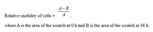
A straight line was drawn on the back of a six-well plate. Hep-2 cells (6.5 × 10⁵) were added to each well and cultured overnight. Then, the cell monolayer was scratched in a direction perpendicular to the line. The control group was cultured in serum-free H-DMEM. The experimental group was cultured in serum-free H-DMEM containing 30 μM OA. Cell migration was observed under a phase contrast microscope at 0 and 48 h. Cell migration was calculated using the following formula:

Transwell experiment

Initially, 65 μL of diluted Matrigel were added in the upper chamber (Matrigel:H-DMEM ratio = 1:5). Then, Hep-2 cells (7 × 10⁴ cells/200 μL) cultured in serum-free H-DMEM for 12 h were added to the upper chamber. The experimental group was cultured in H-DMEM containing 30 μM OA, and the control group was cultured in H-DMEM. Meanwhile, 500 μL of H-DMEM medium containing 15% serum were added to the lower chamber in both groups. Cells were incubated for 48 h in a 5% CO2 incubator at 37°C. The culture medium in the upper chamber was discarded, and the cells in the upper chamber were cleaned with a cotton swab and fixed with paraformaldehyde for 20 min. After staining with crystal violet, the cells were observed under an inverted microscope.

Abbreviations

LSCC              Laryngeal squamous cell carcinoma

OA                 Oleanolic acid

DAPI               4′,6-diamidino-2-phenylindole

NADPH           Nicotinamide adenine dinucleotide phosphate

PBS                Phosphate-buffered saline

PCR               Polymerase chain reaction

RNA               Ribonucleic acid

Declarations

Acknowledgments: None

Author Contributions: Jixuan Liu and Hongyan Ban were involved in drafting and final revision of the manuscript. Yafang Liu and Jinsong Ni were involved in designing and completing the experiment and editing the manuscript.

Competing Interests: The authors declare no competing interests.

Funding: None

Ethics declarations:

References

[1]   Palumbo, A. Jr et al. HMGA2, but not HMGA1, is overexpressed in human larynx carcinomas. Histopathology (2017).

[2]    Cao, D., Fan, S. T. & Chung, S. S. Identification and characterization of a novel human aldose reductase-like gene. J. Biol. Chem. 273, 11429-11435 (1998).

[3]  Cousido-Siah, A., et al. Identification of a novel poly- fluorinated compound as a lead to inhibit the human enzymes aldose reductase and AKR1B10: Structure determination of both ternary complexes and implications for drug design. Acta Crystallogr. D Biol. Crystallogr. 70, 889-903 (2014).

[4]  Endo, S. et al. Kinetic studies of AKR1B10, human aldose reductase-like protein: endogenous substrates and inhibition by steroids. ArchBiochem. Biophys. 487, 1-9 (2009).

[5]  Zhong, L., Shen, H., Huang, C., Jing, H. & Cao, D. AKR1B10 induces cell resistance to daunorubicin and idarubicin by reducing C13 ketonic group. ToxicolAppl. Pharmacol. 255, 40-47 (2011).

[6]  Quinn, A. M., Harvey, R. G. & Penning, T. M. Oxidation of PAH trans-dihydrodiols by human aldo-keto reductase AKR1B10. ChemRes. Toxicol. 21, 2207-2215 (2008).

[7]  Crosas, B. et al. Human aldose reductase and human small intestine aldose reductase are efficient retinal reductases: consequences for retinoid metabolism. Biochem. J. 373, 973-979 (2003).

[8] Chung, Y. T. et al. . Overexpression and oncogenic function of aldo-keto reductase family 1B10 (AKR1B10) in pancreatic carcinoma. Mod. Pathol. 25, 758-766 (2012).

[9]    Heringlake, S. et al. Identification and expression analysis of the aldo-ketoreductase1-B10 gene in primary malignant liver tumours. J. Hepatol. 52, 220-227 (2010).

[10]  Fukumoto, S., et al. Overexpression of the aldo-keto reductase family protein AKR1B10 is highly correlated with smokers’ non-small cell lung carcinomas. Clin. Cancer Res. 11, 1776-1785 (2005).

[11]  Ko, H. H. et al. Expression of AKR1B10 as an independent marker for poor prognosis in human oral squamous cell carcinoma. Head Neck 39, 1327-1332 (2017).

[12]  Jin, J. et al. Aldo-keto Reductase Family 1 Member B 10 Mediates Liver Cancer Cell Proliferation through Sphingosine-1-Phosphate. Sci. Rep. 6, 22746 (2016).

[13]  Ma, J., et al. AKR1B10 overexpression in breast cancer: associa- tion with tumor size, lymph node metastasis and patient survival and its potential as a novel serum marker. IntJ. Cancer 131, E862-E871 (2012).

[14]  Ohashi, T., Idogawa, M., Sasaki, Y., Suzuki, H. & Tokino, T. AKR1B10, a transcriptional target of p53, is downregulated in colorectal cancers associated with poor prognosis. Mol. Cancer Res. 11, 1554-1563 (2013).

[15]  Woenckhaus, M. et al. Smoking and cancer- related gene expression in bronchial epithelium and non-small-cell lung cancers. J. Pathol. 210, 192-204 (2006).

[16]  Pagliuca G, Martellucci S, Degener AM, Pierangeli A, Greco A, Fusconi M, De Virgilio A, Gallipoli C, de Vincentiis M, Gallo A. Role of Human Papillomavirus in the Pathogenesis of Laryngeal Dysplasia. Otolaryngol Head Neck Surg. 2014; 150: 1018-1023

 [17] Matkowskyj, K. A. et al. Aldoketoreductase family 1B10 (AKR1B10) as a biomarker to distinguish hepatocellular carcinoma from benign liver lesions. Hum. Pathol. 45, 834-843 (2014).

.[18] Shen, Y., Liao, D. F. & Cao, D. AKR1B10 in gastrointestinal diseases. Aging (Albany, NY) 7, 221-222 (2015).

[19]  Yoshitake, H., et al. Aldo-keto reductase family 1, member B10 in uterine carcinomas: A potential risk factor of recurrence after surgical therapy in cervical cancer. IntJGynecol. Cancer 17, 1300-1306 (2007).

[20]  Kang, M. W. et al. AKR1B10 is associated with smoking and smoking-related non-small-cell lung cancer. JIntMed. Res. 39, 78-85 (2011).

[21]  He, Y. C., et al. Overexpression of AKR1B10 in nasopharyngeal carcinoma as a potential biomarker. Cancer Biomark. 16, 127-135 (2016).

[22] Li, L. T., Jiang, G., Chen, Q. & Zheng, J. N. Ki67 is a promising molecular target in the diagnosis of cancer. Mol. Med. Rep. 11, 1566-1572 (2015).

[23]  Gioacchini, F. M. et al. The clinical relevance of Ki-67 expression in laryngeal squamous cell carcinoma. Eur. Arch. Otorhinolaryngol 272, 1569-1576 (2015).

[24] Izawa, H., Yonemitsu, N., Shin, T. & Sugihara, H. Histopathological analysis of apoptosis, and expression of p53, bcl-2, bax, and Ki-67 in laryngeal squamous cell carcinomas and dysplasia. Auris Nasus Larynx 26, 317-330 (1999).

[25] Acikalin, M. F. & Oner, U., Tel N, Pasaoglu O, Cakli H, Colak E. Prognostic significance of Ki-67 expression for patients with laryngeal squamous cell carcinoma primarily treated by total laryngectomy. Eur. Arch. Otorhinolaryngol. 261, 376-380. (2009)

[26]  Wei, W. et al. Effects of AKR1B10 gene silence on the growth and gene expression of HCC cell line MHCC97H. Zhonghua Gan Zang Bing Za Zhi 18, 666-671 (2010)

[27]  Schmitz, K. J. et al. AKR1B10 expression is associated with less aggressive hepatocellular carcinoma: A clinicopathological study of 168 cases. Liver Int. 31, 810-816 (2011).

[28]  Xu, J. et al. Mutant p53 promotes cell spreading and migration via ARHGAP44. Sci. China Life Sci. 60, 1019-1029 (2017).

[29]  Wang, D., Yi, Z. & Yang, F. Expression of P53 protein in laryngeal carcinoma and its clinical significance. Zhonghua Er Bi Yan Hou Ke Za Zhi 30, 354-356 (1995).

[30] Li, Y. & Zhang, J. Expression of mutant p53 in oral squamous cell carcinoma is correlated with the effectiveness of intra-arterial chemotherapy. Oncol. Lett. 10, 2883-2887 (2015).

[31] Curran, S. & Murray, G. I. Matrix metalloproteinases in tumour invasion and metas- J. Pathol. 189, 300-308 (1999).

[32] Liu, R. R. et al. Matrix metalloproteinase 2 (MMP2) protein expression and laryngeal cancer prognosis: a meta analysis. IntJClinExp. Med. 8, 2261-2266 (2015).

[33] Li, J., et al. AKR1B10 promotes breast cancer cell migration and invasion via activation of ERK signaling. Oncotarget 8, 33694-33703 (2017).

[34] Takemura, M. et al. Selective inhibition of the tumor marker aldo-keto reductase family member 1B10 by oleanolic acid. JNat. Prod. 74, 1201-1206 (2011).

[35] Zu, X. et al. Aldo-keto reductase 1B10 protects human colon cells from DNA damage induced by electrophilic carbonyl compounds. Mol. Carcinog. 56, 118-129 (2017).

[36] Jung, Y. J. et al. AKR1B10-inhibitory Selaginella tamariscina extract and amentoflavone decrease the growth of A549 human lung cancer cells in vitro and in vivo. J. Ethnopharmacol. 202, 78-84 (2017).

[37] Pillai, K., Pourgholami, M. H., Chua, T. C. & Morris, D. L. Prognostic significance of Ki67 expression in malignant peritoneal mesothelioma. Am. J. Clin. Oncol. 38, 388-394 (2015).

[38] Kropveld, A., Slootweg, P. J., Blankenstein, M. A., Terhaard, C. H. & Hordijk, G. J. Ki-67 and p53 in T2 laryngeal cancer. Laryngoscope 108, 1548–1552 (1998).

Tables

Table 1

Aldosterone reductase family 1 member B10 expression in laryngeal cancer and adjacent tissues

Category

Total

Positive

Negative

Positive rate

P

Laryngeal cancer

87

56

31

64.37%

< 0.001

Paracancerous tissue

77

10

67

12.99%***


Table 2

The relationship of tumor size, lymph node metastasis, tumor differentiation, age, gender, smoking status, and drinking status with the expression of AKR1B10 in laryngeal carcinoma

Clinical indicators

Number

AKR1B10

χ2

P

+

++

+++

Gender

Male

66

23

15

21

7

3.414

0.328


Female

21

8

2

6

5


Age (years)

≤ 60

50

22

10

10

8

7.351

0.062


60

37

9

7

17

4


Tumor size

≤ 2 cm

21

16

2

3

0

18.449

< 0.001


> 2 cm

62

14

15

22

11


Lymph node metastasis

Yes

39

8

8

16

7

10.789

0.01


No

22

14

2

5

1


Differentiation

High

19

10

5

3

1

15.663

0.007


Medium

59

20

10

23

6


Poor

9

1

2

1

5


Smoking

Yes

36

10

10

12

4

1.652

0.195


No

51

21

13

7

10


Long-term drinking

Yes

25

2

6

8

9

31.563

< 0.001


No

62

46

7

5

4

   
+, weakly positive; ++, moderately positive; +++, strongly positive
All variables were analyzed using Fisher’s exact probability test, excluding age, which was analyzed using the R × C χ2 test.


Table 3

Spearman’s correlation analysis of AKR1B10, Ki-67, MTp53, and MMP2 expression and clinical indicators

Category

AKR1B10

MMP2

Ki-67 (%)

MTp53 (%)

Age

0.130

−0.138

−0.156

0.055

Gender

0.071

−0.165

−0.069

−0.112

Tumor diameter

0.474**

0.235*

0.244*

0.196

Differentiation

−0.322**

−0.302**

−0.483**

−0.317**

Lymph node metastasis

0.394**

0.481**

0.260*

0.270*

Long-term drinking

0.385**

0.376**

0.357**

0.374**

Smoking

0.142

0.159

0.053

0.045

MTp53 (%)

0.330**

0.226*

0.306**


Ki-67 (%)

0.353**

0.387**


MMP2

0.404**

     
AKR1B10, aldosterone reductase family 1 member B10; MTp53, mutant p53; MMP2, matrix metalloproteinase 2
*P < 0.05 **P < 0.01


Table 4

Laryngeal cancer specimen information

Clinical index

Number

Gender

Male

Female

66

21

Age (years), range

≤ 60 years old

> 60 years old

40–80

50

37

Tumor differentiation

High

Medium

Poor

19

59

9

Lymph node metastasis

Yes

No

39

22

Tumor size

≤ 2 cm

> 2 cm

21

62

Smoking

Yes

No

36

51

Long-term drinking

Yes

No

25

62

Note: Of the 87 cases of laryngeal cancer, 26 did not feature lymph nodes metastasis, whereas the tumor size could not be determined in four cases.