Dosimetric comparison between helical tomotherapy, volume modulated arc-therapy and Fixed-Field intensity modulated radiation therapy in postoperative adjuvant radiotherapy for cervical cancer

DOI: https://doi.org/10.21203/rs.3.rs-58107/v1

Abstract

Background: To evaluate the dosimetric parameters among three different radiotherapy techniques in patients with postoperative cervical cancer, including the Helical Tomotherapy (HT), the Volume Modulated Arc-Therapy (VMAT), and the Fixed-Field intensity modulated radiation therapy (FF-IMRT).

Methods: Fifteen cervical cancer patients treated with postoperative radiotherapy were re-planning with HT, VMAT and FF-IMRT. The prescribed target dose of the patients was 1.8/45Gy. The paired-samples t-test was used to compare the dosimetric parameters of the planning target and OARs (Organs at risk), and the efficiency of radiation delivery.

Results: Compared with the VMAT and FF-IMRT, the HT plans showed significant improvement in the conformity index (CI) and the homogeneity index (HI). In addition, the HT plans also significantly reduced the volume of high-dose region of the OARS, especially in the V30, V40 of small bowel, rectum and bladder. Meantime, the advantage of VMAT is that it reduced the treatment time and improved the efficiency of radiation delivery obviously, compared with the HT (293.8 ± 12.8s Vs. 557.6 ± 51.9s, P < 0.001) and FF-IMRT (293.8 ± 12.8s Vs. 581.8 ± 26.1s, P < 0.001).

Conclusion: Our result reveals that HT showed better CI and HI for the target and reduced high dose volumes to OARs compared with VMAT and FF-IMRT, but the lower dose volumes to OARs increased slightly. As for the benefit of VMAT, it demonstrated the shortest treatment time. Our results could provide guidance for selecting the appropriate radiation technologies for cervical cancer patients who undergoing postoperative adjuvant pelvic radiotherapy.

Background

Cervical cancer ranks fourth for both diagnosed with cancer and the major cause of cancer-related death among females worldwide[1]. For early-stage cervical cancer cases, radical hysterectomy + pelvic lymph node dissection ± para-aortic lymph node dissection is the standard treatment method. Following primary hysterectomy, the presence of one or more pathologic risk factors may warrant the use of adjuvant postoperative radiotherapy. However, compared with chemoradiotherapy alone, radical hysterectomy combined with postoperative pelvic radiotherapy has been demonstrated to increase the acute/late toxicities including the gastrointestinal (GI) and genitourinary (GU) toxicity[2-4]. According to previous studies, the high-dose volume of small bowel, rectum, and bladder is associated with the GI and GU toxicity after radiotherapy[5-7], and limiting the high-dose radiation delivered to the small bowel, rectum and bladder can reduce the occurrence of acute or late GI and GU toxicities[8, 9]. Therefore, it is critical to explore the feasible and optimal radiotherapeutic techniques to accomplish more highly conformal treatment plans and acquire better OARs sparing for postoperative cervical cancer patients.

With the rapid development of computer technology and radiation equipment of late years, more modern radiation strategies have appeared. Compared with conventional 3D conformal radiotherapy, IMRT is much superior in sparing OARs and optimizing target volume coverage and conformity in cervical cancer[10, 11]. FF-IMRT and VAMT contribute greatly in radiotherapy, followed by HT, a neo-CT based rotational IMRT, which provides better OARs sparing with 51 independent beam directions, delivers a highly conformal dose distribution. Nowadays, HT is frequently used for a variety of diseases[12-15]. However, in cervical cancer research, most of the researches have focused on the potential advantages of HT in radiotherapy of primary cervical cancer [16], there are few published data comparing the planning parameters of three modern IMRT techniques (HT, VMAT, and FF-IMRT) in postoperative cervical cancer patients.

Therefore, our objective in the present study was to assess whether HT plans provide any benefit with regard to the target and OARs for cervical cancer patients undergoing postoperative pelvic radiotherapy.

Materials And Methods

This study complied with the Helsinki Declaration and approval from the Ethics Committee of our center was obtained. All patients provided their informed consents for the publication of their images/data.

Patient characteristics and CT simulation

Fifteen patients with cervical cancer undergoing radical hysterectomy and adjuvant pelvic radiotherapy in our hospital were chosen for this research between August 2019 and November 2019. The selection criteria were staged ⅠB-ⅡA, biopsy-proven squamous cell carcinoma. The 15 eligible patients ranged in age from 45 to 65, the mean and median age was 52.3 and 57 years old, respectively. The thermoplastic pelvic masks were used to immobilize the patients with supine position. All patients were scanned CT simulations of 5 mm slice thickness using a Philips 16-slice Brilliance big bore computed tomography scanner (Philips Medical Systems, Amsterdam, Netherlands), and with comfortably full bladder (after emptying, patients were requested for drinking 1 liter of water 30 to 45 minutes before treatment and holding urine) and a bowel preparation with oral contrast agent prior to simulation. The scan images were performed from the L2 vertebra to the area 5cm below the symphysis pubis.

Target and normal tissue volume definition

Contouring was performed on the platform of Monaco 5.11 (Elekta AB, Stockholm, Sweden) planning system. All outlines were represented by the same radiation oncologist for consistency. Delineation was according to the recommendations of Radiation Therapy Oncology Group (RTOG) 0418 protocol and the International Commission on Radiation Units and Measurements reports (ICRU) Report 62. The clinical target volume (CTV) included the vaginal stump, parametrial soft tissue and pelvic lymph drainage area. The CTV ranged from the L4-L5 vertebra to the inferior margin of the obturator foramen. The planning target volume (PTV) was generated by expanding a uniform 7mm margin from the CTV. The organs at risk (OARs) included small bowel, bladder, rectum, spinal cord and femoral heads.

Treatment planning

To ensure the consistency, all treatment plans were operated by the same radiation physicist. The FF-IMRT and VMAT plans were done with Monaco planning system version 5.11, and executed with Elekta Synergy Linac (Elekta Ltd., Crawley, UK) equipped with 8 MV photon beams and the MLCi2 (40 pairs MLC leaves and each one is 1 cm width at the isocenter). The prescribed dose of PTV was 1.8/45 Gy. The volume of PTV receiving >49.5Gy was limited to <1%. The volume of small bowel receiving >30 Gy was limited to <50%; <50% of the rectum was to receive >30 Gy, <35% of the bladder was to receive >40 Gy, and <5% of the femoral head was to receive >40 Gy. The maximum doses (Dmax) to the small bowel and rectum were both confined to lower than 48 Gy.

Some patients who had positive surgical margins also received brachytherapy using Ir192 source (high-dose rate) once a week, applied 18Gy/3fractions, dosed at the vaginal surface.

HT plans

The HT plans were operated using the tomotherapy planning station with 6 MV x-ray and were implemented on the Tomo HD (Accuray Inc., Madison, USA). Parameters for beamlet calculation were a field width of 2.5 cm, pitch values of 0.287, modulation factor of 3 and normal dose calculation grid.

VMAT plans

All VMAT plans were created using the Monaco 5.11 planning system and one beam with double 360° arcs were used, and there were 100 control points per arc. All VMAT plans were computed using Monte Carlo algorithm.

Fixed-field IMRT plans

Nine evenly distributed coplanar fields with the gantry angles of 200°, 240°, 280°, 320°, 0°, 40°, 80°, 120° and 160° were used, for each setting 20 control points. All FF-IMRT plans were computed using Monte Carlo algorithm. The optimization functions of the FF-IMRT plans were same to those of the VMAT plans. The DMLC (sliding window) technique was used in the FF-IMRT plans.

Plan evaluation parameters

The research analyzed the dose volume histograms (DVHs) obtained from the PTV and other contoured OARs. Dosimetric parameters were quantified from PTV including D98 (the dose received 98% volume of the PTV), D50, D2, the mean dose (Dmean), conformity index (CI) and homogeneity index (HI). CI was used to evaluate the conformity of prescribed dose distribution.

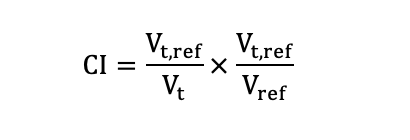
Here, Vt,ref, Vt and Vref denoted the target volume receiving the prescribed dose, the target volume and the total volume covered by the prescribed dose, respectively. The CI ranges from 0 to 1, with a higher CI indicating better conformal dose of the target. According to ICRU report NO.83[17], the HI was calculated as following formula:

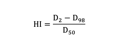
HI was used to indicate the uniformity of dose distribution. The smaller value of HI means a better homogeneity of the target volume.

For the OARs (small bowel, rectum, bladder and femoral heads), data analysis was carried out for the V5 (the OAR volume received the dose of 5 Gy), V10, V20, V30, V40, Dmean and Dmax. Treatment delivery time of each plan was also collected and compared.

Data analysis

The data collected from DVH of different plans were compared with paired t-test using SPSS 19.0 (SPSS, Inc., Chicago, IL, USA). All P values were two-sided, and a P value < 0.05 was considered statistically significant.

Results

The dose distributions of PTV and OARs that were obtained from the 45 treatment plans generated by 15 patients were analyzed. The mean volume of PTV was 982.2  104.8 cc (range 814.1-1134.9 cc). All of the HT, VMAT and FF-IMRT plans were normalized to cover 95% of the PTV volume with ≥100% of the prescribed dose. And the maximal dose constrained in the PTV less than 110% of the prescription dose.

Table 1 displays the dosimetric parameters for PTV. With regard to conformity and homogeneity, the HT plans displayed notably better results. As shown in Table 1 and Figure 1, the CI of HT plans were highest (HT Vs. VMAT: 0.85 ± 0.02 Vs. 0.82 ± 0.03; HT Vs. FF-IMRT: 0.85 ± 0.02 Vs. 0.78 ± 0.03; Both P<0.01), meanwhile the HI of HT plans were much lowest (HT Vs. VMAT: 0.05 ± 0.01 Vs. 0.07 ± 0.01; HT Vs. FF-IMRT: 0.05 ± 0.01 Vs. 0.08 ± 0.01; Both P<0.001). Typical dose distributions for the three techniques obtained in this research are exhibited in Figure 2.

Table 2 showed the volumes of small bowel, rectum, bladder and femoral heads receiving ≥5, ≥10, ≥20, ≥30 and ≥40Gy, and listed the maximal dose of small bowel, spinal cord and rectum. Figure 3 demonstrated a sample DVH in a patient. In general, compared with VMAT and FF-IMRT, HT significantly reduced the volume of high dose level (30 and 40 Gy) for small bowel, rectum and bladder. The average volume receiving ≥ 30Gy reduced by 25.2% and 27.6% (Both P<0.001) for small bowel, 28.8% and 31.5% (Both P<0.001) for rectum and 6.2% and 7.3% (Both P<0.001) for bladder, respectively. The mean volume receiving ≥ 40Gy reduced by 17.2% and 29.1% (Both P<0.001) for small bowel, 23.8% and 26.9% (Both P<0.001) for rectum, 6.3% and 9.5% (Both P≤0.001) for bladder, respectively. The maximal dose of small bowel and rectum were also reduced by 3.2%, 3.5% (P<0.001) and 1.6%, 1.3% (P≤0.001) using HT plans, compared with VMAT and FF-IMRT plans, respectively. As seen in Table 2, compared with VMAT and FF-IMRT, HT has no advantage in the protection of OARS in low-dose radiation, the V5 and V10 of small bowel, rectum, bladder and femoral heads were a little higher in HT plans.

With regard to treatment delivery time: the mean time were 557.6 ± 51.9s, 293.8 ± 12.8s and 581.8 ± 26.1s for HT, VMAT and FF-IMRT plans, respectively.

Discussion

In current study, we compared dosimetric parameters of three different modern radiation techniques, which are HT, VMAT, and FF-IMRT, in postoperative adjuvant pelvic radiotherapy for cervical cancer patients. The data collected from the DVH showed that the HT plans performed better target homogeneity index and conformity index compared with FF-IMRT and VMAT plans. With regard to the normal tissue, HT plans notably reduced the mean dose and maximum dose of small bowel, rectum and bladder, which may contribute to a markedly decrease of the acute/late gastrointestinal and genitourinary toxicities.

Since pelvic external beam radiotherapy (EBRT) with/without brachytherapy is the standard adjuvant treatment approach for some postoperative cervical cancer patients according to pathological results of radical hysterectomy, GI and GU toxicity are the major concern in clinic. Compared with FF-IMRT plans, HT plans decreased the V30 and V40 of small bowel by 27.6% and 29.1%, respectively, in this study. Tan[18] reported that the remarkable reduction of the V30 and V40 of small bowel may bring about a significant risk decrease of acute and late GI toxicity. For rectum, the HT plans decreased by 31.5% and 26.9% in the V30 and V40 in comparison with FF-IMRT plans in the present research. Georg[19] found that the more cut-down dose in rectum in the radiotherapy treatment of cervical cancer conduces to less rectal toxicity. In regards to the GU toxicity, Viswanathan[20] reported that a combination of EBRT and brachytherapy in cervical cancer treatment would make for a high occurrence rate of late GU toxicity, especially in postoperative cervical cancer patients. Therefore, compared FF-IMRT plans, HT plans reduced the volume of bladder in 30Gy and 40Gy by 7.3% and 9.5%, will certainly contribute to less late GU toxicity.

HT plans showed significant advantages over FF-IMRT and VMAT plans in CI and HI of PTV, and also in the volume of high dose region (V30, V40) of the OARs. The linac of HT can rotate 360° continuously with 51 beam angles at the same time optimizing with the couch moving continuously. HT can also use a constant beam width of 1, 2.5 or 5cm to deliver radiation in a form of helical tomoscan. HT is equipped with a pneumatic binary MLC system with rapid leaf transition times. All these advantages above indicate HT plan may lead to a greater degree of intensity modulation and sharper dose fall-off in comparison with FF-IMRT or VMAT plans. In addition to this, the on-board megavoltage computed tomography (MVCT) allows daily setup validation, which can be used to perform adaptive radiotherapy (ART) planning which can eliminate the volume variation of the target and OARs between intra-fractions. And the margin expanding from CTV to PTV could be reduced because the setup errors is reducing by daily setup verification, resulting in a reduction in the dose of the small bowel[21].

Nevertheless, there is a drawback of HT. Vernat[22] and Pasquier[23] reported that HT increased normal tissue volume of low dose region compared with IMRT and VMAT for oropharyngeal cancer and prostate cancer. Xie[24] also reported that HT plan increased the volume of V5 and V10 of lung and heart compared with IMRT and VMAT plans for left-sided breast cancer. Our results are consistent with the results above, the volumes of OARs in low dose radiation region are increased, such as V5, V10 of small bowel, rectum, bladder and femoral heads. However, the acute and late GI and GU toxicity is mainly associated with the volume of the high dose region in pelvic radiation. Therefore, HT has shown great advantages in the dosimetric significance of reducing the occurrence of acute and late GI and GU toxicity, while ensuring high homogeneity and conformity. Therefore, HT technique is a better choice in the postoperative adjuvant pelvic radiotherapy for cervical cancer patients.  

Some literatures reported that VMAT achieved higher dose conformity of PTV and better sparing of OARs with a shorter treatment delivery time than FF-IMRT in comparison with plans of different cancers[25-27]. In our present study, compared with FF-IMRT plans, VMAT were also exhibited better dose distribution of target and better sparing of OARs. Compared with HT and FF-IMRT plans, VMAT plans reduced the delivery time by 47.3% and 49.5%, respectively. Less treatment time may reduce the influence of the uncertainty, the probability of patents’ moving, and patients’ discomfort. Therefore, VMAT may be the appropriate treatment planning strategy in postoperative cervical cancer patients who cannot stay in position for longer time due to physical or mental discomfort.

However, the limited sample size in our study may result in insufficient statistical power to show significance in some of the dosimetric parameters. Therefore, further clinical trials with large sample sizes focusing on the clinical significance of HT correlated with GU and GI toxicity data and oncologic outcomes are essential in the future.

Conclusion

In the three different modern IMRT strategies for the postoperative adjuvant pelvic radiotherapy in cervical cancer patients, our current study showed that the HT plans accomplished the best conformal and homogeneous dose distribution in the aspect of PTV, and also achieved the best sparing of the OARs through reducing the volume of high-dose radiation region. Therefore, while ensuring high homogeneity and conformity of PTV, HT plans might contribute to a significant reduction of GU and GI toxicity. On the other hand, VMAT obviously reduced the treatment time and improved the efficiency of radiation delivery, which could reduce the patients’ discomfort and patients’ involuntary moving while beam on. Our results could offer guidance for choosing suitable radiation technologies for cervical cancer patients who undergoing postoperative adjuvant pelvic radiotherapy.

Declarations

Ethics approval and consent to paticipate

This study complied with the Helsinki Declaration and approval from the Ethics Committee of Harbin Medical University Cancer Hospital (Harbin, China) was obtained. All patients provided their informed consents for the publication of their images/data.

Consent for publication

Not applicable.

Availability of supporting data

Not applicable.

Conflict of interest statement

All the authors declare that there are no conflicts of interest.

Funding

This work was supported by grants from the National Natural Science Foundation of China (No. 81872460 and 81602664), Research project grant of Harbin Medical University Cancer Hospital (JY2016-05), Pandeng Project of National Cancer Center (NCC201808B016).

Author contributions

SS. Y and YL. B designed the study. SS. Y contoured the target and OARs. DY. Y, XY. H, and X. L performed the design of the treatment planning. DY. Y and L. W collected the data. DY. Y and SS. Y wrote and revised the manuscript. YL. B polished the language. All authors reviewed and approved the final version.

Acknowledgments

The authors thank all the people who had participated in this study.

Abbreviations

IMRT - intensity-modulated radiation therapy; RT - radiation therapy; CC - cervical cancers; HT - helical tomotherapy; VMAT - volume-modulated arc therapy; FF-IMRT - fixed-field intensity-modulated radiation therapy; OARs - organs at risk; CI - conformity index; HI - homogeneity index; cCRT - Concurrent chemoradiotherapy; GI – gastrointestinal; GU – genitourinary; 3D-CRT - 3-dimensional conformal radiotherapy;  MUs - monitor units; MLC - multileaf collimator; CT - computed tomography; AJCC - American Joint Committee on Cancer; FIGO - International Federation of Obstetrics and Gynaecology; RTOG - Radiation Therapy Oncology Group; ICRU - International Commission on Radiation Units and Measurements reports; CTV - clinical target volume, PTV - the planning target volume; DVH – dose volume histograms; EBRT – external beam radiotherapy; MVCT – megavoltage computed tomography; ART – adaptive radiotherapy.

References

[1]       Bray F, Ferlay J, Soerjomataram I, Siegel RL, Torre LA, Jemal A. Global cancer statistics 2018: GLOBOCAN estimates of incidence and mortality worldwide for 36 cancers in 185 countries. CA Cancer J Clin. 2018;68(6):394-424.

[2]       Kirwan JM, Symonds P, Green JA, Tierney J, Collingwood M, Williams CJ. A systematic review of acute and late toxicity of concomitant chemoradiation for cervical cancer. Radiother Oncol. 2003;68(3):217-26.

[3]       Collaboration CfCCM-A. Reducing Uncertainties About the Effects of Chemoradiotherapy for Cervical Cancer: A Systematic Review and Meta-Analysis of Individual Patient Data From 18 Randomized Trials. Journal of Clinical Oncology. 2008;26(35):5802-12.

[4]       Green JA, Kirwan JM, Tierney JF, Symonds P, Fresco L, Collingwood M, et al. Survival and recurrence after concomitant chemotherapy and radiotherapy for cancer of the uterine cervix: a systematic review and meta-analysis. The Lancet. 2001;358(9284):781-6.

[5]       Isohashi F, Yoshioka Y, Mabuchi S, Konishi K, Koizumi M, Takahashi Y, et al. Dose-volume histogram predictors of chronic gastrointestinal complications after radical hysterectomy and postoperative concurrent nedaplatin-based chemoradiation therapy for early-stage cervical cancer. International Journal of Radiation Oncology* Biology* Physics. 2013;85(3):728-34.

[6]       Zelefsky MJ, Fuks Z, Hunt M, Yamada Y, Marion C, Ling CC, et al. High-dose intensity modulated radiation therapy for prostate cancer: early toxicity and biochemical outcome in 772 patients. International Journal of Radiation Oncology* Biology* Physics. 2002;53(5):1111-6.

[7]       Logsdon MD, Eifel PJ. Figo IIIB squamous cell carcinoma of the cervix: an analysis of prognostic factors emphasizing the balance between external beam and intracavitary radiation therapy. International Journal of Radiation Oncology* Biology* Physics. 1999;43(4):763-75.

[8]       Coia L, Won M, Lanciano R, Marcial V, Martz K, Hanks G. The patterns of care outcome study for cancer of the uterine cervix results of the second national practice survey. Cancer. 1990;66(12):2451-6.

[9]       John M, Flam M, Caplan R, Rotman M, Quivey J, Steinfeld A, et al. Final results of a phase II chemoradiation protocol for locally advanced cervical cancer: RTOG 85-15. Gynecologic oncology. 1996;61(2):221-6.

[10]     Portelance L, Chao KS, Grigsby PW, Bennet H, Low D. Intensity-modulated radiation therapy (IMRT) reduces small bowel, rectum, and bladder doses in patien ts with cervical cancer receiving pelvic and para-aortic irradiation. Int J Radiat Oncol Biol Phys. 2001;51(1):261-6.

[11]     Fenkell L, Kaminsky I, Breen S, Huang S, Van Prooijen M, Ringash J. Dosimetric comparison of IMRT vs. 3D conformal radiotherapy in the treatment of cancer of the cervica l esophagus. Radiother Oncol. 2008;89(3):287-91.

[12]     Li S, Zhou Q, Shen L-F, Li H, Li Z-Z, Yang Z, et al. Dosimetric Comparisons of Volumetric Modulated Arc Therapy and Tomotherapy for Early T-Stage Nasopharyngeal Carcinoma. BioMed research international. 2018;2018.

[13]     Lee FK, Yip CW, Cheung FC, Leung AK, Chau RM, Ngan RK. Dosimetric difference amongst 3 techniques: TomoTherapy, sliding-window intensity-modulated radiother apy (IMRT), and RapidArc radiotherapy in the treatment of late-stage nasopharyngeal carcinoma (NPC). Med Dosim. 2014;39(1):44-9.

[14]     Cozzarini C, Fiorino C, Di Muzio N, Alongi F, Broggi S, Cattaneo M, et al. Significant reduction of acute toxicity following pelvic irradiation with helical tomotherapy in pati ents with localized prostate cancer. Radiother Oncol. 2007;84(2):164-70.

[15]     Lin JC, Tsai JT, Chen LJ, Li MH, Liu WH. Compared planning dosimetry of TOMO, VMAT and IMRT in rectal cancer with different simulated position s. Oncotarget. 2017;8(26):42020-9.

[16]     Marnitz S, Lukarski D, Köhler C, Wlodarczyk W, Ebert A, Budach V, et al. Helical tomotherapy versus conventional intensity-modulated radiation therapy for primary chemoradiation in cervical cancer patients: an intraindividual comparison. International Journal of Radiation Oncology* Biology* Physics. 2011;81(2):424-30.

[17]     Measurements. ICoRUa. Prescribing, Recording, and Reporting Photon-Beam Intensity-Modulated Radiation Therapy (IMRT). Journal of the ICRU. 2010;10(1):NP.3-NP.

[18]     Tan L, Zahra M. Long-term survival and late toxicity after chemoradiotherapy for cervical cancer—the Addenbrooke's experience. Clinical Oncology. 2008;20(5):358-64.

[19]     Georg P, Pötter R, Georg D, Lang S, Dimopoulos JC, Sturdza AE, et al. Dose effect relationship for late side effects of the rectum and urinary bladder in magnetic resonance image-guided adaptive cervix cancer brachytherapy. International Journal of Radiation Oncology* Biology* Physics. 2012;82(2):653-7.

[20]     Viswanathan AN, Yorke ED, Marks LB, Eifel PJ, Shipley WU. Radiation dose–volume effects of the urinary bladder. International Journal of Radiation Oncology* Biology* Physics. 2010;76(3):S116-S22.

[21]     Engels B, De Ridder M, Tournel K, Sermeus A, De Coninck P, Verellen D, et al. Preoperative helical tomotherapy and megavoltage computed tomography for rectal cancer: impact on the irradiated volume of small bowel. International Journal of Radiation Oncology* Biology* Physics. 2009;74(5):1476-80.

[22]     Vernat SS, Ali D, Puyraveau M, Viard R, Lisbona A, Fenoglietto P, et al. Is IMAT the ultimate evolution of conformal radiotherapy? Dosimetric comparison of helical tomotherapy and volumetric modulated arc therapy for oropharyngeal cancer in a planning study. Physica Medica. 2014;30(3):280-5.

[23]     Pasquier D, Cavillon F, Lacornerie T, Touzeau C, Tresch E, Lartigau E. A dosimetric comparison of tomotherapy and volumetric modulated arc therapy in the treatment of high-risk prostate cancer with pelvic nodal radiation therapy. International Journal of Radiation Oncology* Biology* Physics. 2013;85(2):549-54.

[24]     Xie Y, Bourgeois D, Guo B, Zhang R. Postmastectomy radiotherapy for left-sided breast cancer patients: Comparison of advanced techniques. Medical Dosimetry. 2019.

[25]     Xu Y, Deng W, Yang S, Li P, Kong Y, Tian Y, et al. Dosimetric comparison of the helical tomotherapy, volumetric-modulated arc therapy and fixed-field intensity-modulated radiotherapy for stage IIB-IIIB non-small cell lung cancer. Scientific reports. 2017;7(1):14863.

[26]     Clivio A, Fogliata A, Franzetti-Pellanda A, Nicolini G, Vanetti E, Wyttenbach R, et al. Volumetric-modulated arc radiotherapy for carcinomas of the anal canal: A treatment planning comparison with fixed field IMRT. Radiotherapy and Oncology. 2009;92(1):118-24.

[27]     Vanetti E, Clivio A, Nicolini G, Fogliata A, Ghosh-Laskar S, Agarwal JP, et al. Volumetric modulated arc radiotherapy for carcinomas of the oro-pharynx, hypo-pharynx and larynx: a treatment planning comparison with fixed field IMRT. Radiotherapy and Oncology. 2009;92(1):111-7.

Tables

Table 1. Dosimetric parameters for PTV of three plans

Parameters

IMRT 

VMAT

HT

P*

VMAT Vs. IMRT

HT Vs. IMRT

HT Vs. VMAT

PTV


Dmean (Gy)

46.54 ± 0.22

46.34 ± 0.16

45.91 ± 0.22

<0.001

<0.001

<0.001

HI

0.08 ± 0.01

0.07 ± 0.01

0.05 ± 0.01

<0.001

<0.001

<0.001

CI

0.78 ± 0.03

0.82 ± 0.03

0.85 ± 0.02

<0.001

<0.001

0.005

* P value was computed by paired t test

Table 2. Dose-volume histogram comparisons for the main OARs of three plans

OARs

IMRT 

VMAT

HT

P*

VMAT Vs. IMRT

HT Vs. IMRT

HT Vs. VMAT

Small intestine


V5 (%)

85.77 ± 17.82

86.00 ± 18.25

87.67 ± 18.00

0.485

<0.001

0.006

V10 (%)

78.16 ± 17.25

77.43 ± 17.23

82.21 ± 17.95

0.089

<0.001

<0.001

V20 (%)

63.45 ± 13.28

61.70 ± 13.29

56.26 ± 11.21

0.003

<0.001

<0.001

V30 (%)

45.09 ± 10.70

43.69 ± 10.29

32.66 ± 8.26

0.030

<0.001

<0.001

V40 (%)

23.47 ± 6.91

20.11 ± 6.15

16.65 ± 5.45

<0.001

<0.001

<0.001

Dmax (Gy)

48.96 ± 0.40

48.83 ± 0.38

47.26 ± 0.46

0.270

<0.001

<0.001

Dmean (Gy)

25.63 ± 5.19

24.84 ± 5.05

23.41 ± 4.62

<0.001

<0.001

<0.001

Rectum


V5 (%)

97.76 ± 2.31

97.97 ± 2.43

99.27 ± 1.20

0.166

0.002

0.004

V10 (%)

94.75 ± 4.30

94.76 ± 4.14

95.95 ± 3.40

0.981

0.026

0.007

V20 (%)

84.85 ± 3.04

83.04 ± 3.34

69.95 ± 0.94

0.036

<0.001

<0.001

V30 (%)

60.25 ± 2.91

57.96 ± 5.89

41.28 ± 1.32

0.155

<0.001

<0.001

V40 (%)

27.06 ± 5.37

25.97 ± 4.66

19.79 ± 2.33

0.436

<0.001

<0.001

Dmax (Gy)

47.95 ± 0.52

47.80 ± 0.61

47.17 ± 0.38

0.319

<0.001

0.001

Dmean (Gy)

31.57 ± 0.55

31.05 ± 1.21

28.07 ± 0.60

0.100

<0.001

<0.001

Bladder


V5 (%)

100.00 ± 0.00

100.00 ± 0.00

100.00 ± 0.00

-

-

-

V10 (%)

95.54 ± 3.08

96.79 ± 3.00

97.98 ± 2.37

0.034

<0.001

0.023

V20 (%)

76.64 ± 3.91

76.29 ± 4.49

71.39 ± 1.43

0.491

<0.001

<0.001

V30 (%)

53.60 ± 2.31

53.01 ± 2.43

49.71 ± 1.08

0.319

<0.001

<0.001

V40 (%)

32.55 ± 3.48

31.44 ± 3.78

29.46 ± 2.53

0.035

<0.001

0.001

Dmean (Gy)

31.25 ± 1.19

31.01 ± 0.61

29.71 ± 0.53

0.433

0.001

<0.001

Spinal Cord


Dmax (Gy)

38.59 ± 1.18

36.33 ± 2.66

30.28 ± 1.88

0.003

<0.001

<0.001

Femoral head-L


V5 (%)

83.70 ± 12.52

84.76 ± 11.36

93.74 ± 5.96

0.546

0.001

<0.001

V10 (%)

65.53 ± 19.94

68.72 ± 17.33

77.70 ± 10.30

0.228

0.002

0.002

V20 (%)

41.89 ± 15.66

41.80 ± 14.00

32.02 ± 3.27

0.967

0.028

0.017

V30 (%)

18.88 ± 7.05

20.76 ± 5.38

13.83 ± 4.85

0.288

0.030

0.001

V40 (%)

1.52 ± 1.79

0.52 ± 0.69

0.04 ± 0.17

0.016

0.006

0.025

Dmean (Gy)

17.75 ± 4.21

17.98 ± 3.55

16.92 ± 1.23

0.669

0.378

0.180

Femoral head-R


V5 (%)

82.09 ± 14.48

82.43 ± 14.01

92.93 ± 7.37

0.727

0.001

<0.001

V10 (%)

65.04 ± 20.84

68.11 ± 18.36

77.22 ± 13.46

0.081

0.001

0.003

V20 (%)

42.89 ± 16.14

43.04 ± 15.20

31.05 ± 4.32

0.944

0.013

0.010

V30 (%)

19.19 ± 10.08

22.05 ± 8.66

12.42 ± 5.97

0.101

0.046

0.008

V40 (%)

1.26 ± 1.81

0.65 ± 1.09

0.10 ± 0.28

0.144

0.024

0.078

Dmean (Gy)

17.77 ± 4.85

18.16 ± 4.28

16.61 ± 1.80

0.428

0.303

0.140

* P value was computed by paired t test