The effect of staged surgical treatment for cubitus valgus after non-union of lateral condylar fracture of distal humerus in older children

DOI: https://doi.org/10.21203/rs.3.rs-61027/v2

Abstract

Background: The purpose of this study was to investigate the effect of staged surgery (open reduction/internal fixation and osteotomy) for cubitus valgus after non-union of lateral condylar fractures of the distal humerus in older children.

Methods: From January 2010 to January 2013, 9 patients were treated with two-staged surgery (open reduction/internal fixation and osteotomy). The study included 5 males and 4 females, with a mean age of 12.7 years. The interval from fracture to the first surgery was 8.2 years on average. All patients had symptoms of injury of the ulnar nerve and instability of the elbow. The first surgery included internal reduction, internal fixation, and bone grafting, exposing the elbow through a lateral approach. The procedure included clearing the peripheric callus and proximal distal fracture end cicatrix with rongeur until cancellous bone was exposed, and fixation of the lateral condylar fragment with a hollow screw 4.0 mm in diameter and smooth Kirschner wire. The limb was immobilized in a long arm cast with the elbow at 90 degrees of flexion and the forearm in neutral rotation for 3 weeks, and active exercises were begun after removal. The second surgery, osteotomy of the supracondylar humerus, was completed after 6 months to correct cubitus valgus. Internal fixation from the osteotomy was removed 6 months later.

Results: Six months after the second surgery, follow-up revealed that in all patients the lateral condylar fractures attained clinical union and cubitus valgus was corrected. Elbow function recovered well without arthrochalasis or humeral condylar avascular necrosis. All patients’ ulnar nerve injury symptoms disappeared.

Conclusion: Staged surgery to treat cubitus valgus secondary to lateral condylar fracture non-union in older children serves to first provide elbow stability, then to correct cubitus valgus. Staged treatment may make up for the deficiencies of conventional treatment. However, due to our relatively short follow-up time, the long term effects are unknown.

Background

Humeral lateral condylar fracture is a common elbow injury in young children; 16.9% of distal humerus fractures in children ages 2 to 8 are lateral condylar fractures [1]. Due to epiphyses in children, standard anterior-posterior and lateral x-ray examination of the elbow usually does not fully reveal metaphyseal fractures, sometimes only on the internal oblique view the fracture can be shown [2]. Therefore, clinicians are unable to fully assess the degree of fracture displacement, leading to possible neglect and misdiagnosis of fractures [3, 4]. If a lateral condylar fracture is missed, complications can occur, leading to difficult treatment later. Cubitus valgus deformity due to non-union occurs due to synovial fluid leaking into the fracture site, inhibiting fibrin formation. This leads to a secondary callous formation [1, 5-8]. Additional factors include fracture fragments blocking the joint, continuity stretch of the forearm extensors, and the fragile blood supply of the lateral condylar [1, 5]. The treatment of humeral condylar old non-union fractures with elbow valgus deformity in older children has been controversial [6-12]. Some reports recommended that surgical intervention should be avoided in established nonuions of old humeral lateral condylar fracture, because of the concern that loss of range of motion and avascular necrosis (AVN) after osteosynthesis [6, 10]. While some studies reports good to excellent results with improvement of cubitus valgus and range of motion after surgery in such cases [11-12]. The surgical strategy includes one stage osteotomy and refreshing the fracture site with or without anterior ulnar nerve transposition in patients accompanied with ulnar nerve symptoms [6-12]. However, few articles reported the result of a staged strategy in pediatric patients. The purpose of this study was to investigate the effect of staged surgery (open reduction/internal fixation [ORIF] and osteotomy) without ulnar nerve transposition in older children with humeral condylar old non-union elbow fractures with cubital valgus deformity, and to explore the surgical norms of this type of injury/deformity.

Methods

1.1 General Data

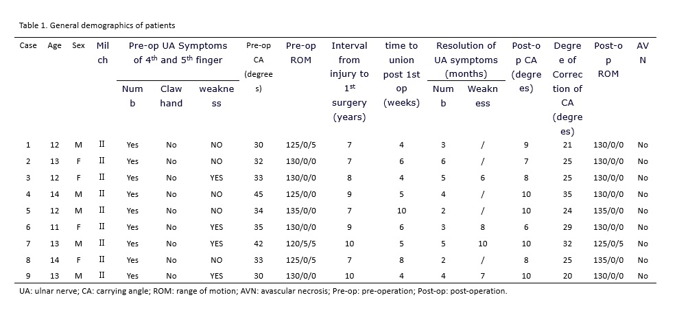
From January 2010 to January 2013, 9 older children (5 males and 4 females) aged average 12.7 years (ranged, 11-14 years) with humeral condylar fracture non-union old elbow valgus deformity were treated. Six were on the left side and three were on the right side. Interval from fractures to the first surgery ranged was 8.2 years on average (range, 7-10 years). The preoperative carrying angle was 35° on average (range, 30°-45°), with 7 cases between 30° and 40° while 2 between 40° and 50°. All patients showed ulnar nerve symptoms. Four cases had functional limitations in the 4th and 5th fingers, manifested by a mild weakness without obvious ulna claw deformity or any fixed deformities in the small joints. Elbow varus stress test showed a lateral instability of the affected elbow in all patients (Table 1).

1.2 Operation method

1.2.1 The first surgical stage was ORIF with bone grafting.

The patients lay on the operating table in a supine position with the affected arm abducted. A tourniquet was applied after induction of general anesthesia followed by skin preparation and draping. After inflation of tourniquet, a lateral longitudinal incision was made, 1/3 of which was distal to the humeroradial joint and 2/3 proximal to the humeroradial joint. The nonunion site of lateral condylar was visualized through the interval between triceps and brachioradialis which was retracted anteriorly and medially. The posterior soft tissue attachment of the fragment should be maintained in case of devascularization of the fragment. The outer periphery callus and fibrous tissue between the fragments were removed to refresh the fracture site, and the hardened bone on the fracture surface was also removed to expose the cancellous bone surface. The fragment was reduced with a point reduction forceps and temporarily fixed with Kirschner wires under direct vision to ensure the anatomic reduction of the fracture. If anatomic reduction was impossible, the fragment should be fixed at a position allowing the best elbow flexion and extension function. Then 4.0 mm diameter hollow screws were used as a final fixation, followed by allograft bone grafting. After the first surgery, a long arm plaster cast was applied to ensure a 90° elbow flexion and neutral position forearm. The cast was removed after 3 weeks. Three weeks after cast removal, functional activity training began.

1.2.2 The second surgical stage was correction of cubital valgus deformity.

Six months after ORIF of the nonunion, the previous internal fixators were removed and a supracondylar medial closing wedge osteotomy was carried out through the proximal part of the original lateral longitudinal incision. After the coronal plane deformity was corrected, the osteotomy was fixed with a lateral reconstruction locking plate and smooth Kirschner wires. External fixation with a cast was unnecessary after the second staged surgery, enabling the patient to begin elbow flexion and extension exercises the second day of recovery. After four weeks, if the elbow function was not fully recovered, patients were permitted to complete passive elbow flexion and extension activities under the guidance of doctors.

Results

Our nine patients had an average of 18.3 months (12.5 to 26.0 months) of follow-up. Approximately three weeks after the first surgery, x-ray examination showed that the original fracture line blurred. After 4 to 10 weeks (average of 6.8 weeks) all non-union fractures healed. After the second surgery, x-ray images of all patients revealed full healing at 5 to 10 weeks, with an average of 7.2 weeks. All patients regained a normal carrying angle of the injured limb, elbow function returned to preoperative status, ulnar nerve symptoms disappeared, and the elbow side pull test was negative. According to the Flynn norms, surgical treatment is considered a failure if elbow functional motion loss is more than 15°, carrying angle valgus angle exceeds 15°, or tie-up in the corner turns over for 5°. However, our clinical results showed that the effect of the staged surgical treatment discussed above was satisfactory (Figure 1).

Discussion

Humeral condylar fracture is a common elbow injury, reaching 16.9% among distal humeral fractures in children ages 2 to 8 years old, with an average of 6 years old. In lateral condylar fractures with no shift or a displacement of less than 2 mm, long arm cast immobilization is effective [1]. However, due to the need to restore the integrity of the articular surface of the distal humerus, internal fixation is considered the optimal choice for most displaced lateral condylar fractures [1-9]. Due to epiphysis, standard x-ray examination of the lateral elbow does not provide a full, clear view of metaphyseal fractures. Therefore, clinicians are unable to properly assess the degree of fracture displacement, leading to possible neglect and misdiagnosis of fractures [2-4]. If a fracture is missed, the treatment becomes difficult and bony abnormalities can occur. Because the fracture surface slopes from the outside to the inside, elbow oblique plain films can show the greatest degree of displacement. Accordingly, a standard x-ray of humeral condylar fractures should include front, side, and oblique views [1-2].

Flynn believes that a fracture of the humeral condylar is considered non-union if the patient has not recovered after 12 weeks of treatment. 2 Non-union occurs when persistent synovial fluid soaks the fracture site, resulting in inhibition of fibrin formation. This leads to secondary barrier callus formation. In addition, other factors contributing to non-union include the continuity stretches of the forearm extensors and the fragile blood supply for the lateral condylar [7-8]. Humeral condylar fracture non-union in children can cause progressive elbow valgus deformity and skeletal dysplasia and is often accompanied by gradual emergence of chronic ulnar neuritis [1]. Moreover, the fracture lines of lateral condylar fractures often extend to the trochlea,  non-union of the fragment or bone absorption may shift the normal position of elbow joint, causing subluxation and instability of the elbow joint [13]. In the case of a non-union, the lateral condylar in the elbow may still allow controlled motion and elbow function may not be impaired. However, in children, ulnar nerve palsy symptoms gradually appear as primary symptoms with lateral condylar fracture non-union [14].

According to reports, the surgery for humeral condylar fracture non-union combined with elbow valgus deformity has a high rate of complications due to the need for ORIF and autologous bone graft. Since the surgery requires the entire complex epicondyle of the humerus bone, an extensor cut is necessary in order to enhance the stability of the lateral condylar, increasing the risk of osteonecrosis of the external humeral condylar [5, 7, 9, 13, 15-16]. In addition, if an abnormal lateral condylar is rigidly stabilized, loss of elbow function is likely to occur. A lateral condylar fracture results in an incomplete elbow pulley surface. Overgrowth of the condylar causes the pulley surface to slope to the radial articular surface, leading to elbow dislocation and lateral elbow instability [13]. Surgical treatment to restore the stability of the elbow without loss of elbow function is difficult. Toh et al. believes that long-term lateral condylar fracture non-union only requires humeral osteotomy line and/or ulnar nerve release pre-surgery and does not immediately require directly addressing the non-union [16]. However, Jakob et al. reported that simple elbow valgus deformity correction cannot completely solve ulnar nerve symptoms [9]. Meanwhile, Dellon recommended initial ulnar nerve release surgery for traumatic ulnar neuritis [17]. According to Dellon’s literature review of over 50 articles reporting more than 2,000 patients, there was no statistical difference in the effect of various treatments for ulnar nerve compression [17]. Abed et al treated nonunion of the lateral humeral condyle using a triple management (fixation of the nonunion site, dome corrective osteotomy, and anterior transposition of ulnar nerve) through a modified para-triceptal approach, the results showed that all patients gained union, all gained excellent or good elbow function according to the Mayo elbow performance score [18]. However, there is still controversy about whether to treat the non-union or not because of complications like stiffness of elbow and avascular necrosis of the fragment [11].

In the past, the main treatment for lateral condylar fractures with non-union was osteotomy surgery to correct elbow valgus deformity [19]. However, osteotomy surgery alone does not address instability of the elbow, and may result in detrimental impact on the patient quality of life. Conversely, the two staged surgery initially manages the stability of the elbow and then focuses on the deformity correction. As such, this approach addresses the deficiencies of conventional treatment modalities. Among our group of patients, the results were satisfactory and largely dependent on the success of the first stage of surgery. The following are key issues in the staged surgical approach:

1) The goal of surgical treatment is to restore the stability of the elbow without losing function. Our experience is that when reducing the distal fragment, shifting the fragment slightly forward will decrease the obstruction of the lateral condylar fragment to the radial head, thus providing the greatest degree of elbow function correction.

2) Hardened scars on the bone surface should be completely removed.

3) The blood supply of the lateral condyle of the humerus should be protected. Studies have shown that lateral condylar epiphyseal vascular supply goes through the posterior part of the articular surface of the condyle to reach the lateral condyle, while protection of the blood supply of the nonunited fragment is the key to successful management [5, 8, 18]. Therefore, the integrity of the soft tissue and vascular supply should be protected.

4) In completing the secondary surgery to correct the valgus deformity in children, the posterior elbow approach should be avoided. The posterior approach requires splitting of the triceps fibers and may impact the function of the elbow. We used the original lateral approach and on-line supracondylar osteotomies to safely manage the cubitus valgus and aesthetic needs.

Conclusions

According to the analysis of more than three years of clinical practice by the authors, staged surgery (ORIF and osteotomy) treatment of humeral condylar fracture non-union old elbow valgus deformity in our population of older children was effective to restore function and aesthetics. Our results suggest that this approach may be one of the best treatment choices for older children with elbow valgus deformity after humeral condylar fracture non-union. However, due to the limited number of cases in this group and short follow-up time, more studies with larger sample sizes and longer follow-ups are recommended.

List of Abbreviations

ORIF: open reduction/internal fixation

Declarations

Ethics approval and consent to participate

This retrospective study was approved by the Ethics Committee of Sichuan Provincial Orthopedics Hospital. Informed consent was waived due to the retrospective nature of this study.

Consent for publication

We had received verbal consent from guardians for publication of clinical details or information.

Availability of data and materials

The data and materials in the current study are available from the corresponding author on reasonable request.

Competing interests

The authors declare that they have no conflict of interest.

Funding

None of the authors received financial support for this study.

Authors’ contributions

XL: Designed and conducted the study, interpreted the data, wrote and edited the manuscript.

LWX: Collected and interpreted the data, edited the manuscript

ZQD: Collected and interpreted the data.

JJY: Collected and interpreted the data.

Final approval of the version to be submitted: XL, LWX, ZQD, JJY.

Acknowledgements

Not applicable

References

  1. Flynn JM, Skaggs DL, Waters PM. Rockwood and Wilkins’ Fractures in Children. Philadelphia: Lippincott Williams & Wilkins; 2014.
  2. Song KS, Kang CH, Min BW, Bae KC, Cho CH, Lee JH. Closed reduction and internal fixation of displaced unstable lateral condylar fractures of the humerus in children. J Bone Joint Surg Am. 2008;90(12):2673-2681.
  3. Ranjan R, Sinha A, Asif N, Ifthekar S, Kumar A, Chand S. Management of Neglected Lateral Condyle Fracture of Humerus: A Comparison between Two Modalities of Fixation. Indian J Orthop. 2018 Jul-Aug;52(4):423-429.
  4. Prakash J, Mehtani A. Open reduction versus in-situ fixation of neglected lateral condyle fractures: a comparative study. J Pediatr Orthop B. 2018 Mar;27(2):134-141. 
  5. Hardacre JA, Nagigian SH, Froimson AI, Bronn JE. Fractures of the lateral condylar of humerus in children. J Bone Joint Surg. 1971;53:1083-95.
  6. Flynn JC. Nonunion of slightly displaced fractures of the lateral humeral condylar in children: an update. J Pediatr Orthop. 1989;691-96.
  7. Flynn JC, Richards JF, Saltzman RJ. Prevention and treatment of non-union of slightly displaced fracture of the lateral humeral condylar in children. J Bone Joint Surg Am. 1975;57:187-92.
  8. Flynn JC, Richards JF. Non-union of minimally displaced fractures of the lateral condylar of humerus in children. J Bone Joint Surg Am. 1971;53:1096-101.
  9. Jakob R, Fowles JV. Observations concerning fractures of the lateral condylar of humerus in children. J Bone Joint Surg. 1975;40:430-36.
  10. MAYLAHN DJ, FAHEY JJ. Fractures of the elbow in children; review of three hundred consecutive cases. J Am Med Assoc. 1958 Jan 18;166(3):220-8. 
  11. Eamsobhana P, Kaewpornsawan K. Should we repair nonunion of the lateral humeral condyle in children?. Int Orthop. 2015;39(8):1579-1585.
  12. Ibrahim MA, Ismail MSAM. Corrective osteotomy and in situ fusion for late-presenting nonunion of lateral condyle fractures of the humerus in adults. J Shoulder Elbow Surg. 2019 Mar;28(3):520-524.
  13. Toh S, Tsubo K, Nishikawa S, et al. Osteosynthesis for nonunion of the lateral humeral condylar. Clin Orthop Relat Re. 2002;405:230-41.
  14. Yin-Chun Tien, Jian-Chih Chen, Yin-Chih Fu, Tsai-Tung Chih, Peng-Ju Huang, Gwo-Jaw Wang. Supracondylar dome osteotomy for cubitus valgus deformity associated with a lateral condylar nonunion in children. J Bone Joint Surg Am. 2005;87:1456-63.
  15. Badelon O, Bensahel H, Mazda K, Vie P. Lateral humeral condylar fractures in children: a report of 47 cases. J Pediatr Orthop. 1988;8:31-4.
  16. Toh S, Tsubo K, Nishikawa S, Inoue S, Nakamura R, Harata S. Long-standing nonunion of fractures of the lateral humeral condylar. J Bone Joint Surg Am. 2002;84:593-8.
  17. Dellon AI. Review of treatment results for ulnar nerve entrapment at the elbow. J Hand Surg (Am). 1989;14(4):688.
  18. Abed Y, Nour K, Kandil YR, El-Negery A. Triple management of cubitus valgus deformity complicating neglected nonunion of fractures of lateral humeral condyle in children: a case series. Int Orthop. 2018;42(2):375-384.
  19. Shaerf DA, Vanhegan IS, Dattani R. Diagnosis, management and complications of distal humerus lateral condyle fractures in children. Shoulder Elbow. 2018;10(2):114-120.