The maxillary sinus, which is the largest of the four sinuses, occupies up a large region of the face [10], and has therefore been the focus of studies in various clinical fields [1–13]. Until now, most studies have been clinical case studies based on sex, but studies have been conducted recently on the growth of the maxillary sinus [2,8,11–13]. According to a study by Jun [4], the maxillary sinus grows until adolescence and the twenties in females and males, respectively. Studies of adults are therefore important considering the lack of literature focusing on this population. While most clinical treatments are performed on adult patients, there have yet to be studies comparing size of the maxillary sinus according to the facial skeletons of adults.
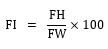
The development and anatomy of the facial skeleton depends on several factors, such as sex, race, socioeconomic status, nutrition, and genetics [9]. These factors are essential for planning orthodontic and other various treatments, and are helpful in predicting potential changes [14]. In particular, facial-skeletal measurements can identify racial differences, and are also useful in the anthropology and forensic science fields [15]. Until now, Angle’s classification has been used for facial skeletal measurements; however, that classification analyzes the relationship between the molars, which may not accurately classify the facial skeleton [9]. A study by Endo [16] indicated that there was no significant difference between sexes in the maxillary sinus size based on Angle’s classification, but there were size differences according to measurements of the tooth face shape. Despite the need to analyzed the maxillary sinus size according to FI classification, no studies have yet been conducted on this topic.
This study classified 60 subjects based on the FI. They comprised 4 mesoprosopic, 14 leptoprosopic, 42 hyperleptoprosopic, and no euryprosopic subjects. The maxillary sinus size was therefore only compared in subjects of mesoprosopic, leptoprosopic, and hyperleptoprosopic types.
About 6.7% of subjects were of the mesoprosopic type. Upon comparing maxillary sinus size, these subjects had the largest RMA-LMA, width, and length. In a study by Jahanshahi[17], which also compared skull size according to FI classification, the distances between the cheekbones, nose width, and mouth width were larger in mesoprosopic than leptoprosopic subjects. Therefore, as for maxillary sinus size, the RMA-LMA, width, and length values of the mesoprosopic type, the midface size of this type was expected to be the largest.
About 70% of the 60 subjects were of the leptoprosopic type. Upon comparing maxillary sinus sizes, these subjects had the largest IOF. Kassab [18] reported that the interpupillary distance, canine arc distance, and incisal width of the central incisor were larger in the leptoprosopic than the mesoprosopic type. Sinavarat[19] reported that there was a strong correlation between the interpupillary distance and canine arc distance. Therefore, considering the maxillary sinus size, the IOF of the leptoprosopic type, which causes a narrow face, was expected to be the largest among the three types.
About 23.3% of the 60 subjects belonged to the hyperleptoprosopic type. Upon comparing maxillary sinus sizes, these subjects had the largest height. Malim [20] reported that the hyperleptoprosopic type had a larger lower part of the face than the leptoprosopic type. The subject with the longest face in the hyperleptoprosopic type was therefore expected to have the largest facial height among the three types.
This study also aimed to determine the association between the facial skeleton and the maxillary sinus. Regression analysis indicated that the FI classification did not affect maxillary sinus size. In addition, the length on the left and the height on both sides were β, indicating that there were differences according to the FI classification. The study by Uchida [21] and Hong [2] indicated that the length and height were correlated with changes maxillary sinus volume. Moore [22] similarly reported that changes in maxillary sinus volume based on age and sex were similar to the changes associated with body growth, such as the height and the development of the wrist bones. It therefore seems possible that the maxillary sinus size changes with the size of the facial skeleton.
In this study, the maxillary sinus size varied based on the shape of the face when classified by FI. The maxillary sinus tended to be wider in those with mesoprosopic type, and tended to be higher in the hyperleptoprosopic type, suggesting a need for clinicians to focus to the shape of the face during clinical treatments. In addition, since the FI classification has revealed an association with the maxillary sinus size, this should be studied further. The results of this study should help to prevent complications during various clinical treatments, and provide valuable data for future research on maxillary sinus growth.