Effect of Massage Before or After Exercise on Delayed-onset Muscle Soreness in a Rat Model

DOI: https://doi.org/10.21203/rs.3.rs-64773/v1

Abstract

Background. Delayed-onset muscle soreness (DOMS), also known as tenderness of touch, refers to the pain caused by muscle mechanical stimulation, such as contraction and stretching. Chinese massage has been widely used in the treatment of sports fatigue and sports injury, but there is controversy in the efficacy. In this experiment, we established DOMS model in rats to observe the prevention and treatment effect of massage, to find the best time for intervention, and to provide scientific basis for the prevention and treatment of exercise fatigue.

Methods. 130 male SD(sprague-dawley, SD) healthy rats were randomly divided into blank group, control group and massage group. Except for blank group, the other rats received DOMS model. Professionals applied kneading and twisting methods on both lower limbs of rats. The expression of IL-2, IL-6 and IL-8 in skeletal muscle of rats were determined by western blot, PCR and ELISA, and the content of serum creatine kinase was determined by ELISA. In addition, we measured the concentration of Ca2+, Ca2+-ATPase in mitochondria of skeletal muscle. The changes of skeletal muscle structure were observed by scanning electron microscopy.

Result. After massage, the expression of IL-2, IL-6, IL-8 and CK decreased compared with control group (P < 0.01), the expression of IL-2, IL-6 and IL-8 in post massage group was lower than that in front massage group (P < 0.01), and the content of CK in front massage group was lower than that in post massage group(P < 0.01). The content of Ca2+ in front massage 24, 48 and 72h group was lower than that in post massage (P < 0.01), the concentration of Ca2+-ATPase in front massage 24h and 72h group was lower than that in post massage group (P < 0.05).

Conclusion. Massage can prevent the injury of muscle and reduce the inflammatory reaction of muscle after exercise. It can also improve the activity of Ca2+-ATPase, enhance the transport of Ca2+ by mitochondria and protect the skeletal muscle.

1. Background

Delayed onset muscle soreness (DOMS) refers to the phenomenon of muscle soreness which often occurs after the body's unadaptable exercise, especially centrifugal exercise, or sudden increase of exercise intensity[1]. It mainly manifests as local tenderness and muscle pain, and often accompany with muscle stiffness, acid swelling, limited joint movement, etc. In severe cases, it may occur acute soft tissue injury related symptoms such as muscle fiber rupture and muscle strength decrease [2]. The symptoms of DOMS are also related to the inflammatory response of sarcomere rupture [3]. At present, the physiological mechanism of DOMS is not clear. In 1902, Hough put forward the hypothesis of "mechanical injury", and thought that DOMS was related to the tear of muscle or connective tissue, as well as the obvious changes of the ultrastructure and contraction structure of skeletal muscle [4]. This is also the first time DOMS has been proposed. The hypothesis of "ischemia spasm" thought that, the pain caused by muscle ischemia after exercise will lead to muscle fiber spasm, aggravate local ischemia and form a vicious circle [5]. Theory of acute inflammatory response hold opinion that Ca2+ trigger a series of inflammatory reactions after mechanical injury [6], and the inflammatory mediators such as neutrophils, eosinophils and basophils are increased [7]. Inflammatory reaction and production of ROS may be the core causes of DOMS, and the interruption of excitation / contraction coupling process of muscle fibers and the disorder of muscle fibers may also be the possible causes [8].

Clinically, the treatment of DOMS mainly include hydrotherapy, ultrasound, vibration, electrical stimulation, laser therapy, anti-inflammatory drugs, acupuncture, etc. But there are different opinions on the evaluation of curative effect [914]. Massage is one of the important methods in treatment of DOMS in traditional Chinese medicine. It has a good effect in reducing pain and improving muscle fatigue [15]. Modern neurophysiology has found that pain is related to the disorder of neurotransmitters such as 5-hydroxytryptamine (5-HT), acetylcholine (Ach), dopamine (DA), β-endorphin (β-EP) and γ-aminobutyric acid (GABA). Research found that massage can reduce the release of 5-HT and DA in peripheral blood, and increase the release in central system [1617], β-EP [18] and GABA [19] also increase. This plays an important role in the process of analgesia.

The dorsal horn of the spinal cord in the nervous system is a very important integration center in the pain afferent system, and also a key part in the mechanism of massage analgesia. In 1965, Melzack and well proposed the "gate control theory", which is the most ideal explanation for dorsal horn neurons. Type A afferent fibers excite SG cells, while type C afferent fibers inhibit. Injury cause Type C afferent tension activity, at this time, the gate is opened. Massage stimulates SG cells by stimulating Type A fibers so as to close the gate, inhibit the activity of dorsal projection neurons (T cells), reduce the transmission of harmful information to the center, and relieve the pain. This explains how massage plays an analgesic role by inhibiting pain signal transmission at the spinal cord level. In addition, massage may promote venous and lymphatic return, decompose inflammatory mediators, improve nutritional metabolism of muscle, repair muscle tissue [20]. Although massage is widely used in treatment of muscle fatigue and injury, there is controversy on efficacy of improving local circulation and assisting muscle function recovery [2123]. At present, most of studies are about the intervention effect of massage on DOMS, while the preventive effect of massage on DOMS is less.Therefore, we established DOMS model in rats, and carried out front and post massage intervention on rats. We measured content of serum creatine kinase (CK), concentrations of mitochondrial Ca2+, Ca2+-ATPase and the expression of IL-2, IL-6, IL-8. We also use scanning electron microscope to observe the changes of rat skeletal muscle. Through the above study to observe the prevention and effect of massage, find the best time for intervention, and provide scientific basis for the prevention and control of sports fatigue.

2. Methods

2.1. animals and groups

130 SPF (specific pathogen free, SPF) male SD healthy rats with an average body weight of 180–200 g (6 weeks old). The animal is provided by Shanghai sipur bikai experimental animal Co., Ltd., license No.: SCXK (Shanghai) 2018-0006. The animals were raised in separate cages with 10 rats in each cage, free diet, natural light, room temperature (20 ± 3) ℃, relative humidity of 40–60%. The rats were used in the animal laboratory for 7 days. According to the random principle, the animals were divided into blank control group, exercise control group and massage group. The massage group was divided into two groups: the experimental group before exercise and the experimental group after exercise. The massage group and the exercise control group were randomly divided into 0 h, 24 h, 48 h and 72 h groups, with 10 rats in each group. The operation process of the experiment strictly complies with the relevant provisions of the regulations of the people's Republic of China on the administration of experimental animals and the guiding opinions on treating experimental animals well.

2.2. Observation index

content of serum CK, concentrations of mitochondrial Ca2+, Ca2+-ATPase and the expression of IL-2, IL-6, IL-8. We also use transmission electron microscope to observe the ultrastructural changes of rat skeletal muscle.

2.3. Main experimental instruments

The antibody was provided by Bio-World technology, Co. Ltd. The gel imaging system comes from Shanghai TianNeng Technology Co., Ltd. (model: 5300). ELISA kit is from bio swamp (China). Use -SYBR ® premier ex Taq Gamma (Takara, rr420a) (Dalian Baosheng biotechnology Co., Ltd. China) carried out quantitative RT-PCR. Primers were synthesized by Shanghai Biotechnology Co., Ltd. (SANGON biotechnology, Shanghai, China). Trizol and cDNA synthesis kits were obtained. Manual strength tester comes from. The skeletal muscle structure was observed with hitachi-650 scanning electron microscope. Ca2+, Ca2+-ATPase test kits are provided by JianCheng company (Nanjing China. product No.: A106-1, A070-4).

2.4. Experimental protocol

2.4.1. DOMS protocol

Except for the blank control group, the other rats were all exercised to in exhaustion. The rats are free to drink and feed the same food. After randomization, the experiment was conducted from 7 to 10 a.m. every day. The experimental animals first swim in a glass cylinder of 100 cm * 60 cm * 70 cm with a weight-bearing capacity of 3% of the weight of the rats. Start a swimming exercise at 9 a.m. In the process of swimming, when the rat's motor coordination decreased significantly, it sank three times in a row for more than 10 s, then take animals out of swimming tank and dry it into the cage with an electric blower at room temperature. The animals in the control group are fed regularly under the same conditions and do not participate in swimming.All the rats were sacrificed by cervical dislocation to obtain the distal colon for future experiments, such as western blot, PCR, enzyme-linked immunosorbent assays (ELISA),and et al.

2.4.2. Massage interventions

The rats in the massage group are fixed on the fixed frame, and the kneading and twisting methods are applied to the lower limbs of the rats for 5 minutes each, and each rat is massaged for 10 minutes. Professional training for massage staff, control force is 4N.The manual strength is 0.4 KG and the frequency is 120–160 times / min.

2.4.3. Western blotting

Fresh rat skeletal muscle tissue was added with RIPA lysate and mixture of protease inhibitor, and the specimen tissue was fully ground. The supernatant was centrifuged and quantified by BCA kit. 10 µg protein samples were separated by SDS-PAGE and transferred to PVDF membrane. After the membrane was sealed by 5% skim milk at room temperature for 2 hours, the first antibody IL-2 (BS60299, Bioworld Technology), IL-6 (MB9296, Bioworld Technology), and IL-8 (BS3479, Bioworld Technology ) and GAPDH (MB001, Bioworld Technology ) were incubated overnight at 4℃, and the membrane was washed with TBST for 3 times, each time for 10 minutes. Add goat anti-rabbit or mouse IgG-HRP second antibody (1:10000) and incubate at room temperature for 2 h. The gel imaging system (Shanghai Tian Neng Technology Co., Ltd., model 5300) was photographed and preserved.

2.4.4. Quantitative real-time polymerase chain reaction(PCR)

Trizol reagent was used to extract 2 µg of total RNA from skeletal muscle tissue, and reverse transcription cDNA was used with revertaid first cDNA synthesis Kit (thermo), -SYBR ® premier ex Taq Gamma (Takara, rr420a) (Dalian Baosheng biotechnology Co., Ltd., China, batch No.: a650t-1) was used for quantitative RT-PCR. Reaction conditions: after 30 seconds pre denaturation at 95℃, 5 seconds at 95℃, and 30 seconds at 60℃, 40 cycles were performed. After the cycle, melting curve analysis was performed to confirm the specificity of amplification. The expression level of GAPDH was used as internal control. Primers were designed by the software primer 5. The primers were synthesized by Shanghai Biotechnology Co., Ltd. and the primer sequence is shown in Table 1.

Table 1

PCR primer sequence

Primer

Sequence (5' to 3')

Base number of alkalis

IL-2-F

IL-2-R

β-actin-F

β-actin-R

IL-6-F

IL-6-R

IL-8-F

IL-8-R

TTGCACTGACGCTTGTCCTCCTTGTCAACA

CCATCTCCTCAGAAATTCCACCACAGTTGC

CTTTTGTGCCTTGATAGTTC

GAGTCCTTCTGACCCATAC

ATT-GTATGAACAGCGATGATGCAC

CCAGGTAGAAACGGAACTCCAGA

CATGGATCTGTCGTAGGGCT

CTGACCAACAGACCAGGGTT

30

31

20

19

25

23

20

20

2.4.5. ELISA

The levels of CK, IL-2, IL-6 and IL-8 were measured by double antibody sandwich method. The purified rat antibody was coated on the microporous plate to make solid-phase antibody. The blood samples of rats and the standards of each factor were added into the microporous of the coated MCAB in turn, and then combined with the antibodies of CK, IL-2, IL-6 and IL-8 labeled by HRP to form the antibody antigen enzyme antibody complex. After thorough washing, the substrate TMB was added to develop the color. The kit is provided by bio swamp (product No.: RA20686, RA20132, RA20607, RA20553). The absorbance (OD value) of the sample was determined by enzyme standard instrument (model: elx800) at the wavelength of 450 nm. The concentrations of CK, IL-2, IL-6 and IL-8 in the sample were calculated by standard curve.

2.4.6. Mitochondrial sampling

Take 1 g of the left gastrocnemius muscle of rat, cut them into pieces on the ice bath, add 5 ml of extraction medium (normal saline), 4℃homogenate for 30 s, and centrifuge for 10 min at 2000r/min. The supernatant was centrifuged at 10000r/min for 15 min. The precipitate obtained by removing the supernatant is mitochondria, which are then suspended in the preservation medium (normal saline) for storage at -20℃.

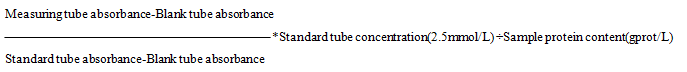
Determination of mitochondrial Ca2+ concentration: The extracted mitochondria were suspended in 1 ml normal saline, grinded by hand in ice bath for 3 minutes with glass homogenizer, and centrifuged at the speed of 2500R/min for 10 min. After centrifugation, the supernatant was mixed with Mtb reagent, alkaline solution, deionized water, 2.5 mmol/L calcium standard solution and protein clarifier. The supernatant was allowed to stand for 5 minutes. The OD value of absorbance at 610 nm was recorded. Calculation formula of calcium content in tissue(mmol/gprot) =

Determination of mitochondrial Ca2+-ATPase: The extracted mitochondria were suspended in 1 ml normal saline, grinded by hand in ice bath for 3 minutes in a glass homogenizer, and centrifuged at a speed of 3500R/min for 10 min. Take 100 µL supernatant to determine phosphorus, and then cool it to room temperature after 20 min water bath at 45℃. The OD value of absorbance at 610 nm was recorded. The amount of 1 µmol inorganic phosphorus produced by protein decomposition of ATP per milligram per hour is regarded as an ATPase activity unit, namely µmolPi/mgprot/hour, Ca2+- ATPase calculation formula =

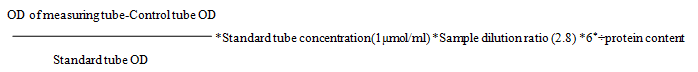
*The reaction time is 10 min, multiplying by 6 is 1 h

Protein content unit: mgprot

2.4.7. Scanning electron microscope of gastrocnemius

Two rats in each group were randomly selected. Within one minute after the rats sacrificed, the left gastrocnemius muscle tissue granules were taken immediately, the size of which was 1*1*1 mm. Fixed solution (1.5-2m1): 4% glutaraldehyde, prepared with PBS (phosphoric acid buffer, pH = 7.2–7.4), stored at 4℃. Muscle tissue granules were fixed, washed with phosphoric acid buffer for 3 times, 10 minutes each time, fixed for 2 hours after 1% osmium acid, washed with phosphoric acid buffer for 3 times, 10 minutes each time, dehydrated with 50%, 70% and 90% ethanol gradients for 15 minutes respectively, dehydrated with 100% ethanol for 3 times, 30 minutes each time. Three times of acetone replacement, three minutes each time, soaking for more than 10 hours, embedding with epoxy resin, polymerizing the embedding plate at 40–60℃ for 48 hours, repairing the ultra-thin section for 50–90 mm, double staining with uranyl acetate and lead citrate for 5–10 min, and observing the changes of myofibrils, myofilaments etc. under scanning electron microscope of Hitachi-650 SEM.

2.5. Statistical analysis

SPSS 22.0 software (IBM SPSS Inc., USA) was used for statistical analysis of the data. Results were expressed as means ± standard error of the mean (SEM). One-way analysis of variance (ANOVA) and Dunnett’s T3 test were performed for statistical analysis. P < 0.05 was considered the level of statistically significant.

3. Result

3.1. Changes of IL-2、IL-6 and IL-8 expression level

3.1.1. Changes of IL-2 expression level

IL-2 plays an important role in inflammatory response and is a key factor in cytokine network. Western blot showed difference in the expression of IL-2 protein between post massage 48 h and 72 h group(P < 0.05); 0 h, 24 h and 48 h, the expression of massage group was lower than that of control group (P < 0.01), and the expression of front massage 72 h group was higher than that of control group. The expression of post massage 48 h group and 72 h group were significantly lower than front massage (P < 0.01) (Fig. 1a, 1b). PCR showed that there was no significant difference in the expression of IL-2 mRNA between post massage 24 h group and 48 h group, and there was significant difference in other experimental groups (P < 0.01); Compared with control group, the expression of front massage 0h、48h、72 h group were lower than control group (P < 0.01), and the expression of post massage group were all lower than control group (P < 0.01),the most significant difference was found in 0 h group;the expression of post massage 0 h and 24 h group was significantly lower than front massage group (P < 0.01) (Fig. 1c). ELISA showed that the level of IL-2 in front massage 0 h, 24 h and 48 h group were lower than control group (P < 0.01);compared with control group, the content of each period in post massage group was lower than control group༌the most significant difference was found in the 48 h group (P < 0.01); the content of each period in post massage group was decreased significantly compared with front massage group in the same period (P < 0.01)༛the content of post massage group was significantly lower than front massage group (P < 0.01) (Fig. 1d).

3.1.2. Changes of IL-6 expression level

Early studies suggested that IL-6 was only expressed in monocyte macrophages, vascular endothelial cells and fibroblasts. In recent years, it has been found that IL-6 can be expressed in most cells, including skeletal muscle cells. Western blot showed that the expression in front massage 0h、24 h group was significantly different (P < 0.01);there was significant difference between post massage 0 h and 72 h group (P < 0.01)༛0 h, 24 h and 48 h, the expression of front massage group was decreased compared with control group (P < 0.01), the expression of front massage 72 h group was increased compared with control group༛the expression of post massage group was decreased compared with control group, and the difference was significant in 72 h group (P < 0.01)༛the expression of post massage 0 h, 48 h and 72 h groups was significantly lower than front massage group, the most significant difference was found in 72 h group (P < 0.01) (Fig. 2a, 2b). PCR showed that there was a significant difference between control 0 h and 24 h group; there was no significant difference between post massage 24 h and 48 h group, but there was significant difference in rest period (P < 0.01); the expression in front massage 48 h and 72 h group was significantly decreased compared with control group (P < 0.01); the expression of post massage group was decreased significantly compared with control group (P < 0.01), 0 h group was the most; the expression of post massage 0 h and 24 h groups was significantly lower than front massage group, 0 h group is the most.(P < 0.01) (Fig. 2c). ELISA showed that there was no significant difference in post massage 24 h and 48 h group, other periods have significant difference (P < 0.01); 0 h, 24 h and 48 h, the content of front massage group was decreased compared with control group (P < 0.01); the content of post massage group was decreased compared with control group, the most significant difference was found in 48 h group (P < 0.01); the content of post massage group in each period was significantly reduced compared with front massage group, the most significant difference was found in 48 h group (P < 0.01) (Fig. 2d).

3.1.3. Changes of IL-8 expression level

IL-8 can promote the release of neutrophils, activate neutrophils, and promote the release of cytotoxic particles, resulting in the inflammatory response of self-tissue damage. Western blot showed that the most significant difference was found in the post massage 0 h and 72 h group (P < 0.01); the expression of front massage group was decreased compared with control group, the most significant difference in the 24 h group (P < 0.01); the expression of post massage group was significantly decreased compared with control group (P < 0.01); the expression of post massage group was significantly decreased compared with front massage group(P < 0.01) (Fig. 3a, 3b); PCR showed that there was no significant difference in post massage 48 h and 72 h group, and significant difference in other periods (P < 0.01); 0 h, 48 h and 72 h, the expression of front massage group was less than control group (P < 0.01), but was not significant in 48 h group (P < 0.05); the expression of post massage group was less than control group, the most significant difference in 0 h group (P < 0.01); the expression of post massage 0 h, 24 h and 48 h groups was significantly lower than front massage group (P < 0.01) (Fig. 3c); ELISA showed that there was no significant difference in post massage 48 h and 72 h group, but significant difference in other periods (P < 0.01); the content of front massage group was less than control group (P < 0.01); the content of post massage group was lower than control group (P < 0.01), the most significant difference was found in 48 h group; the content of post massage group was significantly lower than front massage group (P < 0.01), with the most significant difference in 24 h group (Fig. 3d).

3.2. Changes of serum CK content

CK activity in muscle tissues is higher than other tissues, so serum CK activity has become a common indicator for the diagnosis of some skeletal muscle diseases. ELISA shows that, compared with blank group, the CK content of massage group and control group was increased (P < 0.01); the content of massage group varied significantly at each period,and on declining curve (P < 0.01); the content of front massage group was decreased significantly compared with control group, the difference is significant between 0 h and 24 h groups (P < 0.01); the content of post massage group was less than control group, most significant in0h group (P < 0.01), but the content of 72 h group was more than control group; the content of post massage group was significantly higher than front massage group (P < 0.01),0 h and 48 h group were significantly different (Fig. 4).

3.3. Changes of mitochondrial Ca2+、Ca2+ -ATP concentration

We measured the changes of Ca2+ and Ca2+-ATPase concentration in the mitochondria of the left gastrocnemius muscle of rats. The results showed that compared with the blank group, Ca2+ and Ca2+-ATPase concentration in control group and massage group increased significantly (P < 0.01).

There was a significant difference in the concentration of massage group in each period (P < 0.01), the lowest value was in 0 h group, the highest value was in 24 h group, and the decreasing trend was in 24 h, 48 h and 72 h group. The concentration of Ca2+ and Ca2+-ATPase in massage 0 h group was lower than that in control group (P < 0.01), and the content of 24 h and 48 h group were higher than that in control group (P < 0.01).The concentration of Ca2+-ATPase in massage 72 h group was higher than that in control group (P < 0.05);the concentration of Ca2+ in front massage 0 h group was higher than that in post massage group (P < 0.05), 24 h, 48 h and 72 h group was significantly lower than that of post massage group (P < 0.01);the concentration of Ca2+-ATPase in front massage 24 h and 72 h group was lower than that in post massage group(P < 0.05) (Fig. 5a 5b)

3.4. Ultrastructural observation of skeletal muscle

We observed the changes of myofibrils and filaments of gastrocnemius in rats by scanning electron microscope. In blank group, the structure of sarcomere was intact and the morphology was normal (Fig. 6a). The sarcoplasmic reticulum swelling occurred in different degrees in control group, and contraction, compression, distortion and disintegration were observed in sarcomere and myofibril structure, and edema of myofibrils was found between fibers (Fig. 6b). The swelling of sarcoplasmic reticulum, slight contraction of sarcomere, compression and distortion of some myofibrils were observed in the 0 h and 24 h massage group. 0 h group was damaged more significantly (Fig. 6c, 6d, 6 g, 6 h). 48 h, 72 h group, sarcomere structure was more complete, the morphology was basically normal; the myofibrils were closely arranged, the effect of 72 h group was more significant (Fig. 6e, 6f, 6i, 6j).

4. Discussion

4.1. Analysis of DOMS experimental model

Most of researchers take rats as the objects, and adopt the method of animal experiment to study DOMS. The main exercise methods include downhill running and swimming exhaustion. Swimming is the instinctive exercise of rats, which has a significant effect on the heart and posterior muscle groups [24]. Compared with downhill running, swimming is closer to large amount of exercise in daily life, so we build DOMS model by swimming exhausted exercise. In order to avoid the extra energy consumption caused by cold environment stimulation, we keep the water temperature of the swimming pool between 31–35℃, and static water depth of swimming pool is more than twice the length of rats, so as to prevent rats from using tail to support and rest at the bottom of pool and affect exercise load. For the judgment of swimming fatigue of rats, there are different conclusions, researchers commonly used methods is: rats can't surface again after sinking in water for a certain period (10–60 s, etc.);breathe deeply, rapidly and greatly after leaving the water; eyes are not bright; lie prone; limbs are drooping when held, etc., which can be regarded as fatigue of rats. In practice, the above standards are dangerous. In order to avoid model falling off due to the death of rats, and considering the difference of swimming ability of rats, we think that when the coordination of rats' movement is significantly reduced, repeated sinking occurs, sinking for more than 10 s, and sinking for three times in a row is the fatigue state of rats. We believe that: 1) the judgment of fatigue method of rats is mainly based on the external characteristics, which has certain limitations, and may have the phenomenon of "pseudo exhaustion"; 2) there are differences in swimming ability between rats, different exercise intensity that can be borne, and weight-bearing can affect the results. The above two points need to be paid attention to in the future molding process. We need to pay attention to whether there is a more suitable weight-bearing, and judgment of fatigue state of rats.

4.2. Effect of massage on IL-2、IL-6、IL-8 in DOMS model rats

DOMS can cause muscle tissue damage, and make muscle fibers produce inflammatory reaction, thus increasing the content of inflammatory markers such as neutrophils and eosinophils [2526]. Cytokines are mainly involved in immune response, immune cell differentiation, inflammatory response and tissue repair. Short term or long-term training can affect cytokines.

4.2.1. Effect of massage on IL-2 in DOMS model rats

The effect of muscle injury on IL-2 is complex, and the conclusions are different. The experimental results support that IL-2 increases, decreases and has no obvious change after exercise [2729]. Compared with other inflammatory factors, there are few studies on IL-2. We found that the expression of IL-2 protein and mRNA increased, and the content of serum increased in control group. Massage can significantly reduce the above content. Post massage has a greater impact on IL-2 than front massage. In general, we think 48 h group has the most significant difference. The increase of β-EP after massage is an important mechanism of massage analgesia [30]. Therefore, we believe that massage can improve the level of β-EP, inhibit the release of IL-2, and inhibit mediating effect on inflammation. The pain of DOMS usually reaches the peak at 48–72 hours after exercise[31]. The results of this experiment show that changes of IL-2 in post massage 48 h group are the most significant. Therefore, we speculate that post massage 48 h group has most obvious inhibitory effect on release of IL-2, but more research is needed to prove this hypothesis.

4.2.2. Effect of massage on IL-6 in DOMS model rats

Centrifugal exercise leads to mechanical injury of muscle cells, and tissue fragments act as antigens to activate macrophages. After the injury of skeletal muscle, the permeability of cell membrane changes and the release of IL-6 into the blood can cause the increase of IL-6 expression. Researchers observed the changes of IL-6 after DOMS in traditional Chinese medicine and water bath etc., but the results were different [3233]. We found that expression of IL-6 protein and mRNA increased, and the content of serum increased in control group. The peak time of IL-6 expression was 0 h in control group, and gradually decreased with time. The expression level remained high in 24 h and 48 h group, which was basically consistent with the muscle morphological damage observed by our electron microscope. According to a previous review[34], massage can significantly reduce the content of IL-6, which is one of the most effective treatments for DOMS. We think that IL-6 plays an important role in sports injury and can be used as an important index of DOMS detection. The overall expression of IL-6 in model rats with massage intervention was significantly reduced, and the peak value was decreased, so we analysis that, massage can reduce the injury of skeletal muscle fiber and inflammatory response caused by exercise, inhibit the expression of IL-6, and reduce the symptoms of DOMS: front massage can reduce expression of IL-6, prevent the occurrence of DOMS; post massage can reduce the production of IL-6, thus reduce the symptoms of DOMS, and the prevention and intervention effect of 72 h group is the best. The findings are also in good agreement with the results of electron microscopy.

4.2.3. Effect of massage on IL-8 in DOMS model rats

IL-8 has a two-way regulatory effect on inflammation. Curcumin, Rhodiola, ibuprofen and so on have some influence on IL-8, but there are few studies on this index [3537]. We found that the changes of IL-8 protein level, mRNA expression and serum content in DOMS rats were similar to those of IL-6.Exercise can make body produce stress reaction, activate neutrophils, make skeletal muscle chemotaxis to inflammation, and increase the expression of IL-8.The expression of IL-8 in control group increased significantly, and reached the peak at 0 h. Intense stress leads to immunosuppression, so the expression of IL-8 in the whole group decreased significantly after 72 h of exercise. From the point of the influence of massage before and after exercise on IL-8, we believe that massage can inhibit the activation of neutrophils, reduce muscle injury and inflammatory reaction by inhibiting the expression of IL-8, so as to have a good prevention and intervention on the production of DOMS. The intervention of massage after exercise is better than that before exercise, and the effect of 72 h group is the most obvious. Based on the above analysis, massage can intervene the occurrence of DOMS through the action of cytokines.

4.3. Effect of massage on CK of DOMS model rats

After muscle injury, the leakage of CK enters the lymphatic system through the intercellular fluid, and finally into the circulatory system. Therefore, the muscle tissue injury leads to the increase of serum and muscle enzyme activity. Exercise intensity, muscle injury degree and serum CK activity are in direct proportion [38]. Short-term high-intensity exercise causes mechanical injury of muscle fiber and increases serum CK activity; long-term endurance exercise causes less mechanical injury, and the metabolic change of cell membrane causes normal function loss of muscle cells, increases cell membrane permeability and CK leakage, increase the of serum CK activity. The influence of physical therapy such as electromagnetic field and vibration on CK is controversial [3940]. We found that through massage intervention, serum CK value of model rats in front massage group decreased significantly, which indicated that massage can prevent the muscle damage caused by exercise, reduce the leakage of CK, and increase the clearance of CK in the process of lymph blood circulation, so as to reduce the serum CK activity level [41]. The rule of change is consistent with that of gastrocnemius under electron microscope. We also found that compared with control group, the serum CK in post massage group was lower, but higher than that in front massage group. Therefore, we believe that massage therapy after intensive exercise cannot prevent the damaged cells from leaking CK in time, or even increase the activity of serum CK by accelerating the process of CK entering the blood circulation from lymph. Massage can reduce the activity level of CK, which is a marker of muscle injury, but it needs further study to determine whether massage can recover muscle injury by CK value.

4.4. Effect of massage on Ca2+、Ca2+-ATP in DOMS model rats

Ca2+ is the coupling factor of excitation and contraction of skeletal muscle. Its main function is to promote ATP hydrolysis and provide energy for muscle silk sliding. Mitochondria regulate Ca2+ by internal and external transport, and transport Ca2+ in cytoplasm into mitochondria by the energy produced by Ca2+−ATPase hydrolysis. The uptake of Ca2+ by skeletal muscle mitochondria is affected by the content of Ca2+ in cytoplasm. After exercise, the increase of Ca2+ content in cytoplasm stimulated Ca2+-ATPase hydrolysis to release energy, which enabled mitochondria to take Ca2+ actively and reduced the increase of Ca2+ in cytoplasm to cause muscle damage.

However, when exercise fatigue occurs, the activity of mitochondrial Ca2+-ATPase decreases, which makes the ability of Ca2+ transport into mitochondria decrease, and a large amount of Ca2+ accumulates in cytoplasm, which results in decrease of exercise ability. It is believed that permeability of plasma membrane increases and release of Ca2+ decreases after DOMS injury [42]. Ca2+ exchange through Na+ -Ca2+ and Ca2+-ATPase binding to cytoplasm to support the absorption of Ca2+ by cells [43]. We found that Ca2+ and Ca2+-ATP of mitochondria in control group increased, peaked in 24 h, and then decreased, which was in step with exercise fatigue; The activities of Ca2+ and Ca2+-ATP in massage group were higher than those in control group. We think that massage can interfere with Ca2+ and Ca2+-ATPase: except for 0 h period, the Ca2+-ATPase content of massage group is higher than that of control group. We can think that massage can enhance Ca2+-ATPase activity, then enhance the transport ability of mitochondria to Ca2+, so that protect skeletal muscle tissue. The effect of front massage 24 h group was the most significant. In addition, DOMS leads to the increase of Ca2+ level and Ca2+ accumulation in vacuoles, which may be an effective mechanism for body to reduce Ca2+ content in fibro cytoplasma, so as to maintain the activity of muscle fibers [42].Whether massage can promote the emergence of this mechanism is worthy of further study.

4.5. Effect of massage on the ultrastructure of skeletal muscle in DOMS model rats

The ultrastructural changes of gastrocnemius muscle in DOMS rats were basically consistent with those of other observation indexes. The results of electron microscopy showed that massage group was improved compared with control group, and results of front massage 72 h group were similar to blank group, therefore, we analyzed that the preventive effect of front massage on the structural change of skeletal muscle in DOMS rats was better than post massage.

5. Conclusion

From the results of each index, we find that DOMS is the result of multiple factors and multiple time periods. IL-6 can be used as an important index of DOMS detection. The effect of front massage on CK was better than post massage, and the effect of post massage on IL-2, IL-6 and IL-8 was better than front massage. Combined with the results of Ca2+, Ca2+-ATPase and electron microscopy, we believe that massage can prevent muscle injury caused by large amount of exercise and long-term exercise, reduce the inflammatory reaction of muscle after exercise, and protect skeletal muscle by increasing the activity of Ca2+-ATPase and increasing the transport capacity of mitochondria to Ca2+. Based on comprehensive analysis of experimental results, we believe that post massage 72 h has the best effect on DOMS. However, whether there is a relationship between the inflammatory indexes and whether massage has more influence on transport mechanism of Ca2+ needs further study.

Abbreviations

DOMS: delayed-onset muscle soreness; IL-2: Interleukin-2; IL-6: Interleukin-6; IL-8: Interleukin-8; CK: Creatine Kinase; cDNA: Complementary DNA; ELISAs: Enzyme-linked immunosorbent assays; OD: Optical density.

Declarations

Ethics approval and consent to participate

All experiments were approved by the Laboratory Animal Ethics Committee at Nanjing University of Chinese Medicine (Number: 201908A015) and conducted under the Animal Experiments Guidelines and Animal Care of Chinese Academy of Sciences.

Consent for publication

Not applicable.

Availability of data and materials

The data used to support the findings of this study are available from the corresponding author upon request.

Competing interests

The authors declare that they have no competing interests.

Funding

This study was supported by the Science and technology project of Jiangsu traditional Chinese Medicine Bureau (Nos. YB201849). These funding sources had no role in the design of the study; data collection, analysis, and interpretation of data; or in writing the manuscript.

Authors’ contributions

FL L and QB W designed the experiment. Y Z and ZQ S treated the animals. Q Z, JL G, and J L collected animal blood and tissues and performed laboratory measurements. Q Z and JL G analyzed and interpreted the data. Q Z and QB W were major contributors to the writing of the manuscript. All authors drafted, improved, and revised the manuscript. All authors read and approved the final manuscript.

Acknowledgements

Not applicable.

References

  1. Graven-Nielsen T, Arendt-Nielsen L,“Induction and assessment of muscle pain, referred pain, and muscular hyperalgesia,” Curr Pain Headache Rep, vol. 7, no. 6, pp. 443-451, 2003.
  2. Rynders CA, Weltman JY, Rynders SD, “Effect of an herbal/botanical supplement on recovery from delayed onset muscle soreness: A randomized placebo-controlled trial,” Journal Journal of the International Society of Sports Nutrition, vol. 27, no. 11, 2014.
  3. Manimmanakorn N, Manimmanakorn A, Boobphachart D, et al, “Effects of Zingiber cassumunar (Plai cream) in the treatment of delayed onset muscle soreness,” Journal of Integrative Medicine, 14, no. 2, pp. 114–120, 2016.
  4. Hough, T, “Ergographic studies in muscular soreness,” Boston society of Medical Sciences, vol. 5, no. 3, pp. 81–92, 1902.
  5. Devris, HA, “Quantitative electromyographic investigation of the spasm theory of muscle pain,” American Journal Physical Medicine, vol. 45, no. 6, pp. 119–134, 1966.
  6. Mactintyre DL, “Presence of WBC. decreased strength, and delayed soreness muscle after eccentric exercise, ” Journal of Applied Physiology, vol. 80, no. 3, pp. 1006–1013, 1996.
  7. Kellerer M, Dirk Müller-Wieland, “Differential White Cell Count After Two Bouts of Downhill Running, ” International Journal of Sports Medicine, vol. 19, no. 6, pp. 432–437, 1998.
  8. Jenkins NDM, Housh TJ, Johnson GO, et al., “The effects of anatabine on non-invasive indicators of muscle damage: A randomized, double-blind, placebo-controlled, crossover study, ” Journal of the International Society of Sports Nutrition, 33, no. 10, 2013.
  9. Koeda S, Yoshikawa T, Sato C, et al., “Ultrasound irradiation before high-load exercise reduces muscle rigidity associated with delayed-onset muscle soreness,” Journal of Physical Therapy Science, vol. 31, no. 11, pp. 922–924, 2019.
  10. Ko GWY, Clarkson C, “The effectiveness of acupuncture for pain reduction in delayed-onset muscle soreness: a systematic review,” Acupuncture in Medicine: journal of the British Medical Acupuncture Society, 38, no. 2, pp. 63–74, 2020.
  11. Lu X, Wang Y, Lu J, et al., “Does vibration benefit delayed-onset muscle soreness?: a meta-analysis and systematic review, ” The Journal of Internation Medical Research, vol. 47, no. 1, pp. 3–18, 2019.
  12. Heiss R, Hotfiel T, Kellermann M, et al., “Effect of Compression Garments on the Development of Edema and Soreness in Delayed-Onset Muscle Soreness (DOMS), ” Journal of Sports Science and Medicine, vol. 17, no. 3, pp. 392–401, 2018.
  13. Kawamura T, Suzuki K, Takahashi M, et al., “Involvement of Neutrophil Dynamics and Function in Exercise-Induced Muscle Damage and Delayed-Onset Muscle Soreness: Effect of Hydrogen Bath, ” Antioxidants (Basel), vol. 7, no.10, pp. 127, 2018.
  14. Sellwood KL, Brukner P, Williams D, et al., “Ice-water immersion and delayed-onset muscle soreness: a randomized controlled trial,” British Journal of Sports Medicine, vol. 41, no. 6, pp. 392–397, 2007.
  15. Bervoets DC, Luijsterburg PA, Alessie JJ, et al., “Massage therapy has short-term benefits for people with common musculoskeletal disorders compared to no treatment: A systematic review, ” Journal of Physiotherapy, 61, no. 3, pp. 106–116, 2015.
  16. Field T, Hernandez-Reif M, Diego M, et al., “Cortisol decreases and serotonin and dopamine increase following massage therapy, ” The International Journal Neuroscience, 115, no. 10, pp. 1397–1413, 2005.
  17. Rapaport MH, Schettler P, Breese C, “A preliminary study of the effects of a single session of Swedish massage on hypothalamic-pituitary-adrenal and immune function in normal individuals, ” Journal of Alternative and Complementary Medicine(New York, Y. ), vol. 16, no. 10, pp. 1079–1088, 2010.
  18. Verow H, Dhami M, Howley T, et al., “Spinal manipulative and eta-endorphin: a controlled study of the effect of spinal manipulation and plasma beta-endorphin levels in normal males, ” Journal of Manipulative Physiological Therapeutics, vol. 9, no. 2, pp. 115–123, 1986.
  19. Janssen SP,Truin M,Van Kleef M,et al., “Differential GABAergic disinhibition during the development of painful peripheral neuropathy,” Neuroscience, vol. 184, pp. 183–194, 2011.
  20. Smith LL, Keating MN, Holbert D, et al., “The effects of athletic massage on delayed onset muscle soreness, creatine kinase, and neutrophil count: a preliminary report, ” The Journal of Orthopaedic and Sports Physical Therapy, vol. 19, no.2, pp. 93–99, 1994.
  21. Mancinelli CA, Davis DS, Aboulhosn L, et al., “The effects of massage on delayed onset muscle soreness and physical performance in female collegiate athletes, ” Physial Therapy Sport, vol. 7, no. 1, pp. 5–13, 2006.
  22. Nguyen D, Brown LE, Coburn JW, et al., “Effect of delayed-onset muscle soreness on elbow flexion strength and rate of velocity development, ” Journal of Strength Conditioning Research, vol. 23, no.4, 1282–1286, 2009.
  23. Lawrence MM, Van Pelt DW, Confides AL, et al., “Massage as a mechanotherapy promotes skeletal muscle protein and ribosomal turnover but does not mitigate muscle atrophy during disuse in adult rats,” Acta Physiologica (Oxford, England), vol. 229, no. 3, pp. e13460,
  24. Pierce G N, Kutryk M J B, Dhalla K S, et al., “Biochemical alterations in heart after exhaustive swimming in rats, ” Journal of applied physiology: respiratory, environmental and exercise physiology, vol. 57, no. 2, pp. 326–331, 1984.
  25. Macintyre DL, “Presence of WBC, decreased strength, and delayed soreness muscle after eccentric exercise, ” Journal of applied physiology(Bethesad, Md.: 1985), vol. 80, no. 3, pp. 1006–1013, 1996.
  26. Lieber RL, Fridén J, “Morphologic and mechanical basis of delayed-onset muscle soreness,” Journal of American Academy Orthopaedic Surgeons, vol.10, no. 1, pp. 67–73, 2002.
  27. Lin Y S, Jan M S, Tsai T J, et al., “Immunomodulatory effects of acute exercise bout in sedentary and trainer rats, ” Medicine and science in sport and exercise, vol. 27, no. 1, pp. 73–78, 1995.
  28. Weinstock C, “Effect of exhaustive exercise stress on the Cytokine response, ” Medicine and science in sport and exercise, vol. 29, no. 3, pp. 345–354, 1997.
  29. Kanda K, Sugama K, Hayashida H, et al., “Eccentric exercise-induced delayed-onset muscle soreness and changes in markers of muscle damage and inflammation, ” Exercise Immunology Review, 19, pp. 72–85, 2013.
  30. Wilson R D, “Spinal manipulation and beta-endorphin: a controlled study of the effect of a spinal manipulation on plasma beta-endorphin levels in normal males, ” Journal of Manipulative & Physiological Therapeutics, vol.10, no. 4, pp. 31–32, 1987.
  31. Han K, Kwon O, Jung SY, et al., “Jakyakgamcho-tang in the relief of delayed-onset muscle soreness in healthy adults: study protocol for a randomized, double-blind, placebo-controlled, crossover design clinical trial, ” Trials, vol. 21, no. 1, pp. 211, 2020.
  32. Kawamura T, Suzuki K, Takahashi M, et al., “Involvement of Neutrophil Dynamics and Function in Exercise-Induced Muscle Damage and Delayed-Onset Muscle Soreness: Effect of Hydrogen Bath, ” Antioxidants (Basel), vol. 7, no. 10, pp. 127, 2018.
  33. Shanely RA, Nieman DC, Zwetsloot KA, et al., “Evaluation of Rhodiola rosea supplementation on skeletal muscle damage and inflammation in runners following a competitive marathon, ” Brain Behavior and Immunity, vol. 39, no.7, pp. 204–210, 2013.
  34. Dupuy O, Douzi W, Theurot D, et al., “ An Evidence-Based Approach for Choosing Post-exercise Recovery Techniques to Reduce Markers of Muscle Damage, Soreness, Fatigue, and Inflammation: A Systematic Review With Meta-Analysis, ” Front Physiol, vol. 26, no. 4, pp. 403, 2018.
  35. Drobnic F, Riera J, Appendino G, et al., “Reduction of delayed onset muscle soreness by a novel curcumin delivery system (Meriva®): a randomised, placebo-controlled trial, ” Journal of the International Society of Sports Nutrition, vol. 31, no. 11, 2014.
  36. Nieman DC, Henson DA, Dumke CL, et al., “Ibuprofen use, endotoxemia, inflammation, and plasma cytokines during ultramarathon competition, ” Brain Behavior and Immunity, vol. 20, no. 6, pp. 578–584, 2006.
  37. Kanda K, Sugama K, Hayashida H, et al., “Eccentric exercise-induced delayed-onset muscle soreness and changes in markers of muscle damage and inflammation, ” Exerc Immunol Rev, vol. 12, pp. 72-85, 2013.
  38. Armstrong RB,Ogilvie RW,Schwane JA, “Eccentric exercise induced injury to rat skeletal muscle, ” Journal of Applied Physiology, vol. 54, no. 1, pp. 461–465, 1983.
  39. Lau W Y, Nosaka K, “Effect of Vibration Treatment on Symptoms Associated with Eccentric Exercise-Induced Muscle Damage, ” American Journal of Physical Medicine & Rehabilitation, vol. 90, no. 8, pp. 648–657, 2011.
  40. Zhang J, Clement D, Taunton J, “The Efficacy of Farabloc, An Electromagnetic Shield, in Attenuating Delayed-Onset Muscle Soreness, ” Clinical Journal of Sport Medicine, vol. 10, no. 1, pp. 15–21, 2000.
  41. Wiltshire E, Poitras V, Pak M, et al., “Massage impairs post exercise muscle blood flow and “lactic acid” removal, ” Medicine and Science in Sports and Exercise, vol. 42, no. 6, pp. 1062–1071, 2010.
  42. Cully TR, Murphy RM, Roberts L, et al., “Human skeletal muscle plasmalemma alters its structure to change its Ca2+-handling following heavy-load resistance exercise, ” Nature Communications, vol. 8, pp. 14266, 2017.
  43. Cully TR, Edwards JN, Murphy RM, et al., “A quantitative description of tubular system Ca2+-handling in fast- and slow-twitch muscle fibres,” The Journal of Physiology, vol. 594, no. 11, pp. 2795–2810, 2016.