vMF enhances the sensitivity of MM cells to TMZ in vitro and in vivo
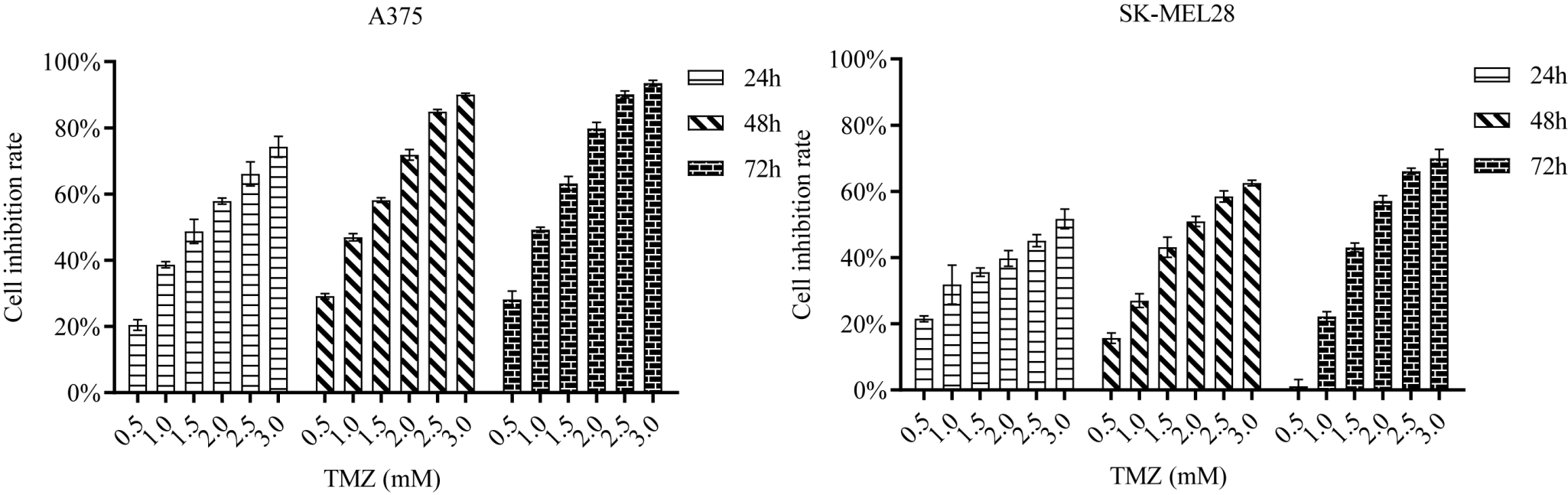
To confirm the appropriate concentration of TMZ for subsequent experiments, the inhibition rates of A375 and Sk-MEL28 cells treated with various concentrations of TMZ were determined in the MTT assay (Fig. S1). As shown in Fig. S1, with an increasing TMZ concentration and application time, the inhibition rates of A375 and SK-MEL28 cells gradually increased. In other words, TMZ inhibited cell growth in a time- and dose-dependent manner. The IC50 values of the A375 and Sk-MEL28 cells were significantly different (see Figure S1), so the TMZ concentration selected in the subsequent MTT assay was different. To confirm whether vMF could enhance the chemosensitivity of MM cells to TMZ, cell inhibition was evaluated with the MTT assay. Briefly, A375 and SK-MEL28 cells were treated with different concentrations of vMF (0.02 and 0.04 μM) and/or TMZ (0.3, 0.6, 0.9, 1.2, 1.5, 1.8, and 2.1 mM or 0.5, 1.0, 1.5, 2.0, 2.5, and 3.0 mM) for 24 h or 48 h (Fig. 1A-B). Compared to TMZ alone, combined treatment with vMF increased the inhibition rate of cells. The IC50 value of the TMZ+vMF group was lower than that of the TMZ group (Tables 1, 2). The results of the MTT assay suggested that the proliferation activity of A375 and Sk-MEL28 cells was decreased significantly by TMZ and that vMF could enhance sensitization to TMZ-induced cytotoxicity. A375 cells were treated with vMF (0.5 or 1 μM) and TMZ (2 mM) for 24 h either alone or in combination, and the number of apoptotic cells was detected by PI/Annexin V-FITC staining (Fig. 1C-D). The results showed that the apoptosis rate increased from 3.37±3.00% in the control group to 28.83±4.02% in the TMZ 2 mM group. Compared with TMZ 2 mM treatment alone, treatment with 2 mM TMZ in combination with 0.5 μM or 1 μM vMF increased the apoptosis rates to 41.37±3.96% and 55.53±5.09%, respectively. The 1 μM vMF group and the control group showed no significant difference in the apoptosis rate (p>0.05). The apoptosis results indicated that vMF can increase TMZ-induced apoptosis in a dose-dependent manner. After treatment, the tumor length and width in nude mice were surveyed every two days, and the tumor volume was calculated. The growth curve of A375 cells in nude mice was drawn with time (day) as the abscissa and tumor volume (cm3) as the ordinate (Fig. 1E). Compared with that in the control group, tumor growth in the TMZ and TMZ+vMF groups was significantly slower (P<0.05 and **P<0.01), and the tumor growth in the TMZ+vMF group was slower than that in the TMZ group (P<0.05). After 21 days of treatment, all groups of nude mice were dissected to remove the xenograft tumors, which were then weighed (Fig. 1E). The tumor weight in the TMZ group and TMZ+vMF group was significantly lower than that in the control group (P<0.05 and **P<0.01), and the tumor weight in the TMZ+vMF group was also lower than that in the TMZ group (P<0.05). The above experimental results indicated that vMF can increase the sensitivity of MM to TMZ in vivo and in vitro.
vMF enhances TMZ-induced DNA damage in melanoma
The protein expression of p-ATR, γ-H2AX, p-ATM, p-chk1 and p-chk2 was detected after A375 cells were treated with vMF (1 and 2 μM) and/or TMZ (1 mM) by western blot analysis (Fig. 2A). Western blotting showed that p-ATR, γ-H2AX, p-ATM, p-chk1 and p-chk2 protein expression in the TMZ group, TMZ+vMF 1 μM group and TMZ +vMF 2 μM group was markedly higher than in the control group (*P<0.05 and **P<0.01). Furthermore, protein expression in the TMZ group, TMZ +vMF 1 μM group and TMZ +vMF 2 μM group increased sequentially. The western blotting results demonstrated that vMF can enhance TMZ-induced DNA damage in a dose-dependent manner. Immunofluorescence staining was used to detect the effect of vMF on the level of the TMZ-induced DNA damage protein γ-H2AX (Fig. 2B). Immunofluorescence staining results suggested that γ-H2AX levels in the control group, TMZ 1 mM group and TMZ 1 mM+vMF 1 μM group increased sequentially. There was no obvious difference between the 1 μM vMF group and the control group. These results indicated that vMF can increase the DNA damage caused by TMZ.
Blocking the ERK pathway downregulates MGMT expression in melanoma
Similar to U0126, vMF can inhibit the MAPK/ERK signaling pathway and reduce MGMT mRNA and protein levels. The protein phosphorylation of ERK1/2 (p-ERK1/2) and MEK1 (p-MEK1) was detected after A375 cells were treated with vMF (0.1, 0.5, 1.0, and 2.0 μM) for 24 h via western blot analysis (Fig. 3C). The western blotting results showed that the expression of p-ERK1/2 and p-MEK1 in the control group, vMF 0.1 μM group, vMF 0.5 μM group, vMF 1.0 μM group and vMF 2.0 μM group decreased sequentially. The results verified that vMF can inhibit the MAPK/ERK signaling pathway. The protein expression of MGMT was detected after A375 cells were treated with vMF (0, 1, 2 and 5 μM) and/or TMZ (1 mM) for 24 h by western blot analysis (Fig. 3A). Fig. 3A shows that vMF can inhibit the expression level of MGMT and that the inhibitory effect increases as the concentration increases. The expression of MGMT mRNA was determined after A375 cells were treated with U0126 (0, 0.5 and 1 μM) (Fig. 3D) or vMF (0, 0.5 and 1 μM) (Fig. 3B) for 2 h, 24 h, 48 h and 72 h by RT-qPCR. From the RT-qPCR results, it can be seen that the expression of MGMT mRNA shows a significant decline with an increase in the U0126 or vMF concentration and the extension of time. The melanomas of the nude mice were stripped, and total protein was extracted. The expression of MGMT, p-ERK1/2, ERK1/2, p-MEK1 and MEK1 was detected in the control, TMZ and TMZ+vMF groups by western blotting (Fig. 3E). The expression of MGMT, p-ERK1/2 and p-MEK1 in the control, TMZ and TMZ+vMF groups decreased in turn. From the experimental results, it can be concluded that vMF can accelerate the depletion of MGMT by TMZ in vivo and that vMF can inhibit the phosphorylation levels of ERK1/2 and MEK1; these results are consistent with those of in vitro experiments.
Overexpression of MGMT attenuates vMF-induced TMZ sensitization
To explore whether vMF enhances the cytotoxicity of TMZ to MM by reducing MGMT, we conducted the following experiment. First, MGMT expression was upregulated in A375 cells by transfection with the pcDNA3.1-MGMT plasmid (Fig. 4A). After transfection for 24 h, A375 MGMT OE cells were treated with 0.2 μM vMF and/or 1 mM TMZ for 36 h, and the expression of MGMT was detected by western blot analysis (Fig. 4B). Western blotting showed that MGMT expression in the MGMT OE-TMZ+vMF group was increased to a greater extent than that in the control-TMZ+vMF group and vector-TMZ+vMF group. Furthermore, the results of MTT assays showed that the IC50 value of the MGMT OE-TMZ+vMF group was markedly increased compared with that of the control-TMZ+vMF and vector-TMZ+vMF groups (Fig. 4C). Our results revealed that MGMT overexpression attenuates vMF-induced TMZ sensitization.
TMZ exposure activates ERK activity in melanoma cells
The protein expression of p-ERK was detected after A375 cells were treated with TMZ (0.5, 1.0, and 2.0 mM) by western blot analysis (Fig. 5). The outcomes showed that the expression of p-ERK1/2 in the control group, TMZ 0.5 mM group, TMZ 1.0 mM group, and TMZ 2 mM group decreased sequentially. The results verified that TMZ exposure can activate ERK activity in melanoma cells.
MGMT expression was positively correlated with p-ERK1/2 in melanoma tissues
The expression and intracellular localization of MGMT and p-ERK in the tissue assay were examined by immunohistochemistry. As illustrated in Fig. 6A, 20 cases were MGMT positive, 28 cases were MGMT negative, 30 cases were p-ERK1/2 positive and 18 cases were p-ERK1/2 negative in the 48 melanoma tissue specimens. Moreover, 19 cases were p-ERK1/2 positive among the 20 MGMT-positive cases, and 17 cases were p-ERK1/2 negative among the 28 MGMT-negative cases (Fig. 6B). The results suggested that the expression of MGMT and p-ERK1/2 in melanoma tissues was positively correlated.