Identification of two novel mutations of ABCD1 gene in pedigrees with X‐linked adrenoleukodystrophy

DOI: https://doi.org/10.21203/rs.3.rs-86131/v1

Abstract

Background: X-linked adrenoleukodysrophy (ALD) is an inherited peroxisomal metabolism disorder, results from the loss-of-function mutation of ATP-binding cassette protein subfamily D1 (ABCD1) gene. The dysfunction of ALD protein, a peroxisomal ATP-binding cassette transporter, results in the excessive saturated very long chain fatty acids (VLCFAs) accumulation in organs including brain, spine and adrenal cortex. X-ALD is characterized as the childhood, adolescent, adult cerebral ALD, adrenomyeloneuropathy (AMN), adrenal insufficiency, and asymptomatic phenotypes, exhibiting a high variety of clinical neurological manifestations with or without adrenocortical insufficiency.

Results: In this study, we reported two cases of X-ALD, which were firstly diagnosed as adrenal insufficiency (Addison’s disease) and treated with adrenocortical supplement. However, both of the cases progressed as neurological symptoms and signs after decades. Elevated VLCFAs level, brain MRI scan and genetic analysis confirmed final diagnosis. In addition, we identified two novel mutations of ABCD1 gene, c.874_876delGAG (p.Glu292del) and c.96_97delCT (p.Tyr33Profs*161) in exon 1 of ABCD1 gene. Sanger sequencing confirmed that the proband’s mother of the first case was heterozygous carrying the same variant. Adrenal insufficiency-only type is very rare, however, it may be the starting performance of X-ALD.

Conclusions: The early warning manifestations should be noticed, and the probability of X-ALD should be considered. This report could be beneficial for the early diagnosis and genetic counseling for patients with X-ALD.

Background

X-linked adrenoleukodystrophy (X-ALD) (OMIM#300100) is a X-linked inherited peroxisome metabolic neurodegenerative disorder, resulted from the defects in ATP-binding cassette protein subfamily D1 (ABCD1) gene. ABCD1 gene locates in chromosome Xq28, contains 10 exons, and codes the adrenoleukodystrophy protein (ALDP), a member of the ABC transporter superfamily. ALDP expresses in the peroxisome membrane, containing 745 amino acids. It plays a key role in VLCFAs transporting into peroxisome for β-oxidation and degradation [1]. The defective ALDP results in the impaired degradation of saturated very long-chain fatty acids (VLCFAs). Excessive VLCFAs accumulate in plasma and tissues including adrenal cortex, cerebral white matter, or spinal cord [2] to exhibit clinical manifestations. Till now, there are more than thousand variants have been reported (X-ALD Mutation Database http://www.x-ald.nl/) [3].

Based on the age of onset and the extent of organs affected, X-ALD is classified into seven subtypes: childhood cerebral, adolescent cerebral, adult cerebral, adrenomyeloneuropathic (AMN), cerebellar, adrenal insufficiency-only (Addison’s disease), and asymptomatic or pre-symptomatic types. X-ALD exhibits a high variety of clinical characteristics [4]. Childhood cerebral ALD (CCALD) and adult-onset AMN are the two most common phenotypes [5]. The clinical symptoms include progressive dysfunction of central and peripheral nervous systems, such asparalysis, behavioral disturbance, abnormalities of sphincter control, sensory loss or paresthesia, with or without adrenal insufficiency. Brain MRI shows a typical pattern of symmetrical enhanced T2WI signal in the bilateral white matter around the lateral ventricle, cerebellum, and the genu of the corpus callosum [6]. Significantly elevated level of VLCFAs is the important diagnostic clue for the individuals with ALD. There are three parameters of VLCFAs to analyze, the concentration of C26:0, the ratio of C24:0/C22:0, and the ratio of C26:0/C22:0 [5]. Final diagnosis relays on the genetic analysis to detect ABCD1 mutation.

Adrenal insufficiency-only type is very rare, especially in females less than 1% [78]. Some individuals were diagnosed as Addison’s disease without symptoms of cerebral or myelopathy ALD for decades. However, the early-onset Addison’s disease may gradually progress to exhibit neuropsychiatric symptoms, showing a time-dependent fellow-up [9].

In this study, we reported two pedigrees of X-ALD patients firstly diagnosed as Addison’s disease, and gradually exhibited variable clinical neuropsychiatric manifestations in their late twenty’s, and showed rapid progression and poor prognosis. In addition, we identified two mutations of ABCD1 gene, c.874_876delGAG (p.Glu292del) and c.96_97delCT (p.Tyr33Profs*161). Further study of genotype-phenotype correlation analysis is needed to provide deeper insights into X-ALD.

Methods And Materials

Patients’ recruitment

Three participants (two family members from a pedigree and one sporadic individual) were recruited. The diagnosis of X-ALD depends on clinical symptoms, biochemical parameters, neurological imaging and genetic analysis. This study was approved by the ethics committee of the Affiliated Hospital of Qingdao University.

Genetic Analysis

Genomic DNA was extracted from peripheral blood sample using the QIAamp Blood DNA Mini Kit (QIAGEN, USA) according to the manufacturer’s protocol. After amplification using 2X polymerase chain reactions (PCR) MasterMix polymerase (Tiangen, China) by ABI9700 PCR (Life technology, USA), the products were captured and purified with Panel probe (illumine Inc. USA), then directly sequenced on the ABI 3500 automated DNA sequencer (Life technology, USA). Mutations of ABCD1 were detected using next generation sequencing (NGS) and subsequently confirmed using Sanger sequencing.

Case Reports and Results

Case 1

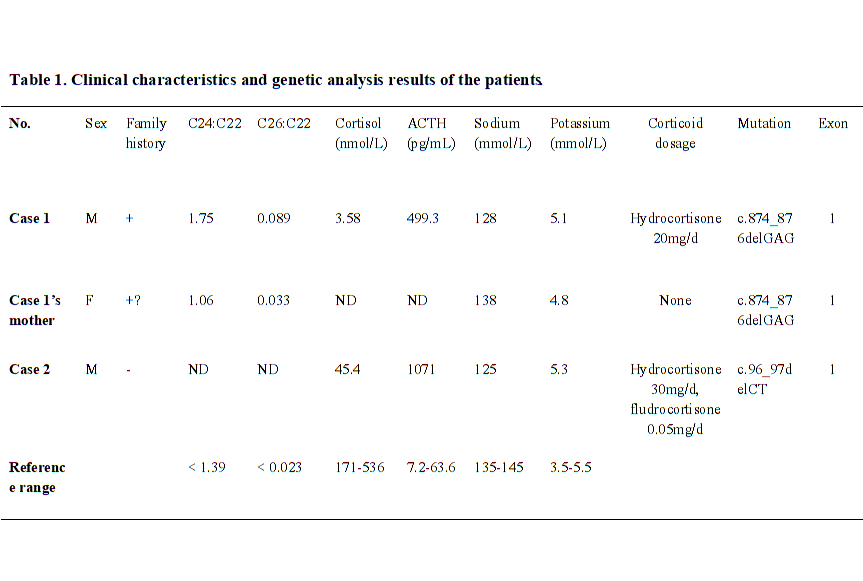
A 27-year old male was presented in hospital with weakness, fatigue, poor academic performance, exhibiting skin and mucous membrane pigmentation, especially in areolae, joints and axilla for two decades. The cortisol at 8am was reduced 3.58 nmol/L (range 171–536 nmol/L), and adrenocorticotropic hormone (ACTH) was increased 499.30 pg/mL (range 7.2–63.6 pg/mL). After diagnosed as primary adrenal insufficiency (Addison’s disease), he was administrated with glucocorticoid replacement (hydrocortisone 20 mg per day) for one year, and the skin pigmentation and weakness were improved. In recently half a year, the patient acted mild cognitive impairment, slurred speech, ataxia, and rapidly progressed as walking instability, incontinence, and paroxysmal blurring vision. Physical examination detected scanty scalp hair, Vgrade muscle strength, hypomyotonia, tendon hyperreflexia (+++), positive Barbinski’s sign and Gordon’s sign. The light and deep reflex were normal. The heel-to-knee test, alternating movement test, and Romberg’s test were negative. The respiratory, cardiovascular, and abdominal examinations were unremarkable. Brain MRI showed symmetrical distribution of hyperintensity long T1 and long T2 signal in of bilateral splenium of corpus callosum, and widened fissures and sulci (Fig. 1A). Serum VLCFA levels were significantly increased (Table 1). Genetic sequencing detected heterozygous mutation of c.874_876delGAG in ABCD1 gene Exon 1, leading to the deletion of translation product glutamate and accumulation of VLCFAs. Sanger sequencing of ABCD1 gene was performed on his family members (Fig. 2A and 2B). His mother was confirmed as the heterozygous point mutation for the same variant. The serum VLCFA levels of his mother were elevated (Table 1). Until now, his mother did not show manifestations of cerebral disorders, myeleterosis, or adrenal insufficiency. His maternal grandmother died at her 55-year-old due to encephalopathy. However, family members were unable to provide valid information, and no blood samples could be obtained for testing. Other siblings of his mother did not showed neuropsychiatric manifestations. According to the clinical manifestations and genetic analysis, a pedigree of X-ALD was constructed. The patient was administrated with hydrocortisone 20 mg per day, mecobalamine, and compound vitamin B. During the fellow-up, the proband showed rapidly progressed neuropsychiatric manifestations including blurred vision, instability of gait, and incontinence within six months.

Case 2

A 31-year-old male was presented with adrenal crisis, exhibiting severe fatigue, anorexia, nausea and vomiting five years ago. The patient had significantly generalized skin pigmentation especially in the gingiva, areolae, and creases of the hands. Laboratory examination revealed severe hyponatremia (serum sodium 108 ~ 125 mmol/L, normal range 145–155 mmol/L), markedly decreased cortisol level (24.83-45.40-35.13 nmol/L) and increased ACTH level (1522-1071-1750 pg/mL) at 8am-4 pm-0am, respectively. The adrenal CT scan showed bilateral adrenal atrophy. At first, the patient was diagnosed as adrenal insufficiency, without exhibiting central or peripheral neuropsychiatric symptoms. After therapy with hydrocortisone (20 mg 8am,-10 mg 4 pm), symptoms of fatigues, vomiting were improved. The patient showed incorrigible severe hyponatremia, only corrected into normal by treated combination with fludrocortisone (0.05 mg per day). The serum sodium levels were maintained in normal range for five years. However, in recent eight months, the patient showed significantly aggravated walking instability, cognitive dysfunction, intellectual disabilities, emotional disturbance, memory loss and incontinence. Neurological physical examination revealed positive Barbinski’s sign and Hoffman’s sign, hyperactive tendon reflexes, and abnormal heel-to-knee-to-toe test. He spoke unclearly with dysarthria, while the vision and hearing were normal. Brain MRI revealed long T1 and long T2 signals in white matter of bilateral frontal lobes, and around the anterior of lateral ventricle, and corpus callosum (Fig. 1B). The patient was sickness and showed poor learning ability during childhood. Gene sequencing showed a heterozygous deletion mutation c.96_97delCT in ABCD1 gene (Fig. 1C and 1D), leading to the frameshift and premature transcription termination of amino acid p.Tyr33Profs*161. His immediate family members (parents and his sister) refused to perform genetic sequencing. His brother also showed sickness in childhood, and died at his eight-year-old. His maternal uncle was weak and fatigued in his youth, and showed disturbance of consciousness from 25-year-old, and died at 28-year old. None of the other family members exhibited neuropsychiatric symptoms and signs of X-ALD. The pedigree of X-ALD was suspected. During the fellow-up, the patient was treated with hydrocortisone and fludrocortisone, and serum sodium level was maintained in normal range. However, he showed rapidly progressed symptoms of incontinence and consciousness dysfunction.

Discussion

X-ALD is a demyelinating and neurodegenerative disorder, due to the loss-of-function mutation of ABCD1 gene. ABCD1 gene codes the peroxisome transporter protein ALDP, which plays an important role in regulating peroxisome oxidation and degradation of VLCFAs [5]. The dysfunction of ALDP, a fatty acid transporter, results in the impaired VLCFA oxidation and excessive VLCFAs accumulation in tissues [5, 10]. Excessive VLCFAs in cytoplasm leads to mitochondrion dysfunction and ER stress, finally caused cell death. In addition, accumulated VLCFAs arouses inflammation, results in demyelinating and neurodegeneration.

In this study, we reported two Chinese X-ALD pedigree. Both of the probands were diagnosed as adrenal insufficiency at beginning without neuropsychiatric symptoms for decades, but developed into neurological disorders at their late 20’s, and showed rapidly progression and poor prognosis. Two novel mutations in exon 1 of ABCD1 gene was detected and suggested to be associated with the disease.

According to the previous reports, the majority is cerebral ALD among those seven types of ALD [11]. CCALD is early onset with a feature of rapidly progressed neuro-inflammatory of cerebral demyelination. Neurological symptoms of AMN commonly occur from the ages of 20 s, with manifests as a chronic progressive paraparesis, accompanied with sensory and motor disturbances [12]. Due to the variable clinical manifestations, some are difficult to make the diagnose at early stage [13]. Adrenocortical insufficiency was the reported as the first clinical manifestation of 38% X-ALD, greater than the group of spinal cord 25% and cerebral disorders 14.5% [9]. To be noticed, some of those male patients diagnosed as adrenal insufficiency may develop with cerebral or myeloneuropathy ultimately, and show a rapid progressive neurological symptoms and poor prognosis. On the contrast, female ALD patients rarely show adrenocortical insufficiency or cerebral ALD. Most of female patients suffer from lately-onset myelopathy [10, 14]. We reported two cases both were firstly diagnosed as Addison’s disease, and then the therapy focused on the adrenocortical replacement. However, after the neurological symptoms and signs become apparent, both of the cases were rapidly progressed. The therapy including fatty acid restriction or neurotrophic treatment did not show therapeutic effect. Therefore, at diagnosis of congenital adrenal insufficiency, the probability of ALD should be fully considered especially for male patients [8, 15]. To make the early diagnosis, family history, MRI scan of brain or spinal cord, VLCFAs measurement, and genetic screening may be helpful for those patients with subtle early neurological manifestations.

We identified two novel point mutations in exon 1 of ABCD1 gene in the pedigree of first case (c.874_876delGAG, p.Glu292del) and in second case (c.96_97delCT, p.Tyr33Profs*161). The genetic sequencing demonstrated nucleotides deletion, resulting in those frameshift mutations. Based on the clinical symptoms and positive neurologic signs, family history, imaging and biochemical characteristics, we suggest those mutations as pathogenic variants. However, the function of ALDP was not further studied, and the biopsy of brain, spinal cord, and adrenal gland were not done. Based on the elevated plasma VLCFAs, the deleterious change of ALDP impaired the cellular beta-oxidation of fatty acid [16].

There are more than thousand mutations have been reported in ALD database (http://www.x-ald.nl/). Among them, 66.9% of them are pathogenic mutations, 8.93% are synonymous, 3.14% are benign, 18.73% are VUS (variant of undetermined significance), and 1.74% are status unknown mutations detected during the screening of newborns (Fig. 3A). More than 78% are point mutations, other types including 12.37% deletion, 2% del-insertion, 3.92% insertion, and 3.14% duplication (Fig. 3B). Most of the mutations locate in the exon (92.94%), others in the intron (6.18%), 5’UTR (0.61%) and 3’UTR (0.26%), respectively [4]. Though some variations may not affect protein translation or function, they still represent polymorphisms. The genotype-phenotype correlationship have not been identified clearly [1718]. In the database, nearly half of the mutations were reported anchoring in exon 1, which encodes the transmembrane domain (TMD) of the protein, contains ligand-specific binding sites and plays an important role in the localization of ALDP [19]. Another clustering of mutations occurs in exon 6, which encode the ATP binding domain of ALDP [20]. ALDP locates in the peroxisome membrane, to transport VLCFAs from cytosol into the peroxisome for degradation. Pathogenic ABCD1 mutations may induce defective the stability of ALDP transmembrane structure region and ATPase activity, to affect the localization of VLCFAs to the peroxisome correctly. As a result, VLCFAs cannot be transported into peroxisomes for β-oxidation, then accumulate mainly in the white matter, neuron axons of central nervous system, and adrenal cortex. Excess VLCFAs accumulation leads to destructive progression by triggering oxidative stress and mitochondrial dysfunction, chronic inflammation, lipid-induced neuron apoptosis, and degenerative changes in the nervous system, and finally leading to neurodegeneration and adrenal insufficiency [3, 21].

Therapeutic strategies include adrenocortical replacement, some of the adrenal insufficiency patients may require both glucocorticoid and mineralocorticoid to maintain water electrolyte balance. For severe adrenal insufficiency, or even adrenal crisis, timely initiation of steroid replacement is essential. Nutrition intervention, for instance, low-fat diet and Lorenzo's Oil may reduce VLCFAs level [22]. In this way, the risk of development with cerebral disorders may be reduced. However, they lack the evidence to improve the symptoms and progression. Another efficiency approach is hematopoietic stem cell transplantation (HSCT), especially in the early stage of the disease [23]. Transplantation with autologous bone marrow transfected in vitro with ABCD1gene modification has been performed [2425]. Bone marrow transplant has been reported in CCALD therapy, most of the patients achieved improved prognosis. However, the pre-existed severe symptoms may influence the outcome after transplantation [26]. In animal experiments, Abcd1 mice show reduction of the mitochondrial biogenesis driven by PGC-1α/PPARγ pathway [27]. Oxidative stress is involved in the physiopathological mechanism of neurodegenerative cascade and mitochondrial depletion [28]. Pioglitazone, a PPARγ/PGC-1α axis activator may modulate the anti-inflammatory and anti-oxidant response. Recently, pioglitazone is under phase II Clinical trials for adrenoleukodystrophy patients, reveals a potential for clinical translation [29]. Therapy options are limited, in addition, some therapeutic approaches relay on the early diagnosis and intervention. Therefore, gene sequencing analysis may be an important tool for newborn screening and genetic counseling.

Conclusions

In this study, we reported two novel mutations of ABCD1 gene in men with X-ALD. These two cases were both considered as adrenal insufficiency at first, and finally diagnosed with X-ALD when neurological manifestations occurred after several years. In these two pedigrees, we identified two novel mutations of ABCD1 gene, c.874_876delGAG (p.Glu292del) and c.96_97delCT (p.Tyr33Profs*161). Screening test for ALD, such as VLCFA concentration and brain MRI scan, should be considered in the early-onset Addison’s disease patients with family history to screen X-ALD early.

Abbreviations

X-ALD: X-linked adrenoleukodystrophy

ABCD1: ATP binding cassette subfamily D member 1;

ALDP: adrenoleukodystrophy protein;

VLCFAs: Very long chain fatty acids;

AMN: Adrenomyeloneuropathy;

ACTH: Adrenocorticotropic hormone;

MRI: Magnetic Resonance Imaging;

ER: endoplasmic reticulum;

ROS: reactive oxygen species;

Declarations

Ethic approval and consent to participate:

 All procedures performed in this study involving human participants were in accordance with the ethical standards of the Affiliated Hospital of Qingdao University with the 1964 Helsinki declaration and its later amendments or comparable ethical standards.

Consent for publication:

The clinical data, images and individual details were obtained from the probands and their family members. All the participants gave their written consent for publication.

Availability of data and materials:

All data analyzed during this study are included in this published article.

Competing interests:

All authors declare that they have no competing of interest.

Funding:

This work was supported by the grant from the China Postdoctoral Science Foundation funded project (Grant No.2018M640615). The content of the article has not been influenced by the sponsors.

Authors’ Contributions:

Bingzi Dong and Wenshan Lv carried out the molecular genetic studies, the design of this study, and drafted the manuscript. Lili Xu participated in the sequence alignment and coordination of this study. Yuhang Zhao, Xiaofang Sun and Bingfei Cheng provided helpful suggestion. Yangang Wang conceived the study, and take responsibility of this study. All authors read and approved the final manuscript.

Acknowledgements:

We are grateful to the patients and their family members for their participation in this study. All authors have no conflicts of interest related to this work.

References

  1. Kemp S, Huffnagel IC, Linthorst GE, Wanders RJ, Engelen M. Adrenoleukodystrophy-neuroendocrine pathogenesis and redefinition of natural history. Nat Rev Endocrinol. 2016 Oct;12(10):606-15
  2. Berger J, Forss-Petter S, Eichler FS. Pathophysiology of X-linked adrenoleukodystrophy. Biochimie. 2014 Mar;98:135-42
  3. Kemp S, Pujol A, Waterham HR, et al., ABCD1 mutations and the X-linked adrenoleukodystrophy mutation database: role in diagnosis and clinical correlations. Hum Mutat. 2001 Dec;18(6):499-515
  4. Engelen M1, Kemp S, de Visser M, et al., X-linked adrenoleukodystrophy (X-ALD): clinical presentation and guidelines for diagnosis, follow-up and management. Orphanet J Rare Dis. 2012 Aug 13;7:51
  5. Chu SS, Ye J, Zhang HW, et al. Eight novel mutations in the ABCD1 gene and clinical characteristics of 25 Chinese patients with X-linked adrenoleukodystrophy. World J Pediatr. 2015 Nov;11(4):366-73
  6. Moser HW, Loes DJ, Melhem ER, et al. X-Linked Adrenoleukodystrophy: Overview and Prognosis as a Function of Age and Brain Magnetic Resonance Imaging Abnormality. A Study Involving 372 Patients. Neuropediatrics, 2000, 31(05): 227-239
  7. Engelen M, Barbier M, Dijkstra IM, et al. X-linked adrenoleukodystrophy in women: a cross-sectional cohort study. Brain. 2014 Mar;137(Pt 3):693-706
  8. Horn MA, Erichsen MM, Wolff AS, et al. Screening for X-linked adrenoleukodystrophy among adult men with Addison's disease. Clin Endocrinol (Oxf). 2013 Sep;79(3):316-20.
  9. Huffnagel IC, Laheji FK, Aziz-Bose R, et al. The Natural History of Adrenal Insufficiency in X-Linked Adrenoleukodystrophy: An International Collaboration. J Clin Endocrinol Metab. 2019 Jan 1;104(1):118-126
  10. Engelen M, Kemp S, Poll-The BT. X-Linked Adrenoleukodystrophy: Pathogenesis and Treatment. Curr Neurol Neurosci Rep. 2014 Oct;14(10):486.
  11. Jiang MY, Cai YN, Liang CL, et al. Clinical, biochemical, neuroimaging and molecular findings of X‐linked Adrenoleukodystrophy patients in South China. Metab Brain Dis. 2015 Dec;30(6):1439-44
  12. Tran C, Patel J, Stacy H, et al. Long-term outcome of patients with X-linked adrenoleukodystrophy: A retrospective cohort study. Eur J Paediatr Neurol. 2017 Jul;21(4):600-609.
  13. Paláu-Hernández S, Rodriguez-Leyva I, Shiguetomi-Medina JM. Late onset adrenoleukodystrophy: A review related clinical case report. eNeurologicalSci. 2019 Jan 10;14:62-67
  14. Hsieh S, White PC. Presentation of primary adrenal insufficiency in childhood. J Clin Endocrinol Metab. 2011 Jun;96(6):E925-8
  15. Eng L, Regelmann MO. Early Onset Primary Adrenal Insufficiency in Males with Adrenoleukodystrophy: Case Series and Literature Review. J Pediatr. 2019 Aug;211:211-214.
  16. Huffnagel IC, van de Beek MC, Showers AL, et al. Comparison of C26:0-carnitine and C26:0-lysophosphatidylcholine as diagnostic markers in dried blood spots from newborns and patients with adrenoleukodystrophy. Mol Genet Metab. 2017 Dec;122(4):209-215
  17. Wiesinger C, Eichler FS, Berger J. The genetic landscape of X-linked adrenoleukodystrophy: inheritance, mutations, modifier genes, and diagnosis. Appl Clin Genet. 2015 May 2;8:109-21
  18. Wang C, Liu H, Han B, Zhu H, Liu J. A novel ABCD1 gene mutation causes adrenomyeloneuropathy in a Chinese family. Brain and Behavior. 2019 Oct;9(10):e01416
  19. Procko E, O'Mara ML, Bennett WFD, et al. The mechanism of ABC transporters: general lessons from structural and functional studies of an antigenic peptide transporter. The FASEB Journal, 2009, 23(5): 1287-1302
  20. Niu YF, Ni W, Wu ZY. ABCD1 mutations and phenotype distribution in Chinese patients with X-linked adrenoleukodystrophy. Gene. 2013 Jun 10;522(1):117-20
  21. Turk BR, Theda C, Fatemi A, Moser AB. X-linked Adrenoleukodystrophy: Pathology, Pathophysiology, Diagnostic Testing, Newborn Screening, and Therapies. Int J Dev Neurosci. 2019 Nov 25. pii: S0736-5748(19)30133-9.
  22. Aubourg P, Adamsbaum C, Lavallard-Rousseau MC, et al. A two-year trial of oleic and erucic acids (“Lorenzo’s oil”) as treatment for adrenomyeloneuropathy. N Engl J Med. 1993;329(11):745–52
  23. Eichler F, Duncan C, Musolino PL, et al. Hematopoietic Stem-Cell Gene Therapy for Cerebral Adrenoleukodystrophy. N Engl J Med. 2017 Oct 26;377(17):1630-1638
  24. Cartier N, Hacein-Bey-Abina S, Bartholomae CC, et al. Hematopoietic stem cell gene therapy with a lentiviral vector in X-linked adrenoleukodystrophy. Science. 2009;326(5954):818–23
  25. Kühl JS, Kupper J, Baqué H, et al. Potential Risks to Stable Long-term Outcome of Allogeneic Hematopoietic Stem Cell Transplantation for Children With Cerebral X-linked Adrenoleukodystrophy. JAMA Netw Open. 2018 Jul 6;1(3):e180769.
  26. Miller WP, Rothman SM, Nascene D, et al. Outcomes after allogeneic hematopoietic cell. Transplantation for childhood cerebral adrenoleukodystrophy: the largest single-institution cohort report. Blood. 2011;118(7):1971–8
  27. Lu JF, Lawler AM, Watkins PA, Powers JM, Moser AB, Moser HW, et al. A mouse model for X-linked adrenoleukodystrophy. Proc Natl Acad Sci U S A. 1997;94(17):9366–71
  28. Powers JM, Pei Z, Heinzer AK, Deering R, Moser AB, Moser HW, et al. Adreno-leukodystrophy: oxidative stress of mice and men. J Neuropathol Exp Neurol. 2005;64(12):1067–79
  29. Pujol A. Novel Therapeutic Targets and Drug Candidates for Modifying Disease Progression in Adrenoleukodystrophy. Endocr Dev. 2016;30:147-60