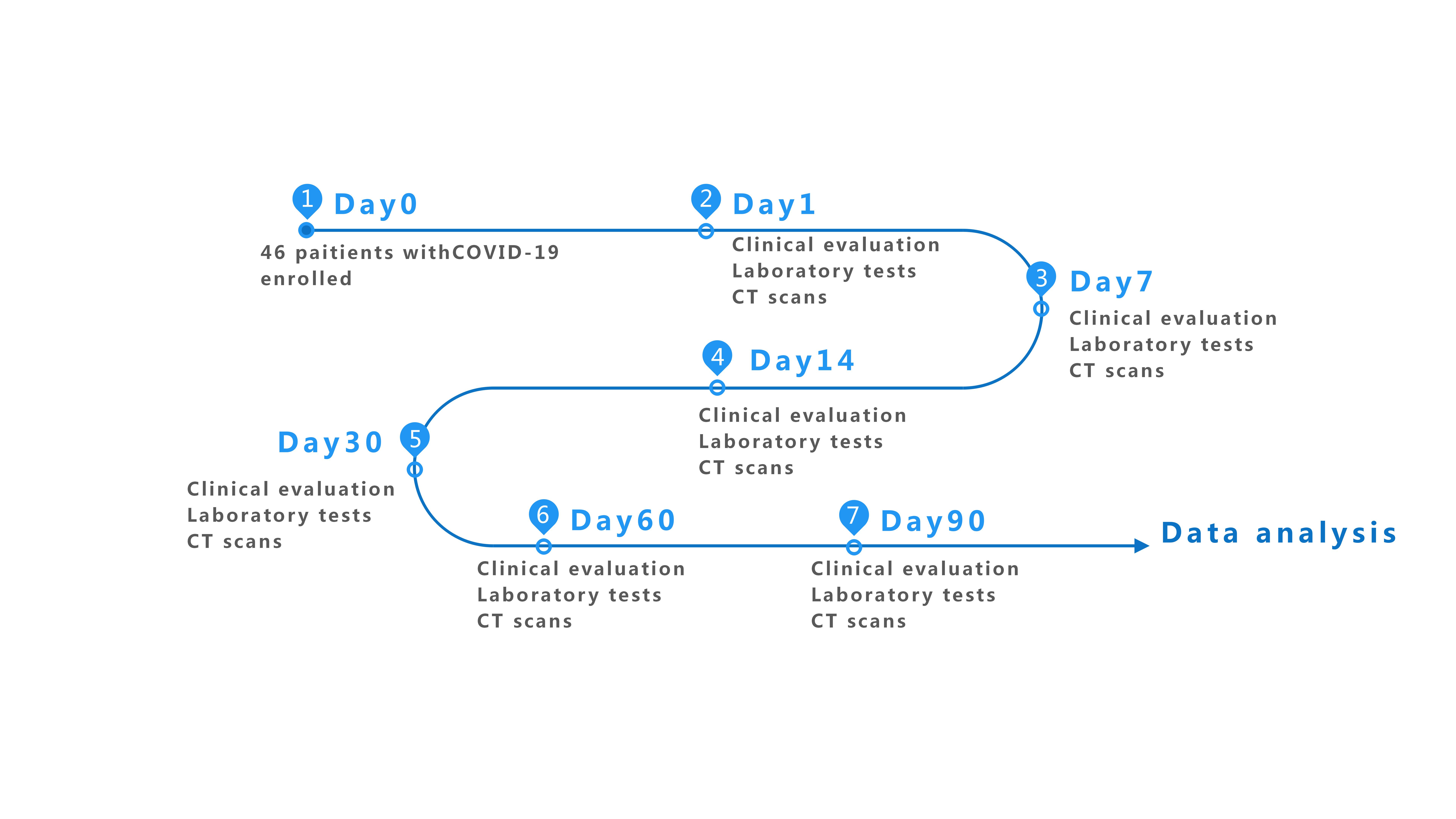
At present, most patients with COVID-19 have been discharged after appropriate medical treatments, but there remains concern that lung injury, including pulmonary fibrosis, might lead to long-term impairment following infection. In our study, we paid attention to the follow-up of these recovered patients with COVID-19. Clinical features between patients with mild-to-moderate or severe fibrosis were compared, and we attempted to identify monitors and predictors for the development of pulmonary fibrosis by combining follow-up clinical data with disease progression during 90 days .
In this study, 35 recovered patients with COVID-19 were followed up, of which 76.29% (26/35) displayed severe fibrosis on CT images. This finding further suggested that fibrosis might be a familiar comorbidity in COVID-19. Laboratory testing and clinical evaluation were performed to identify the progression of the disease. Although many routine studies have shown that routine blood indexes (leuko- and lymphopenia), liver enzymes (mildly elevated LDH and ALP), and inflammatory indicators (increased levels of CRP and IL-6) have good diagnostic values and are associated with pneumonia and extensive lung damage[4, 16, 17], most of them show no obvious changes due to mild inflammatory reactions during recovery. This is not consistent with an associated study, which showed that compared with control individuals, levels of PCT, ESR, and all fibrosis indicators (TGF-β, CCL18, PIIIP, HA, LN, and CIV) were increased in patients with COVID-19. PIIIP, HA, LN, and CIV have been applied widely as serum markers of hepatic fibrosis in clinical practice[18, 19]. Recent reports have indicated that levels of these indicators are also increased in patients with idiopathic pulmonary fibrosis (IPF), which reflected its prognosis and severity[20]. Pulmonary fibrosis is the end-stage pathological changes of many lung diseases characterized by the proliferation of fibroblasts, excessive deposition of extracellular matrix (ECM) and unsuccessful reconstruction of the destructed alveolar epithelium, as well as inflammatory damage and normal lung structure destruction. PIIIP, HA, LN, and CIV are associated with the ECM, suggesting their potential as diagnostic biomarkers for pulmonary fibrosis[21]. In our study, although there were no obvious differences between the mild-to-moderate fibrosis group and the severe fibrosis group because of the small sample size at the initial observation point, HA, LN, and CIV gradually decreased with the improvement of fibrosis during the 90 days’ follow-up. These results further indicate their potential as monitoring biomarkers for pulmonary fibrosis in patients with COVID-19, yet larger sample's cohort studies, as well as further prospective studies, are needed to prove this point.
In addition to the above mentioned four fibrosis indicators, numerous reports have indicated that TGF-β (anti-inflammatory cytokine) and CCL18 (chemokine) are closely related to extensive lung injury and pneumonia fibrosis[22, 23]. In our study, baseline serum levels of the two markers were obviously increased in patients with COVID-19. During the follow-up period, elevated levels of serum TGF-β and CCL18 were associated with pulmonary dysfunction and poorer prognosis of pneumonia fibrosis. COVID-19 is ascribed to “cytokine storm” characterized by largely uncontrolled inflammatory responses [24]. Nevertheless, few published reports have displayed circulatory concentration of TGF-β and CCL18 in patients with COVID-19, and most focus on pro-and anti-inflammatory cytokines such as IL-6, TNF-α, IL-1β, and so on. In this respect, our findings supplement the doctrine of a cytokine storm in COVID-19. In addition, our study also illustrated that circulatory levels of TGF-β and CCL18 were capable of differentiating severe fibrosis patients from mild subjects with COVID-19, suggesting their potential as evaluation and monitoring biomarkers.
Generally, it is considered that TGF-β is a central medium of the repair phase of tissue damage, adjusting immunity and inflammation response and promoting scar formation. Series of studies on acute pulmonary injury thoroughly evaluated the role of TGF-β during the late phases of tissue repair and demonstrate that it is associated with the development of pulmonary fibrosis[25]. It plays a critical role in epithelial–mesenchymal interactions during alveolarization and lung branching morphogenesis, and involve in the regulation of multiple cellular processes, such as the differentiation of alveolar epithelial cell, the proliferation of fibroblasts, excessive deposition of ECM, and growth inhibition of epithelial cells[26]. In patients with COVID-19, large numbers of neutrophils are infiltrated into the lung tissues by the virus and release stored TGF-β resulting in apoptosis of numerous cells, such as pneumocytes, bronchial epithelial cells, T-lymphocytes, and even neutrophils themselves [27]. Consequently, it induces more macrophages migration and infiltration into the lungs, with the resultant production and secretion of large amounts of TGF-β, which leads to tissue remodeling of lung injury in COVID-19[28, 29]. These may be the main possible sources of massive increases in TGF-β.
CCL18 as a CC-chemokine is derived from alveolar macrophages and acts as a chemoattractant. Some studies reported that CCL18 could promote collagen production and induce chemotaxis of lung fibroblasts in vitro[30]. In IPF, serum CCL18 is increased and negatively correlated to pulmonary function[23, 31], which is consistent with our results in COVID-19. Another prospective study also demonstrated that CCL18 was an independent factor related to the mortality of IPF patients (HR 1.98, 95% CI 2.49–25.51, P < 0.05)[32]. Similarly, elevated CCL18 levels are associated with poorer prognosis of pneumonia fibrosis[23, 33]. Therefore, we speculated that the uncontrolled and sudden increases of TGF-β and CCL18 in COVID-19 (possibly accompanied with other inflammatory cytokines) may participate in progression to fibrosis. Therefore, TGF-β and CCL18 blockade may be beneficial for COVID-19 patients.
Certainly, there are several limitations in the present study. Initially, the sample size of 35 patients was relatively small, which could have lead to some potentially useful biomarkers or clinical characteristics being missed. Furthermore, only severe COVID-19 patients were included in this study, and fibrosis indexes in asymptomatic or mild patients were not further followed up. This might have introduced a selection bias for some data. Finally, control composition only included the subjects without pneumonia. Patients with other lung diseases, especially IPF, were not included. Thus, the diagnostic and predictive utility of these indicators mentioned in this study were not fully evaluated.