Bacterial Cultivation
The phyllosphere bacteria were from Atma Jaya Catholic University of Indonesia culture collections in cryopreservation. These bacteria were from previous research and recovered from Psidium guajava, Averrhoa carambola, and Anredera cordifolia leaves [8,9]. Bacteria were grown in Luria-Bertani Agar (LA) and were incubated at 28°C for 48 hours. After that, single colony was picked and grown in King’s B medium and incubated at 28°C for 48 hours.
Pathogenic bacteria used were B. cereus ATCC 14579, S. aureus ATCC 29213, E. faecalis ATCC 33186, P. aeruginosa ATCC 27853, S. typhimurium, V. cholerae. All pathogenic bacteria were from cryopreservation and were streaked onto LA then incubated 37°C overnight.
Primary Screening of Anti-Quorum Sensing Activity
The monitor strain C. violaceum was grown separately in 50 mL of LB broth medium and incubated at 28°C, 120 rpm for 48 hours. Phyllosphere bacteria were streaked onto LA in a straight line then incubated at 28°C for 24 hours. After that, 100 μL of monitor strain (OD600=0.132) were put into 2 mL semisolid agar (0.75% agar) for overlay on top of the phyllosphere isolates which had been streaked before. These plates were incubated at 28°C overnight. A positive result indicated by inhibiting violacein pigmentation (opaque zone) of the C. violaceum around the streak of the phyllosphere isolates [10].
Production of Crude Extract
Isolates that had given positive result from the primary screening of anti-quorum sensing activity were extracted by using liquid-liquid extraction. The bacterial culture were inoculated into 100 mL of Luria Bertani Broth (LB) then incubated in orbital shaker incubator at 28°C for 48 hours 120 rpm. After that, centrifuged at 13888 xg for 15 min and cell-free supernatant was harvested and mixed with an equal volume of ethyl acetate. The solvent layer was harvested and evaporated in a rotary evaporator. After that, extract evaporated in an oven vaccum overnight to obtain the crude extract. To this, 1% of Dimethyl Sulfoxide (DMSO) will be added to obtain a final concentration of 5, 10, and 20 mg/mL stock (w/v) and kept at -20°C [11].
Antibacterial Activity Assay
The crude extracts that had been obtained were tested against pathogenic bacteria such as B. cereus, E. faecalis, and S. aureus, P. aeruginosa, S. typhimmurium, and V. cholerae using agar well diffusion method. Pathogenic bacteria were streaked continuously on Brain Heart Infusion Agar (BHIA). Then, the extracts were applied 50 μL of 5, 10, and 20 mg/mL solution to the well. Streptomycin (Merck; 10 mg/mL) were used as positive control, whereas DMSO was used as negative control. The plates were incubated at 37°C for 24 hours. This assay was performed in triplicate [12].
Detection of Anti-Quorum Sensing Activity
The crude extracts were tested for anti-quorum sensing activity against C. violaceum by agar well diffusion method. C.violaceum was streaked on LA with a sterile cotton swab. Then the extracts (50 μL) with various concentration (5, 10, and 20 mg/mL) were applied to the well. DMSO was used as a control. The plates were incubated at 28°C for 24 hours. Anti-quorum sensing activity was observed through a turbid halo zone against a background of violacein pigment. This assay was performed in triplicate [10]
Quantification of Antibiofilm Activity
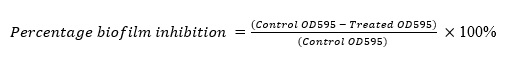
The pathogenic bacteria were inoculated into BHIB and incubated overnight. After that, for biofilm inhibition test, 100 µL of crude extracts and 100 µL of bacterial cultures (OD600=0.132) were transferred into 96-well microtiter plates (polystyrene) then incubated at 37°C for 24 hours. Meanwhile for biofilm destruction test, 100 µL of bacterial culture were transferred into 96-well microtiter plates then incubated. After that, 100 µL of crude extracts will be added and incubated at 37°C for 24 hours. Then planktonic cells and media were discarded. Adherent cells were rinsed gently twice with distilled water and allowed to air dry. The biofilms were stained by 200 μL of 0.4% (w/v) crystal violet solution for 30 minutes. After that, the dye were discarded and the wells were rinsed twice with distilled water. The wells were air dried and then 200 µL of ethanol were used to solubilize the crystal violet. The optical density were determined at 595 nm using a microplate reader. BHIB was used as blank and bacterial cultures without extracts were used as control. This test was performed triplicate [13]. (see Formula 1 in the Supplementary Files)
Microscopic Observations
This step was done using Scanning Electron Microscope (SEM) at Dexa Laboratories of Biomolecular Sciences (DLBS). First, B. cereus and S. typhimurium were grown in BHIB and incubated overnight. Then, bacteria were spotted to a steril cover glass and incubated overnight to form biofilm. After that, crude extracts were spotted into the biofilm and incubated at 37°C overnight. At the last step, the results were analyzed using SEM at DLBS [14].