Animal care and use statement
The protocols were designed to minimize pain or discomfort to the animals. All experimental procedures were in accordance with the European Community guiding principles on the care and use of animals (86/609/CEE; Official Journal of the European Communities no. L358; December 18, 1986), French Decree no. 97/748 of October 19, 1987 (Journal Officiel de la République Française; October 20, 1987), and the recommendations of the Cenomexa ethics committee (#23767).
Experiments were performed on adult Sprague Dawley female rats (Janvier Labs, Le Genest-Saint-Isle, France) at eight–ten weeks of age (average weight 260 – 280g).
Female rats were housed (two rats/cage) in secure conventional rodent facilities on a 12-hr light/dark cycle with constant access to food and water.
Our two procedures were composed of two main experimental groups:
SCI control group: animals received SCI.
SCI + STM group: animals received SCI and rTSMS treatment during 15 days.
For the two procedures histological analyses have been performed at 15 and 60 days after SCI. Functional analyses including plantar and locotronic tests have been performed at 15, 30 and 60 days after SCI.
For the second procedure only (contusive SCI), MRI experiments have been performed at 7, 21 and 42 days after SCI.
A total of 104 rats have been included in the entire study (procedure 1 and procedure 2).
A general overview of these experiments is presented in Figure 1.
Experimental design
Procedure 1
Surgical procedure
In order to assess the effects of the rTSMS treatment and to compare them to those described in mice, first we performed penetrating SCI in rats 9,16.
To do so, rats received 30 min before surgery a subcutaneous injection of buprenorphine hydrochloride (0,3 mg/mL). Then, rats were anesthetized with 2% of isofluorane during the entire surgery (Iso-Vet, Osalia, Paris, France). Animal's body temperature has been kept steady at 37°C with a heating pad during entire surgical intervention. After being shaved and disinfected with betadine solution, the dorsal skin of the rats was incised, the superficial fat gently shifted, and the muscle tissue dissected to expose laminae T9–T11. Posterior part of vertebrae was countersank in order to create an ample space for lesion. SCI were performed at T10 level as described previously 9,16. After laminectomy, the dura mater was removed and a complete transection of the spinal cord was performed with 25-gauge needle. After surgery, rats underwent daily check, none of them showed neither skin lesion, infection, nor autophagy throughout the study.
For this procedure 56 rats have been used (Figure 1):
- 2 groups of 8 rats for the immunohistological experiments 15 days after SCI
- 2 groups of 20 rats for functional tests and for immunohistological experiments 60 days after SCI
Procedure 2
Surgical procedure
In Humans, SCI are mainly due to contusive injuries, thus in a second time we investigated the effects of rTSMS in a contusion model in rats. A moderate/severe lesion model has been chosen due to the fact that 60% of the lesions in Humans are incomplete 14.
The surgery was performed as described above except for the lesion step.
Indeed, after laminectomy, the dura mater was removed and contusion injury was applied using a force-controlled spinal cord impactor (IH-0400 Impactor, Precision Systems and Instrumentation LLC, USA). The applied force was set to 175 kdyn. The spinal cord displacement induced by the impact was measured for each animal. After surgery, rats underwent daily check, none of them showed neither skin lesion, infection, nor autophagy throughout the study.
For this procedure 56 rats have been used as described above for the procedure 1 (Figure 1).
rTSMS treatment
rTSMS was delivered with a commercially available figure of eight double coil featuring an air cooling system connected to a Magstim rapid2 stimulator used for focal cortical and peripheral stimulations (Magstim, Whitland, UK). The coil was positioned in close contact with the back of the animal at the site of injury. The size of the area stimulated has been defined according to the manufacturer’s device manual. The area stimulated was 1.5 cm2. The position of the coil was maintained using an articulated arm stand. The exact position of the coil was defined using the mark located in the middle of the coil. rTSMS treatment was applied at a frequency of 10 Hz, 10 min per day during 14 days. Stimulation protocol consisted of 10 s stimulation followed by 20 s of rest. Rats were kept under anesthesia with 2% of isoflurane during stimulation; the equivalent anesthesia were used for untreated animals. Peak magnetic intensity at the experimental distance was 0.4 T.
Functional test
Locotronic test: Foot misplacement apparatus
Experiments have been performed as described previously by Chalfouh et al. (Intellibio, Nancy,
France) 9. The equipment consists of a flat ladder on which the animal can move from the starting zone towards the arrival zone. On both sides of the ladder, infrared sensors allow the visualization and recording of the displacement of the animal. The location and precise length of time of all the errors are recorded, in distinguishing the errors from front legs, back legs and tail. Based on all data recorded; number of back legs errors, total back legs errors time and total crossing time were provided by the software and compared between groups of animals.
All the rats were pre-trained on the ladder, one week prior to injury to provide baseline data.
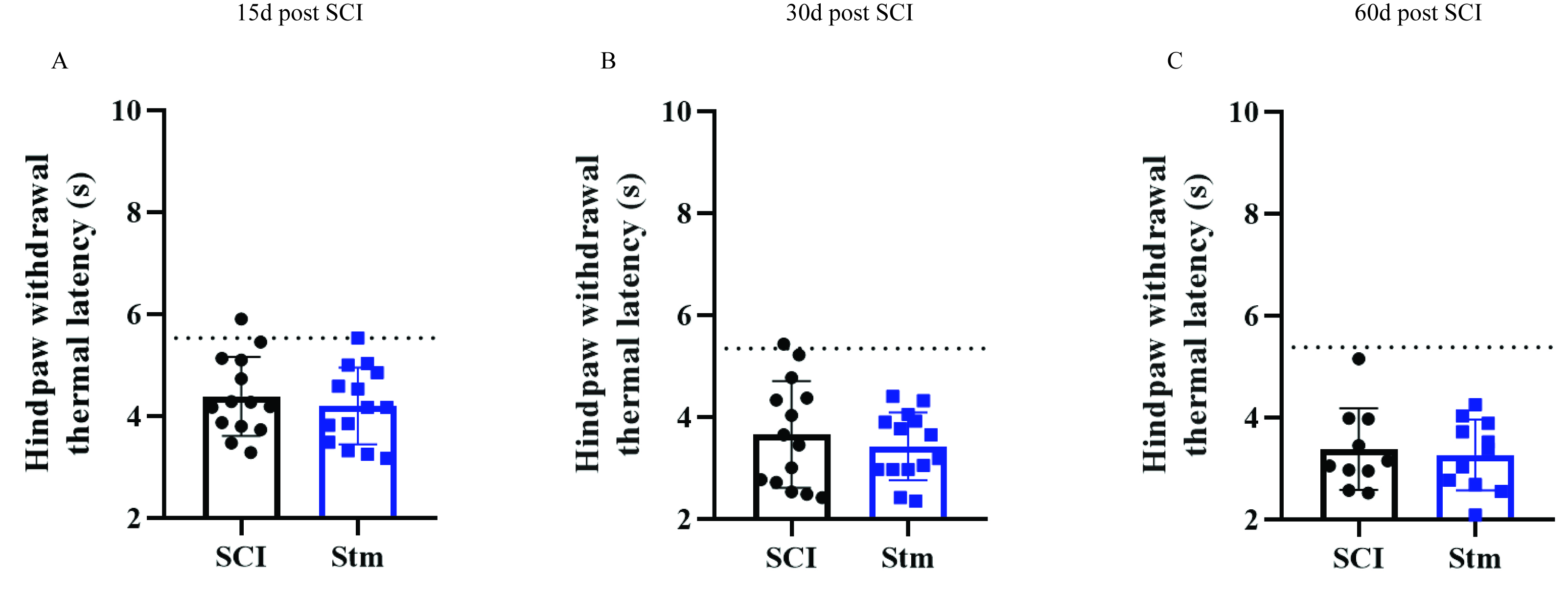
Hargreaves apparatus
The Hargreaves test instrument used in this study was a plantar test (Ugo Basile, Italy). A radiative heat source was placed beneath the animal and pointed at the plantar surface of the hindpaw. The time between the onset of the thermal radiation stimulus and the appearance of paw withdrawal was recorded as the hindpaw withdrawal thermal latency 18. Baseline parameters have been obtained with an additional group of non-injured animals.
MRI Imaging
In-vivo imaging experiment were performed on rat to follow on the same rats the evolution of the spinal cord structure overtime after SCI. Analysis of the spinal cord structure was achieved by the MRI BioSpec Advanced II (Bruker, Germany), with a magnetic field of 4.7 Teslas, monitored with ParaVision software.
The rats were anesthetized by intraperitoneal injections (Thiopental, 1g/20mL, Panpharma). To perform the MRI recordings, the rats were placed in supine position. The vertebra T9 was putted down on the marker of the antenna in order to have the area of interest in the field of view of the MRI. Two cardiac electrodes were placed on the rats to follow their cardiac constants.
A comprehensive analysis including T2*-weighted gradient echo, in axial and sagittal sections, was carried out with the parameters presented in the Table.1.
The sequence performed on axial sections is a multi-slice gradient which allowed to provide a T2* map for tissues characterization.
The sequences of images were then analyzed through the:
- ParaVision software, to determine the surface quantization of hyposignal and hypersignal, but also identify tissues structure on sagittal images;
- Osirix software, to determine the volume of spinal cord section and realize a 3D representation on axial images.
Tissue preparation and sectioning
Animals were deeply anesthetized with sodium pentobarbital (120mg/kg body weight) and perfused transcardially with PBS followed by ice-cold 4% formaldehyde in PBS. Dissected spinal cords were further post-fixed in 4% PFA in PBS at 4 °C overnight and cryoprotected in
30% sucrose (Life Technologies, Carlsbad, CA) for at least 48 h. After embedding in Tissue-
Tek OCT compound (Sakura, Tokyo, Japan), the spinal cords were cut sagittally to 20 μm thickness. Sections were collected accordingly to stereological principles (five sections per slide) and stored at −20 °C until further use.
Immunohistochemistry
Spinal cord sections were blocked with 10% Normal Donkey serum (Jackson ImmunoResearch, Cambridge, UK), 0.3% Triton-X100 (Sigma-Aldrich) in PBS, then incubated overnight at room temperature in a humidified chamber with primary antibodies diluted in blocking solution. The following primary antibodies were used: Rabbit anti-Platelet-derived growth factorβ (PDGFRβ, Abcam, ab32570), Mouse anti-Glial fibrillary acidic protein (GFAP Cy3-conjugated Sigma-Aldrich, C9205 and GFAP unconjugated Sigma-Aldrich, G3893), Rabbit anti-ionized calcium-binding adapter molecule 1 (Iba1, Wako, 019-19741, Osaka, Japan) and Mouse anti-neurofilament 200kD (NF200, Millipore, MAB5256).
After washing, antibody staining was revealed using species-specific fluorescence-conjugated secondary antibodies (Jackson ImmunoResearch). Sections were counterstained with 4′,6-diamidino-2-phénylindole (DAPI; 1 μg/mL; Sigma-Aldrich) and coverslipped with Vectashield mounting media (Vector Labs, Burlingame, UK).
Image acquisition analysis
Representative images of the lesion site and spinal cords were acquired using the Zeiss Apotome2 microscope set up. For tissue analysis sagittal sections have been used. For each experimental group and staining, 6 to 17 animals were analyzed. The image processing and assembly was acquired with Image J software.
Quantification of immunohistochemically stained areas
On sagittal sections, the GFAP negative (GFAP-), PDGFRβ positive (PDGFRβ+), NF200 negative (NF200-) and DAPI negative (DAPI-) areas were measured at the epicenter of the lesion and section rostral and caudal to the injury site, thus a minimum of 3 sections (60μm) per animal have been measured. For Iba1 staining, analysis of the area in which Iba1 positive amyboid cells were present has been measured. Analysis of Iba1 intensity measurement was performed on rectangle of 6 μm × 2 μm. Iba1 intensities were collected after threshold standardization.
Statistical analysis
Data are presented as means ± standard deviation (SD). Comparison of means were performed using two-tailed Mann-Whitney tests for all the experiments. In all tests, P < 0.05 were considered statically significant.