Synthesis Of Catalysts
The commercially available kaolin and bentonite clay were purchased from Loba Chem Pvt. Ltd, India and used without further purification for the impregnation with Zn2+and Al3+ nitrate salt and heteropolyacids were used. The wet impregnation method was successfully employed for the metal-ions loading process (Sen Gupta and Bhattacharya, 2008). The 5% weight load on the metal ions, however, a weight loss of 1-2 % weight was observed when drying at 80 ° C due to loss of moisture content in the clay sample. The synthesized catalysts were characterized by nitrogen adsorption and desorption, FTIR, powder X-ray diffraction (PXRD), SEM with EDX mapping, and XPS studies.
Bet Surface Area Analysis
Pore volume, pore size distribution, and BET surface area are essential characteristics of porous material to act as a catalyst or catalyst support. The catalytic activity of clay material could be correlated with modification of surface area and pore volume before and after loading of metal ions (Al3+ and Zn2+) and heteropolyacids (PWA and PMA). The impact on porosity modification obtained from nitrogen adsorption-desorption studies. BET adsorption-desorption studies are extensions of Langmuir theory applied for multilayer adsorption on a homogeneous surface. The study is very helpful for finding gas-solid adsorption systems. The BET study is enabling the determination of the specific surface area of the catalysts. In addition to BET, the BJH model has been used to determine the average pore volume and pore diameter of porous materials. Since nitrogen gas is a non-reactive on acid or base surfaces, nitrogen adsorption-desorption isotherm used as a tool to build the BET and BJH model for the determination of surface area and pore details. In Figure 1a and 1b, the results of the nitrogen adsorption and desorption isotherms of kaolin and bentonite impregnated with Zn2+, PWA, PMA and Al3+ compared to pure kaolin and bentonite, respectively. The average pore diameter calculated from the BJH model and the average pore distribution are given in supplementary information Figure S1. The calculated BET surface area and average pore diameter and pore volume calculated using the BJH model are summarized in Table 1.
The typical adsorption and desorption of N2 gas with respect to relative pressure (P/Po) have been recorded. It is interesting to note that, Kaolin supported catalysts showed Type II adsorption isotherm (Figure 1a) and bentonite supported catalysts showed Type IV adsorption pattern with clear Hysteresis loop (Figure 1b). The type II and IV adsorption pattern indicates the presence of both macro- and mesoporous nature of clay materials (Kuila and Prasad, 2013). The formation of hysteresis loop indicates capillary condensation of gas molecules in the mesopores (Wang et al., 2020). In Figure 1b, it is very interesting to note that pore widening is occurring while impregnation of 5wt% of PMA, PWA and Zn2+ on the bentonite support. On the other hand, the impregnation of metals and heteropoly acids did not show a significant effect on the porous nature of the kaolin support. The uptake of N2 occurring at relative pressure of 0.48-0.52 indicates micropore filling. Average pore size distribution in supported kaolin and bentonite catalysts showed in supplementary information Figure S1. The average pore distribution showed the presence of the micro- and mesoporous nature of the clay materials. Impregnation with metals and heteropoly acids has a clear impact on surface area and pore size (Table 1).
Table 1
BET Surface area, pore volume and pore diameter of Kaolin and Bentonite catalysts
|
Catalyst
|
BET surface area (m2/g)
|
Pore volume (cm3/g)
|
BJH average pore diameter (nm)
|
|
Kaolin
|
6.602
|
0.0483
|
2.43
|
|
Al-Kaolin(5 wt%)
|
16.309
|
0.0522
|
1.66
|
|
Zn-Kaolin(5 wt%)
|
12.504
|
0.0512
|
2.76
|
|
PWA-Kaolin (5 wt%)
|
11.557
|
0.0435
|
2.14
|
|
PMA-Kaolin (5 wt%)
|
19.449
|
0.0605
|
1.22
|
|
Pure Bentonite
|
14.533
|
0.0317
|
1.66
|
|
Al-Bentonite (5 wt%)
|
19.803
|
0.0316
|
1.66
|
|
Zn-Bentonite (5 wt%)
|
23.684
|
0.0820
|
1.88
|
|
PWA-Bentonite (5 wt%)
|
18.303
|
0.0605
|
1.22
|
|
PMA-Bentonite (5 wt%)
|
23.949
|
0.0832
|
1.22
|
In our previous studies on bentonite and kaolin clay using BET and BJH revealed that bentonite exhibits a surface area of 19.1 m2/g with a pore volume of 0.071 cm3/g and a pore diameter of 15.79 nm. Similarly, the kaolin clay showed a surface area of 5.1 m2/g with an average pore volume of 0.0200 cm3/g and a pore diameter of 17.36 nm. In the present study, the pure kaolin (Figure 1a) showed BET surface area 6.602 m2/g with pore volume 0.0483 cm3/g and pore diameter 2.43 nm. However, it is interesting to note that ion exchange with Al3+, Zn2+, PWA and PMA showed notable changes in surface area and porosity, as discussed earlier (Table 1). The impregnation of 5 wt% loading of metal ions or heteropolyacids is increased in surface area from 12.504, 16.309 and 19.449 m2/g, and pore volume increased from 0.0483, 0.0522 and 0.0605 cm3/g for Zn2+, Al3+ and PMA respectively. In contrast pore diameter is decreased from 2.43 to 2.14, 1.22 and 1.66 nm for ion exchanged kaolin catalysts. Similarly, the pure bentonite clay (Figure 1b) showed BET surface area 14.533 m2/g with pore volume 0.0317 cm3/g and pore diameter 1.66 nm. Like the effect of loading on kaolin, the specific area, pore volume, and pore diameter of the bentonite catalysts increased (Table 1). For example, especially for PWA, Zn2+ and PMA an increase in surface area from 14.533 to 23.949 m2/g, pore volume from 0.0317 to 0.0832 cm3/g, respectively. As compared to pure kaolin and bentonite, 5wt% PMA kaolin and bentonite showed a tremendous increase in the surface area and pore volume and marginal variation was observed in surface area and porosity for Zn2+, PWA and Al3+- exchanged clay catalysts.
Xrd Pattern Of Kaolin And Bentonite Catalysts
The powder XRD patterns of pure kaolin and bentonite, as well as ion exchanged analogues, have been recorded at 2 theta angles in the range of 10 and 80 °. The pure kaolin, bentonite and ion-exchanged catalysts are presented in Figure 1c and 1d. XRD analysis can be used to determine mineralogical composition of the clay catalysts. To find the effect of impregnation on the crystalline nature, the diffraction pattern of a catalyst supported by 5% metal ions and HPA was compared with pure kaolin and bentonite (Wang et al., 2020). Bentonite is consists of 7-39, 0.4 to 21, 30-75, and 5-20% kaolinite, montmorillonite, quartz, and cristobalite respectively. However, the main crystalline phases of bentonite are montmorillonite and quartz. Bentonite forms lattice structures consisting of a single plate located between alumina and silica plates. Owing to the layer system, montmorillonite may be expanded, and contract upon impregnation with metal ions. On the other hand, the crystalline nature could be completely collapsed due to exceeding the loading capacity limit of impregnation. The loading of metal ions and heteropolyacids could be expected to occupy the inter-layer space of montmorillonite that can be reflected by the size of its intermediate space (d001 and d020) of montmorillonite (Naswir et al., 2013). The diffraction pattern of supported bentonite clay Zn2+, PWA, PMA and Al3+ catalysts was confirming a sharp peak at 2θ value of 26.80° that is attributed to the reflections of (001) plane of bentonite clay (Figure 1c). The peaks were found at 19.90° and 24.96° at all samples, revealing the presence of a narrow amount of quartz with bentonite catalysts. The decreased in the (d001 and d020) interval indicates an increased in interlayer space owing to the impregnation of metal ions respectively (Jeya et al., 2019; Bouraie et al., 2017). Moreover, XRD pattern of pure and catalyst supported with bentonite catalysts showed similar crystalline pattern representing that the impregnation is not affected the bentonite framework.
The crystalline nature of kaolin clay and Zn2+, PWA, PMA, and Al3+ catalyst containing kaolinite was confirmed by the presence of a sharp peak at 2θ value of 29.10° at which they are responsible for the reflections of (001) plane of clay (Figure 1d). The peak at 62.50 ° at 2θ the reflection by a (060) plane indicates well-crystallized kaolin. The diffraction peak at 19.25 ° and 25.10 ° was also observed in all samples, revealing the presence of a small amount of quartz along with kaolin catalysts (Shukla et al., 2008; Jeya et al., 2017). Since the diffraction patterns of 5wt% of Zn2+, PWA, PMA and Al3+ loaded clay is almost similar to that of pure kaolin, it is assumed that the impregnation process does not make any significant impact on the crystalline nature of the clays. Similar XRD patterns also suggest that loading up to 5.0wt% does not affect the crystalline nature of the catalyst. In addition, the retaining of porous nature of supported clay catalysts have been confirmed by N2 adsorption and desorption studies.
Ft-ir Analysis Of Supported Clay Catalysts
The FTIR spectra of raw kaolin and wet impregnation samples of 5wt% Zn, PWA, PMA and Al-kaolin samples in the wave number range of 4000-400 cm−1 are shown in Figure 1e (Whole FT-IR spectra given in the supplementary information, Figure S2). The infrared spectra of the kaolin absorption band at 820 cm−1 and 860 cm−1 are attributed to Si-O-Al vibrations, and the band at 960 cm−1 is assigned to Al-OH bend vibration. The broad peak at 1060 cm−1 corresponds to Si-O –Si in plane vibration and at 1130 cm−1 allocated to asymmetric Si-O-Si stretching vibration (Olaremu, 2015). The band appears at 3700 cm−1, associated with hydroxyl group consistent with the kaolin layer. However, the band at 1100 cm−1 and 800 cm−1 to 760 cm−1 did not gain prominence, as they were intertwined with a strong band of silica in favor of kaolin (Aher et al., 2021a).
The FT-IR spectrum of bentonite and supported bentonite catalysts containing Al3+, Zn2+, PWA and PMA are shown in the supplementary information, Figure 1f (Whole FT-IR spectra given in the supplementary information, Figure S3). The IR spectra of pure and supported bentonite recorded in the region of 4000 – 400 cm−1, and the bands are comparable and similar to the bentonite structure. The bands at 1058 cm−1 and 900 cm−1 correspond to the Si-O stretching vibration and OH bending mode of vibration, respectively (Al-Sabagh et al., 2015). The bands appearing at 460-532 cm−1 correspond to Mg-O stretching vibration (Morgan et al., 2004). A broad band appears at 3424 cm−1, it is corresponding to OH asymmetric and symmetric stretching vibrations of M-OH, and a band at 1638 cm−1, which is similar to bending vibrations of d-OH. The band appears at 3700 cm−1, corresponding to the hydroxyl moiety of the bentonite layer (Russell et al., 1994). The infrared spectrum of PWA/Bentonite exhibits identical bands at 2800 and 2872 cm−1, which correspond to the C-H stretching of the alkyl chain. The band appears at 1360 cm-1 and corresponds to carbonyl moiety symmetric stretching vibrations. The existence of characteristic groups of Zn2+, Al3+, PWA, and PMA impregnated in kaolin and bentonite is very difficult to ascertain from FT-IR due to overlapping of peaks.
Sem-edx Mapping Analysis
SEM analysis combined with EDX elemental mapping used to find elemental composition and the presence of impregnated catalytic sites in clay framework. Figure 2a-c and 3a-c summarises the impregnated kaolin and bentonite catalysts respectively analyzed by EDX analysis to find out the percentage of elemental composition (whole SEM-EDX images, elemental mapping given in supplementary information, Figure S4 and S5). Similarly, the estimated composition of supported kaolin and bentonite catalysts summarised in Table S1 (supplementary information) and Table 2 respectively. Figure 2a and Table S1 show the mass and atomic percentage of Zn2+ in the Zn2 + impregnated kaolin catalyst and the weight percent composition of Zn2+ with respect to Al3+ and Si4+ is found to be 0.16±0.03%. The EDAX spectrum showed (Figure 2a) showed presence of Zn2+ and values of elemental mapping signifies that the impregnation of Zn2+ is less. On other hand, according to Figure 2b indicates EDAX spectrum of PWA loaded kaolin catalyst. The wet impregnation of PWA in kaolin is found to be very significant and the% mass of W6+ is found to be 3.43±0.10% when compared to Al3+ and Si4+ and theoretically we have synthesized 5wt% of PWA containing kaolin with respect to the weight of W6+ in PWA. The mass% of W6+ is found to be close with theoretical % of W6+. On the contrary, the composition of Mo6+ from PMA loaded with kaolin showed 0.18±0.02% of Mo6+, which is found to be much lower compared to the theoretical value (Figure 2c).
Apart from the elemental composition, SEM images clearly showing the surface morphology of the supported catalysts is similar to pure kaolin. There is no change in the morphology of the catalyst proves that phosphotungstic acid and phosphomolybdic acid species are scattered well into the hexagonal holes (Aher et al., 2021a). Further, it is confirmed by X-ray diffraction analysis and BET surface area analysis.
Figure 3a is SEM-EDX analysis of Zn2+ loaded bentonite support. EDX analysis revealed that the mass and elemental composition of Zn2+ is 3.73±0.16 and 1.18±0.05% respectively (Table 2). It is interesting to note that the amount of Zn2+ loading is found to be higher when compared with kaolin supported analogue. Although similar experimental conditions have been followed, the bentonite is showing excellent loading capacity, especially in supported clay catalysis. We have clearly seen that with respect to catalytic activity towards depolymerization of post-consumer PET wastes using bentonite impregnated with Zn2+ and Al3+ performed excellently while comparing kaolin-supported analogues (Jeya et al. 2017; Jeya et al. 2020). The SEM-EDX spectrum of PWA loaded bentonite is shown in Figure 3b and Table 2, respectively. The SEM-EDX analysis confirmed the presence of W6+ and the composition of PWA-bentonite showed 14.74±0.12 wt% of W6+ in bentonite clay. It strongly indicates the successful impregnation of PWA in bentonite catalysts (Aher et al., 2021b). EDX analysis also revealed the presence of other elements present in bentonite clay, namely, Na+ in 2.01±0.03, Mg2+ in 1.13±0.02, Al3+ in 8.56±0.05, Si4+ in 19.12±0.07, Ca2+ in 1.60±0.03 and Fe2+/3+ in 4.90±0.08% (Siredar et al., 2018). Figure 3c is the EDX spectrum of PMA loaded bentonite. The percentage of mass of Mo6+ was found to be 1.06±0.03%, while compared to the kaolin support, bentonite was found to be the best catalytic support with a higher metal ion loading capacity (Aher et al., 2021b). Comprehensively, the mass and atomic % values signify that amount of PWA and PMA loading on bentonite support is higher compared to PWA and PMA supported kaolin catalysts. FT-IR results in clear agreement with SEM-EDX studies, this indicates continued layer broadening of bentonite. In addition, the BET surface area analysis showed increasing surface area of bentonite and kaolin support upon PMA and PWA loading. The higher loading capacity of bentonite resulted in higher catalytic activity, which eventually afforded excellent yield of a pure glycolyzed and aminolyzed single product, BHET and BHETA.
EDX analysis of pure bentonite showed that the presence of O, Na, Mg, Al, Si, Ti, Fe and similar components have been present in 5wt% of impregnated bentonite catalysts with Zn2+, PWA and PMA and SEM images along with elemental mapping and EADX spectrum given in Figure 8a-c respectively (Munir et al., 2019). The mass and elemental composition of impregnated bentonite catalysts are summarized in the supplementary information Table 2.
Table 2
SEM- EDX mapping Mass percentage and atomic percentage of bentonite catalysts
|
Elements
|
5wt% Zn-Bentonite
|
5wt% PWA-Bentonite
|
5wt% PMA-Bentonite
|
|
Mass%
|
Atomic %
|
Mass%
|
Atomic %
|
Mass%
|
Atomic %
|
|
O
|
51.69±0.19
|
67.08±0.25
|
47.33±0.15
|
68.57±0.21
|
54.38±0.15
|
69.05±0.19
|
|
Na
|
2.38±0.04
|
2.15±0.04
|
2.01±0.03
|
2.03±0.03
|
3.06±0.04
|
2.70±0.03
|
|
Mg
|
2.52±0.04
|
2.15±0.03
|
1.13±0.02
|
1.07±0.02
|
1.45±0.02
|
1.21±0.02
|
|
Al
|
10.62±0.07
|
8.18±0.05
|
8.56±0.05
|
7.36±0.04
|
9.70±0.05
|
7.30±0.04
|
|
Si
|
22.00±0.10
|
16.26±0.08
|
19.12±0.07
|
15.78±0.06
|
22.61±0.08
|
16.35±0.06
|
|
K
|
2.14±0.04
|
1.14±0.02
|
0.48±0.01
|
0.28±0.01
|
0.45±0.01
|
0.24±0.01
|
|
Fe
|
4.45±0.10
|
1.65±0.04
|
4.90±0.08
|
2.03±0.03
|
5.39±0.08
|
1.96±0.03
|
|
M*
|
3.73±0.16 (Zn)
|
1.18±0.05 (Zn)
|
14.74±0.12 (W)
|
1.86±0.02 (W)
|
1.06±0.03 (Mo)
|
0.22±0.01 (Mo)
|
*M – Metal loadings from Zn, W from PWA, and Mo from PMA
Analysis Of The Morphology Of Supported Catalysts
The SEM images (supplementary information, Figure S6) and Figure 2d-f show the surface morphologies of the kaolin before and after wet impregnation of Zn2+, PWA and PMA respectively. SEM images revealed that no notable modifications in the morphology on pure kaolin (Figure S6) and ion- exchanged metal ions (Figures 2d, 2e and 2f) have been observed. However, at a closer look at higher magnification (7000X, 2 µM) the layered structure of clay can be clearly seen in Fig. 2d-f. The clear appearance of layered structure could be due to broadening of inter layer distance because of impregnation of PMA and PWA, which is higher molecular size (Winiarsk et al., 2018).
Similarly, the SEM images of pure bentonite and Al3+ impregnated bentonite (supplementary information, Figure S7) and Zn2+, PWA, PMA, and Al3+ loaded bentonite are clearly shown in Figure 3d – f, respectively. The SEM images showed the morphology of bentonite clearly consist of stacked layered structure (Winiarsk et al., 2018; Aher et al., 2021b). Due to the layered structure, the SEM imaging showed the effects of impregnation on clay supports. In pure bentonite clay (Fig. S7a), the layered nature of clay is not clearly visible even at higher magnification (3000X, 5 µM). Whereas Figure 3d and S7b are Zn2+ and Al3+ incorporated bentonite showed rough surface and deformed layered structure. However, the XRD studies confirmed that the crystalline nature of bentonite is retained even after metal ion loading. More interestingly, the PWA loaded bentonite showed small crystallite PWA crystals deposited on the surface (Figure 3e). The resulting surface morphology of the PWA/bentonite catalyst is completely different from that of pure bentonite. The SEM images clearly showed that PMA is homogeneously dispersed on the surface of the bentonite (Figure 3f).
Xps- Analysis And Elemental Composition
The chemical composition of Zn2+, Al3+ and HPA loaded kaolin and bentonite was recorded by X-ray photoelectron spectroscopy (XPS). The XPS spectra of Kaolin and bentonite supported Zn2+, PWA and PWA catalysts are shown in Figure 2g-i and 3g-i, and binding energy (eV), metal to Al3+ ratio and atomic composition is summarised in Table S2 and S3 of supplementary information, respectively. Figures 2g and 3g are the XPS spectra of Zn2+ loaded kaolin and bentonite, respectively. Zn2+ loaded kaolin as well as bentonite support showed two peaks at 1022 and 1045 eV are the binding energies of Zn2p3/2 and Zn2p1/2, which is related to the spin-orbit splitting of about approximately 22 eV. The metal-to-Al3+ ratio of bentonite showed a higher content of Zn2+ loading compared to catalysts supported by kaolin. Due to this higher loading capacity, the bentonite supported catalysts showed higher catalytic activity towards depolymerisation of PET wastes in glycolysis and aminolysis reaction. All XPS spectra showed a symmetric peak approximately at 533 eV that was O2− connected to Si and attributed to SiO2 based materials and summarized in the Supplementary Material, Figure S8 to S19.
Figure S13b and S19b showed binding energy peaks of Al3+ in Zn2+ loaded kaolin and bentonite. XPS analysis revealed the presence of binding energies of 2pA and 2pB for Al3+ approximately at 74.72 and 75.75 eV in kaolin and bentonite-supported catalysts. Figures 2h and 3h are the peaks of W6+ binding energy in phosphotungstic acid (PWA). The presence of W6+ in PWA supported on kaolin and bentonite is confirmed by the binding energy peaks at 36.06 and 38.19 eV attributed to spin-orbit coupling 4f7/2 and 4f5/2 states. The mineral composition of W6+ to Al3+ is found to be 0.07 (7%) and 0.11 (11%) with respect to Al3+ for kaolin and bentonite loaded with PWA, respectively. Thus, the presence of a higher loading of PWA in bentonite was attributed to the higher catalytic activity. In the case of, kaolin and bentonite loaded with phosphomolybdic acid (PMA) showed Mo6+ spin orbit coupling binding energy peaks approximately at 233 and 236 eV, which is attributed to transition states 4d5/2 and 4d3/2 (Figure 2i and 3i). Similarly, bentonite showed higher phosphomolybdic acid (PMA) loading is observed from the elemental composition of Mo6+, which is estimated to be 0.04 (4%) and 0.13 (13%) for PMA loaded kaolin and bentonite respectively.
Figure S13e and S13f is spin-orbit coupling peaks of Al3+ in Al3+ loaded kaolin and bentonite respectively. The binding energy peaks at 75.50 and 74.50 eV, which corresponds to Al 2pB and Al 2pA. Similarly, pure kaolin and bentonite showed binding energies of Al3+ at 75.60 and 74.60 eV, which corresponds to the spin-orbit coupling states of Al 2pB and Al 2pA.
Depolymerisation of PET waste using glycolysis and aminolysis
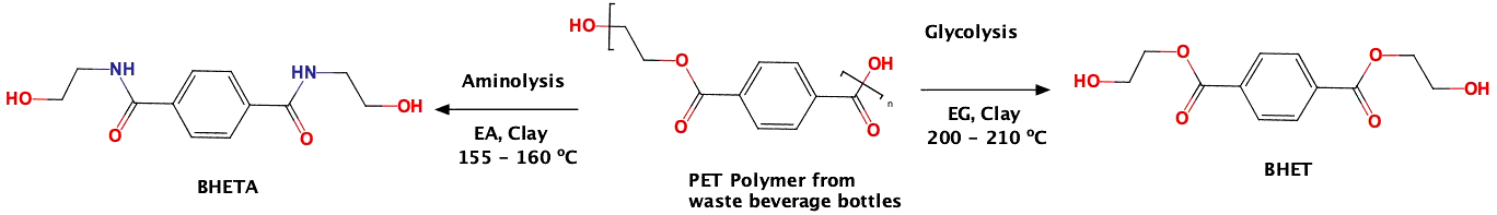
Glycolysis and aminolysis are the common depolymerisation reaction applied for PET wastes. The presence of ester linkage in the PET polymer chain enables the hydrolysis using acids or bases to give terephthalic acid. However, the ester linkage could be cleaved using methanol to give dimethylterephthalate (DMT) as the final product through the transesterification reaction. However, the methanolysis process needs a high-pressure reactor to overcome the pressure built by a low-boiling methanol solvent. By using a similar transesterification methodology, ethylene glycol or ethanolamine has been used to depolymerise PET waste to generate bis(2-hydroxyethylene)terephthalate (BHET) or bis (2-hydroxyethylene) terephthalamide (BHETA), respectively. However, a Lewis or Bronsted acid catalyst required to activate the ester carbonyl group and trigger the transesterification reaction and higher reaction temperature is required for the dissolution of PET chips in ethylene glycol for the complete depolymerisation of PET.
Thus, in the present investigation, an economical catalyst support, clay material has been opted to prepare Lewis and Bronsted acid supported catalyst. Ethylene glycol has been used as a glycolyting agent as well as solvent. The depolymerisation reaction has been carried out in the presence of 10wt% of the clay catalyst.
In both cases, Figures 4a and 4b, the glycolysis and aminolysis of PET waste using kaolin and bentonite clay loaded with Zn2+, PWA and PMA, respectively. The use of ethylene glycol (EG) and ethanol amine (EA) as a common depolymerizing agent for glycolysis and aminolysis process. The glycolytic depolymerisation lead to monomer, bis(hydroxyethyl)terephthalate (BHET) and aminolytic depolymerisation lead to monomer, bis(hydroxyethyl)terephthalaimide (BHETA). In both depolymerisation reactions, we have observed complete depolymerisation of PET wastes. The final product isolated from the reaction mixture is highly pure is confirmed by 1H and 13C NMR, in which the peaks correspond to PET oligomers were absent. The reaction time is 6-7 hrs for glycolysis, whereas the aminolysis required only 4-5 hrs for the complete depolymerisation. The reaction was monitored by TLC using an ethylacetate to hexane ratio at 20:80. After completion of the reaction, the reaction mixture was diluted with hot water (100 mL) and filtered to the catalyst. After filtration, the reaction mixture was heated to 60 ˚C for 15 min and kept at 5 ˚C for 24 h for the glycolysis reaction and after heating the reaction mixture kept room temperature for 24 h for the aminolysis reaction mixture. In the case of glycolysis, we identified issues associated with the isolation of the final product from water because the product is water soluble. However, the final product is obtained completely by extraction with chloroform and the final yield is calculated. In the case of aminolysis, the final product BHETA is obtained as pale yellowish needle crystals from the reaction mixture at room temperature itself. We have tried with many diamines for aminolysis, ethanol amine is worked better at low temperature and afforded excellent yield of BHETA. The crystals of BHETA filtered, dried and yield of the reaction calculated. Both 1H and 13C NMR, and mass spectral analysis showed BHETA is a single pure product.
Figures 4a and 4b are the result of the glycolysis and aminolysis reaction. In the case of glycolytic depolymerization reactions, Zn2+ and PWA loaded bentonite-supported clay catalysts gave 92 and 93% of BHET, respectively, when compared with kaolin-supported analogues. On other hand, the aminolytic depolymerisation reaction revealed that Zn2+ and PWA loaded bentonite afforded 94 and 96% of BHETA respectively in comparison with kaolin supported analogues. Similarly, the glycolysis reaction was performed with ethylene glycol as the transesterification catalyst. In both reactions, the bentonite clay support found to be the better catalytic support as it is supported by BET surface area analysis, SEM-EDX, and XPS analysis.
Catalysts Recycling Studies
The PWA and Zn2+ loaded kaolin and bentonite catalysts were subjected to recycling studies using glycolysis as a model reaction. At the end of each reaction cycle, the spent catalysts were filtered following washed with water and dried in the oven before the next reaction cycle. The yield of the depolymerized product obtained from the subsequent reaction cycle was compared (Figure 4c). The study revealed that the catalyst can be reusable up to six cycles with marginal loss in yield. Among the catalysts, PWA and Zn2+ loaded bentonite catalysts showed better results. Higher catalytic activity of bentonite catalyst could be correlated with higher surface area of bentonite, which eventually can take more PWA and Zn2+ as supported by BET, SEM -EDX and XPS studies. The slower leaching of PWA from the surface and interlayer spaces is reflected in the yield of the BHET formed.
SPECTRAL ANALYSIS OF DEPOLYMERISED PRODUCTS
All depolymerized products would have been characterized by 1H, 13C NMR, and mass spectroscopy to confirm the structure.
1 H NMR spectrum
The four aromatic hydrogens present in BHET have appeared (Supplementary information, Figure S20) as singlets 8.08 ppm confirm the formation of the glycolyzed product, the BHET monomer. The 1HNMR run for the crude product showed singlet aromatic protons that confirmed the complete depolymerization of PET wastes (Sarkar et al., 2014). In the case of BHET, the two hydrogens attach to carbon next to the ester carbonyl oxygen appeared as a triplet at 4.47 ppm. Another two hydrogens attached to carbon next to OH has appeared as triplet at 3.97 ppm. The former appeared downfield due to the presence of electron-withdrawing carbonyl oxygen attached. The OH proton appears as a broad singlet at 3.70 ppm due to the exchangeable nature of its kind. The deuterated CDCl3 solvent appeared as singlet 7.26 ppm (Jeya et al., 2017; Jeya et al., 2020). Similarly, the 1H NMR of the crude depolymerized product obtained from the aminolysis of PET using 2-amino ethanol is given in the Supplementary information (Figure S21). The main aminolyzed product N1,N4-bis(2-hydroxyethyl)terephthalamide (BHETA) is a symmetrical molecule. Hence, the four aromatic hydrogens present in the central aromatic ring of BHETA were appeared as singlet 8.07 ppm confirming the formation of aminolysed product BHETA and complete depolymerisation of PET wastes. The two N-H protons of the amide linkage attached to the methylene group are observed as a triplet at 8.74 ppm and the -OH protons of the alcoholic group are observed at 2.64-2.65 ppm as a triplet (Ghosal et al., 2019). In the case of BHETA, the two hydrogens attach to carbon next to amide carbonyl oxygen, and -NH appeared as a triplet at 3.48-3.51 ppm. Another two hydrogens attached to carbon next to OH has appeared as triplet at 3.65-3.68 ppm. Therefore, presence of –N-H and–O-H protons confirms the 2-amino ethanol attached to terephthalaoyl group to form the BHETA.
13 C NMR spectrum
13C NMR spectrum of BHET is shown in supplementary information (Figure S22). The ester carbonyl carbon of BHET appeared at 166.16 ppm is showing single ester carbonyl present the structure and no other carbonyl carbons were recorded, which confirmed the presence of BHET as a single product. Since BHET is a symmetrical molecule, here are two aromatic carbons that appeared at 133.80 and 129.70 ppm. The peak at 133.80 ppm corresponds to C-1 and C-4 attached to ester carbonyl, and another peak at 129.70 corresponds to C-2, C-3, C-5 and C-6 attached with hydrogen appeared as singlet with high intensity. The -CH2- carbons of the aliphatic chain attached to the ester carbonyl oxygen appeared at 67.0 ppm for BHET. The variation in –CH2- the shift is owing to two -CH2- attached to the chain. Hydroxyl group attached -CH2- carbons appeared at BHET at 61.10 ppm, respectively (Jeya et al., 2017; Jeya et al., 2020) and we were not able to account for the peak appeared at 63.70 ppm. The 13C NMR spectrum of the aminolysed product, BHETA is shown in the supplementary information (Figure S23). The amide carbonyl carbon of BHETA appeared at 167.50 ppm, showing that the amide carbonyl has the structure that confirmed the symmetrical nature of the BHETA single product. Since, BHETA is a symmetrical molecule, there are two aromatic carbons that appeared at 137.10 and 127.60 ppm. The peak at 137.10 ppm corresponds to C-1 and C-4 attached to amide carbonyl, and another peak at127.60 ppm corresponds to C-2, C-3, C-5 and C-6 attached with hydrogen appeared as singlet with high intensity. Two -CH2- carbons of the aliphatic chain attached to amide -NH- appeared at 42.7 ppm for BHETA. The second hydroxyl group connected to carbon -CH2 appeared at 60.1 ppm of BHETA.
Additionally, functional groups such as hydroxy and amide or ester carbonyl groups present in BHET (Figure S24) and BHETA (Figure S25) were analyzed through FT-IR. In addition, the molecular weight of BHETA confirmed by the ESI-MS of the aminolysed product (Figure S26). The mass spectrum showed a molecular ion peak at m/z 253.15 that indicated the molecular weight of BHETA.