Study area
The study was carried out in Serere district, south-eastern Uganda, in 2017. The district is made up of two rural counties (Kasilo and Serere), eight sub-counties (Bugondo, Kadungulu, Pingire, Labor, Atiira, Kateta, Chere and Serere/Olio) encompassing 254 cattle-owning villages. Tick collections were conducted in Kadungulu sub-county. Serere district was selected because it has a large number of small scale livestock producers (1-50 cattle per herd) whose potential to commercialise livestock production is primarily constrained by ticks and tick-borne diseases. Six of the 254 villages were randomly selected for this study (Fig. 1).
Cattle herd and individual animal selection
Farmers in Serere district predominantly keep short-horn East-African Zebu cattle in communal village herds. Given that any animal sampled from each of these villages is likely to be infested with ticks, the number of villages (n=6) and animals selected (n=240) need not to be based on any rigorous statistical methods. However, to estimate the chance of finding R. decoloratus ticks, we conducted a power calculation assuming a prevalence of 5% at P=0.01; we expected to find 18 R. decoloratus ticks in a sample size of 18,000 ticks [17]. In this study we sampled 19,007 ticks. Cattle were included in this study if they had not been sprayed against ticks for the past two weeks, were young [1-2 years] and non-fractious. Young non-fractious animals were preferred for inclusion because they were easier to restrain and pose very low risk of injury to themselves or personnel. An average of 40 cattle was sampled from each of the six selected villages. All cattle sampling sites were geo-referenced prior to tick collection.
Tick collection and identification
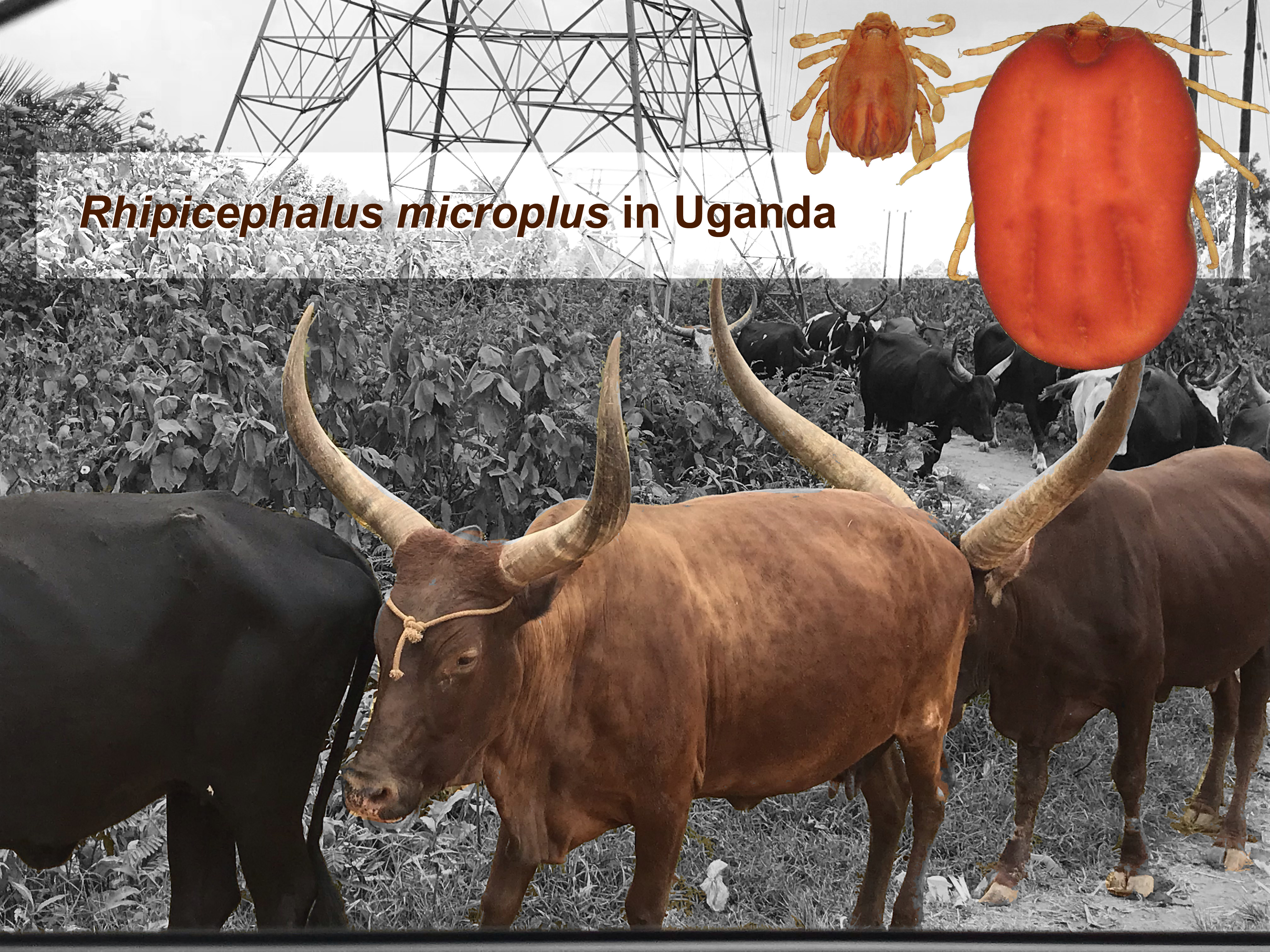
Selected cattle were physically restrained before half-body tick collections were carried out. Each of the collected ticks was morphologically identified to the genus level before they were preserved in 70% ethanol. The tick samples were then transferred to Makerere University for further species identification using taxonomic keys [18]. Five representative R. microplus specimens were selected for molecular species confirmation based on 12S ribosomal RNA (12S rRNA), 16S ribosomal RNA (16S rRNA) and the internal transcribed spacer 2 (ITS2) gene sequences [4, 19]. A taxonomically and molecularly confirmed R. microplus specimen was photographed under a stereo microscope (Olympus model SZX7, Japan)
DNA extraction
Prior to tick DNA extraction, each tick was cleaned in five one minute steps, each step involving centrifugation at 10,000 rpm in freshly prepared 1.5 ml of Phosphate Buffered Saline (PBS). Individual clean ticks were immersed under liquid nitrogen for 5 min and thereafter crushed with a sterile mortar and pestle to create a tick homogenate. DNA was then extracted from each tick homogenate using DNeasy kit (Qiagen, 19300 Germantown Road, Germantown, MD - USA) following the manufacturer’s guide. The presence and quality of DNA were checked by resolving 5µl of the extracted DNA on a 1 % agarose gel and viewing them under an ultraviolet transilluminator (Wagtech International, Thatcham, UK). The remaining DNA was stored at -20°C until use in the downward amplification steps.
DNA amplification
PCR amplification was performed on 12S rRNA, 16S rRNA and ITS2 genes using primers (Table 1) and thermocycling conditions as previously described [4, 19]. Each reaction was prepared into a final volume of 50µl containing; 1x-reaction buffer (670 mM Tris–HCl, pH 8.8, 166 μM (NH4)2SO4, 4.5 % Triton X-100, 2 mg/mL gelatin) (Bioline, Humber Road, London, UK), 0.25mM of each dNTP, 0.25 mM each of forward and reverse primers, 1.56U BioTaq DNA polymerase (Bioline, Humber Road, London, UK), 1.25 mM MgCl2, 32.2 µl of PCR grade water and finally 5 µl of the template DNA.
The 16S ribosomal RNA gene was amplified in a thermo-cycler (Personal Thermocycler, Biometra, Germany) with initial denaturation of 94oC for 5 min followed by 30 cycles at 94oC for 30 sec, 48oC for 45 sec, 72oC for 45 sec and a final extension at 72oC for 7 min. Amplification of the ITS2 and 12S ribosomal RNA was performed using similar thermocycling conditions to those of 16S at annealing temperature of 55oC and 52oC respectively. PCR products were resolved on 2% agarose gels. The resultant PCR products were sized against a 1kb DNA molecular ladder (Bioline, Humber Road, London, UK). The expected PCR product sizes ranged between 300-1,200 bp. PCR products were purified using QIAquick PCR purification kit (Qiagen, 19300 Germantown Road, Germantown, MD- USA) and commercially Sanger sequenced (Inqaba Biotec, 525 Justice Mahomed St, Muckleneuk, Pretoria, 0002, South Africa).
Gene sequence analysis
Each of the 12S rRNA, 16S rRNA and ITS2 tick gene sequences from this study were queried in a BLASTn search with default settings (NCBI BLASTn software version 2.6.10) [20] to reveal their identity. The query sequence identity was assigned/matched based on the hits (tick species sequences returned) with the highest identity scores (≥80%) and most significant E-values (closest to 0.0). The identified query sequences from this study were annotated and submitted to GenBank under accession numbers: MK332390, MK332391, KY688455, KY688455, KY688459, KY688461 and KY688467.
Annotated gene sequences from this study were each analysed in a Multiple sequence alignment (MSA) with their corresponding reference gene sequences downloaded from GenBank using the ClustalW algorithm in MEGA version 10 [21]. The MSA files were used to infer nucleotide similarity between sequences from this study and their corresponding nucleotide reference sequences from the GenBank. Each of the data sequence sets were analysed in MSA using the ClustalW algorithm and trimmed in MEGA software version 10 [22, 23]. Phylogenetic analysis for each nucleotide sequence set was performed using the MEGA maximum likelihood method utilising the Tamura 3 parameter + Gamma distribution with 1000 bootstrap [22] as the best fit model to infer phylogenetic relatedness among the gene sets.
To evaluate the 12S rRNA, 16S rRNA and ITS2 gene sequence divergence of newly typed Ugandan R. microplus ticks and their corresponding reference sequences from the GenBank, pairwise genetic distances were calculated in MEGA software version 10 [22] using default settings for each gene.