Chemicals and media
TCP (99%, powder) and 2-HP (99%, powder), purchased from Yiji chemical company (Shanghai, China); Concentrated stock solutions of the compouds (10 g/L) was prepared in sterilized water. Methanol (HPLC-grade), obtained from Shanghai Chemical Reagent Co. Ltd. (Shanghai, China). All other reagents (analytical-reagent grade), purchased from Shanghai Chemical Reagent Co. Ltd. (Shanghai, China).
Cupriavidus sp. DT-1 was isolated and characterized in the previous study; E. coli SM10 (λ pir) pUT- mini-Tn5-gfp (Ampr); E. coli HB101 pRK2013 (Kmr) and all other molecular biological reagents were obtained from Genscript Biological Science and Technology Co. Ltd. (Nanjing, China).
Medium for bacterial culture: Luria–Bertani (LB) medium contained (g/L): tryptone 10.0, yeast extract 5.0 and NaCl 10.0, pH 7.0. Medium for degradation experiment : The mineral salts medium (MSM) contained (g/L): NH4NO3 1.0, K2HPO4 1.5, KH2PO4 0.5, NaCl 0.5, MgSO4 0.2, pH 7.0. All the medium were sterilized by autoclaving at 121.3℃ for 30 minutes.
Inoculum preparation for degradation studies
Strain DT-1 was cultured to exponential phase in LB medium at 30℃ at 180 rpm, and collected by centrifugation at 6000 g for 5 min at room temperature. The cell precipitation was washed twice with sterilized MSM and adjusted to approximately 2 × 10⁸ CFU/mL. For the degradation experiments in liquid culture, the cells were inoculated to approximately 1 × 10⁷ CFU/mL.
Degradation of TCP and 2-HP in liquid culture
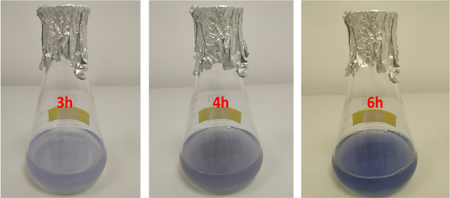
Degradation experiment was carried out in 100 mL MSM containing 50 mg/L TCP. Culture was incubated at 30℃ at 180 rpm for 24 h after inoculation of the strain, and regularly sampled for the determination of the concentrations of TCP and 2-HP every 2 h, 3 mL each time. All samples were in triplicate. Changed the carbon source in the MSM medium from 50 mg/L TCP to 500 mg/L 2-HP and inoculated the cells. Culture was incubated and sampled for determination of the concentrations of 2-HP and the growth of DT-1 at the same conditions as above.
Growth of strain D-2 in MSM culture
Diluents ranging from 10− 4-10− 1 were obtained by tenfold gradient dilution. According to dilution plate counting method, 0.2 mL diluent was spread on LB plate and cultured at 30℃ for 48 h. Plates with a colony number from 30–300 were selected for counting. All samples were in triplicate.
Extraction and Analytical methods
High-performance liquid chromatography (HPLC) was used to analyze the concentration of TCP in liquid samples. 3 mL sample was centrifuged for 10 min at 15,000 g, and the supernatant was filtrated through a 0.2 µm fiber filter. Samples were freeze-dried, mixed with 2 mL methanol, and HPLC analysis. The liquid chromatograph was Shimadzu LC-201A with SPD-M20A UV-detector (190–800 nm), column was Agilent C-18 (250 mm × 4.6 mm, 5 µm), The mobile phase containing methanol/water (80:20, v/v) was delivered at a flow rate of 1.2 mL/min at 25℃. The detection wavelength was 230 nm. Concentration of TCP was determined by comparison with values in the calibration curve established by concentrations between 0.1 and 100 mg/L. The limits of detection (LOD) and quantification (LOQ) were 0.016 and 0.057 mg/L, respectively. The recovery and RSD were 96.5%-101.5% and 1.10%-2.84% at concentrations ranging from 0.1 to 100 mg/L.
Concentration of 2-HP was detected following the method above. The calibration curve established by concentrations between 0.1 and 100 mg/L. The recovery and RSD were 94.6%-103.8% and 1.37%-2.66%. The limits of detection (LOD) and quantification (LOQ) were 0.021 and 0.075 mg/L, respectively.
To identify the metabolites of 2-HP, MSM containing 500 mg/L 2-HP as the sole carbon source was inoculated, and cultured at 30 ℃ at 180 rpm. Samples were collected from the culture and centrifuged for 10 min at 15,000 g. The supernatant was filtered through a 0.2 µm fiber filter, freeze-dried, and dissolved in 1 mL methanol, then identified by HPLC–MS (Finnigan TSQ Quantum Ultra AM, Thermal, USA). The HPLC system was implemented as described above. MS was operated in the electron spray ionization mode with a negative polarity, and scanning at a mass range from 30 m/z to 400 m/z.
Degradation of TCP by strain DT-1 in soil
Soil samples used for the experiment were collected from Anhui Normal University and a farmland which had no previous exposure to TCP. Samples were air-dried, sieved to 2 mm, and homogenized immediately after collection. For analyzed the physical and chemical properties of the soil, 20 g sample was weighed, dried at 105℃, and measured its water content. Soil total nitrogen and nitrate nitrogen were determined with continuous flow analyzer by kjeldahl method (Bulluck et al., 2002). Organic matter was determined by potassium dichromate volumetric method (Ciavatta et al.,1991). Select characterisitics of the soil samples were showed in Table 2.
Table 2
Select characterisitics of the soil samples.
| From | pH | Water (%) | Total N(%) | Organic matter (%) | Total K (cmol/kg) | Total Na(cmol/kg) |
| campus | 6.4 | 14.4 | 0.13 | 2.27 | 0.13 | 0.17 |
| farmland | 6.9 | 13.9 | 0.37 | 4.46 | 0.18 | 0.24 |
For the degradation research, glass beaker (200 mL) microcosms, each containing 100 g soil, were spiked with TCP (50 mg/kg soil). For the set of inoculated beakers, MSM medium (4.0 mL) containing strain DT-1 was added at the final concentration 1 × 10⁷ cells/g of soil. On the contrary, MSM medium (4.0 mL) without DT-1 was added for the set of non-inoculated beakers. Each soil sample was incubated at 30℃ under sterile condition. During incubation, the soil microcosms were weighed regularly, the weight loss was compensated by the addition of sterile water. Soil samples (5 g) were collected for analysis of TCP concentration every 5 d for 35 d. Triplicate soil samples were prepared for two sets of treatments.
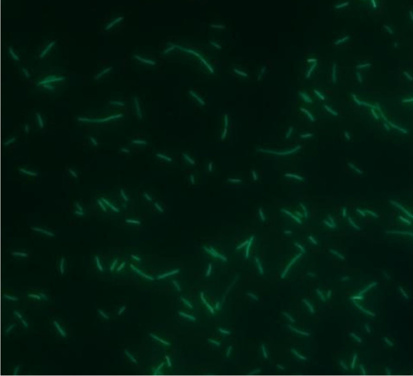
Gfp -tagged of strain DT-1
Gfp gene which can encod bioluminescent protein is one of the most useful biomarkers available for monitoring purposes (Errampalli et al., 1999; Elvang et al., 2001). Strain DT-1, E. coli SM10 (λ pir) pUT- mini-Tn5-gfp, E. coli HB101 pRK2013 was cultured in LB liquid medium for 12 h, respectively. Cells were collected by centrifugation at 6000 g for 5 min at room temperature and washed twice with sterilized water. 5ML of each three bacterial suspension was intensive mixing, centrifuged at 6000 g for 5 min. The cell precipitation was dissolved with 20 µL LB medium and the mixed bacterial suspension was spread on the filter membrane. Then the membrane was placed on a LB plate, cultured at 30℃ for 24 h. Cells were washed and dissolved in saline and spread on LB plate which contained 100 mg/L ampicillin and kanamycin, cultured at 30℃ for 24 h. Bacterial colonies on the plate were the transformants of strain DT-1 which contained gene gfp.
Content analysis of strain DT-1- gfp in soils
Soil (1 g) was dissolved in 9 mL sterile water and 1 mL soil suspension was diluted 104 times, 0.2 mL diluted suspension was spread on LB plate and cultured at 30℃ for 48 h. Plates were placed under an ultraviolet lamp to count the colonies which emitted green fluorescence. All samples were in triplicate.