3.1 Characterization of magnetic nanoparticles
3.1.1 Fourier Transform Infrared Spectroscopy (FTIR)
The equipment used to characterize the magnetic nanoparticles and eugenol was Perkin-Elmer FTIR, model Spectro One. KBr disc method was used to record the spectra in spectral region between 4000 and 400 cm−1. The infrared spectra (FTIR) of the free eugenol, magnetite pristine, and EUG·Fe3O4 are shown in Fig. 1.
In the FTIR spectrum of Fe3O4, it is possible to verify a band of vibration at 602 cm−1 typical of bonding Fe-O. The bands at 3443 cm−1 and 1633 cm−1 are attributed to the O-H stretching and vibration resulting from the absorption of water molecules [8].
For the spectrum of free eugenol, a characteristic band is observed around 3462 cm−1, referring to the O-H bond in 2838 cm−1 the absorption peak corresponds to the stretching of the C-H bond. In the 1600-1500 cm−1 region, the peaks refer to the C=C bond, and the sharp peaks in the 1200-700 cm−1 region are due to the C=C bonds of the aromatic ring [7, 19].
In the spectrum of EUG·Fe3O4, in addition to the peaks remaining constant, a more intense peak is observed around 600 cm−1, referring to Fe-O binding confirming the functionalization of eugenol with Fe3O4 [3].
3.1.2 X-ray diffraction (XRD)
Bruker Optics D2 Advance USA equipment was used for the characterization using X-ray diffraction (XRD) to determine the crystalline phases of the samples. Fig. 2 shows the XRD referring to the magnetite, and magnetite-functionalized eugenol, which demonstrates the characteristic peak of these nanoparticles.
The figure above show XRD patterns of magnetic nanoparticles synthetized in this work. In the diffractogram of the Fe3O4 pristine, it is possible to verify the crystal planes at 2θ ≈ 30º, 35º 45º, 57º, and 62º, which corresponds to the characteristic diffraction interference of magnetite [7]. For the XRD of EUG Fe3O4, it can be observed that the functionalization of eugenol with magnetite caused a decrease in the intensity of the diffraction peaks. The partial suppression of the peaks indicates that eugenol presence slightly affected the crystal structure of the magnetite [3, 8].
3.1.3 Scanning electron spectroscopy (SEM) and Energy dispersive X-ray spectroscopy (EDS)
The morphology of magnetite functionalized eugenol and the elemental analysis of the nanoparticle was obtained by Scanning electron microscope (SEM) (Sigma 300 VP Carl Zeiss), and Energy dispersive X-ray spectroscopy (EDS) (Quantax 200-Z10, Bruker). Fig. 3 (a-b) shows the SEM image and EDS (c) of the eugenol and magnetic nanoparticles (EUG·Fe3O4).
The morphological structure and size of the magnetic eugenol nanoparticles were measured according to the SEM image. Fig. 3 (a) and (b) show SEM images of EUG·Fe3O4 at different scales.
As can be observed in the SEM images, the Fe3O4 deposited on the eugenol surface has a spherical shape, and the nanocomposite produced exhibited a morphology nanoflower-like and size particle of 200 nm [3, 20, 21].
Through the EDS analysis (Fig. 3c), it was possible to verify the presence of Fe (11.15%), C (46.97%), and O (41.31%) as the main chemical elements in the sample. Furthermore, the low relatively S percentage (0.57%) can be attributed to the residue of the iron salt (FeSO4) used in the reaction.
3.1.4 Hydrodynamic size of the magnetite-functionalized eugenol
The size distribution of EUG·Fe3O4 1:1, EUG·Fe3O4 1:5, and EUG·Fe3O4 1:10 were analyzed using the Zetasizer®, nano-ZS model ZEN 3600, Malvern.
The average particle size distributions for EUG·Fe3O4 1:1, EUG·Fe3O4 1:5 and, EUG·Fe3O4 1:10 were 2.32, 185.3, and 190.1 nm, respectively, as shown in Fig. 4 (a-c). From hydrodynamic size profiles, it is possible to assume that the average size of nanoparticles rises directly with the amount of the Fe2+ employed in synthesis. The increase of the size distribution of magnetite-functionalized eugenol could be related to the tendency of formation aggregates due to the critical diameter of nanoparticles [22].
3.2 Cytotoxicity evaluation
3.2.1 3-(4,5-dimethylthiazol-2-yl)-2,5-diphenyl tetrazolium bromide (MTT) assay
The MTT test 3-(4,5-dimethylthiazol-2yl)-2,5-diphenyl tetrazolium bromide is a colorimetric test used to assess cell viability. Fig. 5 (a-b) represents the results of the MTT assay after 24 h treatment of HaCat and A375 cell lines, at a concentration of 2x10⁴ well, cultured in a 96-well plate and treated with free EUG, and EUG·Fe3O4 with different concentrations and proportions of incorporated magnetite, in triplicates.
For the MTT assay, it is possible to observe a significant viability decrease in the HaCat cell line with 10 µg.mL−1 of the free EUG, while for A375 cells was necessary only 3 µg.mL−1. Meanwhile, for all treatments of magnetite-incorporated eugenol, those confirming that the functionalization of eugenol offers protection against cytotoxic in healthy cells damage caused for free oil.
However, tumor cells were more sensitive to treatments of the magnetite-functionalized eugenol (EUG·Fe3O4 1:1, EUG·Fe3O4 1:5 and, EUG·Fe3O4 1:10).
Figure 6 (a-b) shows the viability of HaCat and A375 cells treated with magnetite pristine after 24 h of contact, respectively.
After 24 h of treatment at concentrations of 1, 3, 10, 30, and 100 µg.mL−1, Fe3O4 pristine showed different toxicity against HaCat and A375 cell lines compared to free EUG. For the keratinocytes cell line, a significant decrease in viability is observed only at the highest concentrations tested (30-100 µg.mL−1), which correspond to about 80% viable cells.
In contrast, for melanoma lineage from the lowest concentration, the cell viability rate decreases, in a dose-dependent manner, presenting statistical significance, as shown by Fig. 6b, in all dilutions of the magnetite [8].
3.2.2 Colorimetric viability assay from the neutral red vital dye
Figure 7 (a-b) shown the results of the cellular toxicity of the free eugenol and magnetite-functionalized eugenol employing the neutral red colorimetric assay under the same treatment conditions of the HaCat and A375 cell lines described in the cell culture item of the materials and methods assignment.
Through the neutral red (NR) colorimetric assay, a significant decrease in the viability of HaCat cells was observed at the highest concentrations (30 and 100 µg.mL−1) in all treatments, except for EUG·Fe3O4 1:1, which showed cytotoxic effects only in the concentration of 100 µg.mL−1.
However, in human melanoma cells, the action of nanoparticles was more evident, demonstrating that the materials offer better specificity against the tumor lineage, for instance, the highest concentration of free eugenol, EUG·Fe3O4 1:1, EUG·Fe3O4 1:5, and EUG·Fe3O4 1:10 reduced the number of viable cells to 46, 64, 54, and 44%, respectively.
Figure 8 (a-b) shows the viability of HaCat and A375 cells treated with magnetite pristine after 24 h of contact using the NR assay, respectively.
Confirming the MTT results (Fig. 5), only the highest concentrations of magnetite pristine shows a decrease in the cell viability of HaCat lineage. In contrast, Fe3O4 nanoparticles exhibited lower cell viability and the largest cytotoxicity to melanoma cells when compared to healthy cells. For these in vitro assays, all dilutions of magnetite caused the decrease of viability with statistical significance (p < 0.05*, and p < 0.001***, respectively), as can observed in the graph (Fig. 8b).
3.2.3 Lactate dehydrogenase assay
The effect of magnetic nanoparticles on cell membrane integrity was measured using the lactate dehydrogenase assay, as shown in Fig. 9 (a-b).
The LDH assay allows the assessment of cell damage caused by the release of the enzyme lactate dehydrogenase from lysed cells by rupture of the cell membrane. For the HaCat lineage, the LDH assay confirmed low cytotoxicity for all treatments and dilutions with the maintenance of membrane integrity similarly the negative control, with a significant difference only at the highest concentration of EUG·Fe3O4 1:10 (p <0.01**) [23].
The lactate dehydrogenase results on the A375 cell line demonstrated that only the highest concentrations of all treatments caused slightly enzyme release for the extracellular medium (considered statically significant, p <0.05* and p <0.01**) [24].
The release of LDH by cell lines (HaCat and A375) after 24 h of contact with different dilutions of magnetite pristine is shown in Fig. 10 (a-b).
The results of the LDH assay show that treatment of magnetite does not induce the release of enzyme LDH from the intracellular to the extracellular medium maintaining the cell integrity of the two cell lines tested.
A significant decrease in survival after the incubation time with the different treatments was found only at the highest concentrations tested, and even more expressive results can be observed in the tumor cell line.
Similar results of Fe3O4 pristine cytotoxicity in human epidermal keratinocytes were reported by Amin et al. [25]. According to experimental data, iron oxide nanoparticles are safe and biocompatible in high concentrations. Meantime, the cytotoxicity on the human melanoma cell line did not show a linear dependence with the proportion of Fe3O4 incorporated in eugenol and dilutions of treatments (EUG·Fe3O4 1:1, EUG·Fe3O4 1:5, and EUG·Fe3O4 1:10). However, A375 cells demonstrate sensitivity to treatments, even at the lowest concentration tested.
Farcas et al [26] developed a study to assess the cytocompatibility/cytotoxicity of Fe3O4 microparticles. The results reveal that the toxic effects on healthy cell lines and melanoma occur in a dose-dependent manner, and the mechanism involved in the decrease in cell viability is the induction of the apoptotic pathway.
It is shown in the literature that eugenol exhibits antitumor effects against several cell lines through different mechanisms. Regarding the cytotoxicity of eugenol, as it can be observed a decrease in the cell viability of a dose-dependent manner.
Al Wafai et al. [27] reported that MCF-7 cell lines are chemosensitive at low concentrations of eugenol. Furthermore, it was observed that cytotoxicity occurred not only due to mitochondrial dysfunction but also due to loss of plasma membrane integrity. Additionally, the apoptotic molecular mechanism of EUG for melanoma also has been supported and mentioned by several studies [28, 29].
3.2.4 IC50 values of HaCat and A375 cell lines
The IC50 is the concentration of nanoparticles required for resulting in a 50% decrease in cell numbers compared to untreated controls. The results of IC50 for the different treatments and cell lines are shown in Table 1.
Table 1
IC50 evaluation of different compounds in HaCat and A375 lineages
|
Treatments
|
HaCat
|
A375
|
|
Fe3O4
|
226.08 µg.mL−1
|
147.51 µg.mL−1
|
|
EUG
|
79.08 µg.mL−1
|
55.65 µg.mL−1
|
|
EUG·Fe3O4 1:1
|
184.11 µg.mL−1
|
242.22 µg.mL−1
|
|
EUG·Fe3O4 1:5
|
346.84 µg.mL−1
|
125.22 µg.mL−1
|
|
EUG·Fe3O4 1:10
|
183.03 µg.mL−1
|
112.87 µg.mL−1
|
According to the IC50 values (Table 1), the free EUG was considered more toxic against the A375 cell line (55.65 μg.mL-1) than for the keratinocyte cell lines (79.08 μg.mL-1). Furthermore, it is also observed that tumor cells were more susceptible to treatments with magnetic nanoparticles when compared to normal cells, except for EUG·Fe3O4 1:1, which showed higher cytotoxicity for the HaCat cell line (IC50 =184.11 µg.mL-1).
In addition, it is possible to verify that the magnetite-functionalization with eugenol raised the IC50 value of free eugenol, suggesting that the synergistic effect of the compounds decreases the cytotoxicity of the free oil to healthy cells.
Recently, Shahabadi et al. [7] developed magnetic nanoparticles coated silica and functionalized with eugenol for biological activity. The compound showed IC50 of 150.21 µg.mL-1 for U-87MG (human glioblastoma astrocytoma), 50.26 µg.mL-1 for A-549 (human lung carcinoma), and 25.34 µg.mL-1 for A-2780 (human ovarian carcinoma).
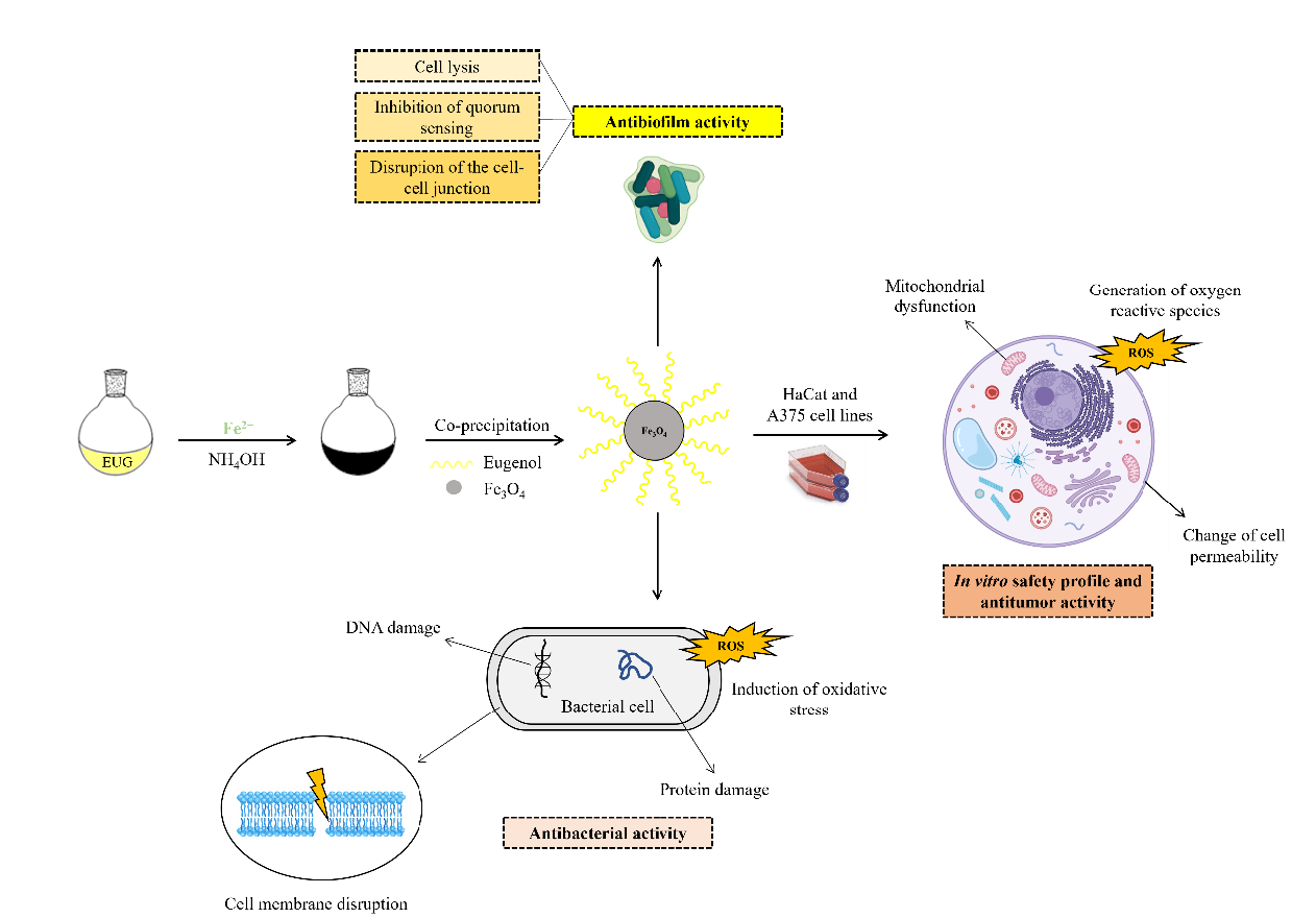
Diverse studies have been proposing some mechanisms of cytotoxicity of the eugenol in tumor cells, for instance, Jaganthan et al. [30] and Ghost et al. [31] report that effect against colon cancer and melanoma occurs by antiproliferative effect and induction of apoptosis. Regarding iron oxide nanoparticles, the mechanisms described are the generation of reactive oxygen species, inhibition of chemoresistant proteins, impairment of mitochondrial function, damage of DNA, and increase of apoptotic signals [32, 33].
The safety profile study performed using the HaCat lineage demonstrates that the nanoparticles proved to be biocompatible due to cell viability remaining around 80% according to ISO 10993-12 2009 [34].
3.3 Antibacterial and Antibiofilm activity
3.3.1 Disc diffusion
After the incubation, the inhibition zones were measured with digital caliper. The results of free eugenol, magnetite pristine, EUG·Fe3O4 1:1, EUG·Fe3O4 1:5 and EUG·Fe3O4 1:10 are shown in Table 2.
Table 2
Inhibition zone diameters formed after treatment (mm).
|
Bacterial strain
|
EUG
|
Fe3O4
|
EUG·Fe3O4 1:1
|
EUG·Fe3O4 1:5
|
EUG·Fe3O4 1:10
|
|
S. aureus
|
10.0 ± 1 mm
|
-
|
7.0 ± 1 mm
|
-
|
-
|
|
P. aeruginosa
|
7.0 ± 1 mm
|
-
|
7.0 ± 1 mm
|
-
|
-
|
|
E. coli
|
8.0 ± 1 mm
|
-
|
6.0 ± 1 mm
|
-
|
-
|
|
E. faecalis
|
6.0 ± 1 mm
|
-
|
-
|
-
|
-
|
According to the results, it is possible to verify that free eugenol has a broad spectrum against pathogenic microorganisms. In this regard, the bacterial strain S. aureus demonstrated the most susceptible, and E. faecalis is the most resistant. On the other hand, among the magnetic compounds, only EUG·Fe3O4 1:1 showed antimicrobial activity. The presence of an inhibition zone indicates a biocidal effect of the compounds that may implicate disruption of the bacterial cell membrane [35]
The sensibility to treatments depends on some factors, as the concentration of the substance, size particle, solubility, and bacterial concentration. As demonstrated on the topic of hydrodynamic size of the magnetite-functionalized eugenol (Fig. 4), the amount of the iron precursor employed in the reaction increased the size of magnetic nanoparticles [3], which is possibly related to the results obtained in the study of antibacterial activity. A larger surface area and smaller diameter particle enable better interaction with bacterial cells, increasing permeability and adhesion in the cell membrane, facilitating rupture and release of intracellular content [36].
Previous studies reported different mechanisms to the antibacterial activity of eugenol, which includes alteration of fatty acids, changes in morphology, and the cytoplasmatic membrane. Further, it can also cause ions transport modification, production of reactive oxygen species [37], and binding of the hydroxyl group with proteins, inhibiting bacterial enzymatic action [38, 39].
While the action mechanisms of MNPs can be attributed to the production of free radicals, such as hydrogen peroxide (H2O2) and superoxide anion (O2˙), causing intense oxidative stress, resulting in the degradation of vital substances for bacterial survival, such as lipids, proteins, and nucleic acids [40, 41].
3.3.2 Minimal inhibitory and bactericidal concentration
With the addition of the reagent, it was possible to verify visible microbial growth and determine the MIC. The MBC was observed after the incubation of seeded Nutrient agar plates. The MBC and MIC were demonstrated in Table 3.
Table 3
Minimal inhibitory and bactericidal concentration (mg.mL−1)
|
Bacterial strain
|
EUG
|
Fe3O4
|
EUG·Fe3O4 1:1
|
EUG·Fe3O4 1:5
|
EUG·Fe3O4 1:10
|
| |
MIC
|
MBC
|
MIC
|
MBC
|
MIC
|
MBC
|
MIC
|
MBC
|
MIC
|
MBC
|
|
S. aureus
|
1.25
|
5.0
|
-
|
-
|
2.5
|
5.0
|
5.0
|
-
|
5.0
|
-
|
|
P. aeruginosa
|
1.25
|
2.5
|
-
|
-
|
0.62
|
0.62
|
5.0
|
-
|
5.0
|
-
|
|
E. coli
|
0.62
|
2.5
|
-
|
-
|
5.0
|
5.0
|
5.0
|
-
|
5.0
|
-
|
|
E. faecalis
|
2.5
|
-
|
-
|
-
|
5.0
|
-
|
5.0
|
-
|
5.0
|
-
|
The MIC values of free EUG for bacterial strains ranged from 0.62 mg.mL−1 to 2.5 mg.mL−1, with the MBC measured to be approximately double the MIC. For the treatment with Fe3O4 pristine, no inhibitory/bactericidal activity against Gram-positive and Gram-negative bacteria used in this study it was observed.
The antimicrobial assay demonstrated that MIC values of EUG·Fe3O4 1:1 nanoparticle are different among tested bacterial strains. For E. coli and E. faecalis, the MIC value was the highest among the four tested species (5.0 mg.mL−1). The MIC value for S. aureus was 2.5 mg.mL−1, while for the P. aeruginosa, the lowest concentration able to inhibit the bacterial growth was 0.62 mg.mL−1. Similar results were reported by Negut et al. [42] when using Nigella sativa functionalized Fe3O4 nanoparticles in different microbial strains.
No difference was observed in MIC values between EUG·Fe3O4 1:5 and EUG·Fe3O4 1:10 for all strains tested. However, these nanoparticles did not show the MBC values. The higher MIC values for magnetic nanoparticles with the highest amount of magnetite (EUG·Fe3O4 1:5 and EUG·Fe3O4 1:10) may be related to the lower amount of eugenol present in the sample (mass: mass ratio), which can be justified by the absence of biological activity of the nanoparticle pristine (Fe3O4). Likewise, Mohamed et al. [43] reported that plain Fe3O4 showed low antibacterial activity against P. aeruginosa (PAO1) and clinical isolates, considering the high MIC values.
3.3.3 Quantification of biofilm biomass
After the treatment, the biomass biofilm was quantified by crystal violet assay. The results inhibit the growth of biofilm using free eugenol and EUG·Fe3O4 1:1 is showed in Fig. 11.
The experiments demonstrate that free eugenol exhibits a low antibiofilm effect, while for EUG·Fe3O4 1:1 causes a significant decrease in biofilm biomass when compared to the positive control. A synergistic effect of magnetite and eugenol nanoparticles can be observed for P. aeruginosa, corroborating the results presented in Table 3.
The antibacterial and antibiofilm activity of eugenol is widely known in the literature. The antibacterial effects are associate to increase cell permeability, disruption of the cytoplasmic membrane, and modification of shape cell [43, 44]. Meanwhile, antibiofilm activity occurs through cell lysis, disruption of the cell-cell junction, and inhibition of the quorum detection system [45, 46].