Subjects
Twenty-one patients with epilepsy and thirteen controls were recruited from the Neurology Department of Nanfang Hospital, Southern Medical University. Twenty-one patients with epilepsy were classified as patients with primary epilepsy group (n=10) and patients with seizure secondary to autoimmune encephalitis group (n=11) according to the cause of epilepsy. Moreover, 21 patients with epilepsy consisted of 11 patients with status epilepticus and 10 patients without status epilepticus. All patients were diagnosed by their supervisory doctors according to criteria established by the International League Against Epilepsy[11]. As controls, none of these subjects had a history of epilepsy, other neurological diseases, or exposure to antiepileptic drugs. The study was approved by the ethics committee of the Nanfang Hospital, Southern Medical University.
Animals
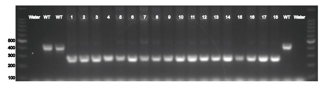
Male C57BL/6 mice (6-8 weeks of age) were purchased from the Experimental Animal Center of Southern Medical University (Guangzhou, China). The IDO1 KO mice were obtained from the Jackson Laboratory (Bar Harbor, ME, USA) and WT littermates were produced by heterozygous mating. The results of gene identification of IDO1-/- mice were presented in the Supplementary Fig. S1. Animals were maintained in specific pathogen-free facilities with a temperature of (22 ± 1℃) and a 12h light/12h dark cycle and were offered free access to standard food and water. All animal procedures in this study were approved by the Institutional Animal Care and Use Committee of Southern Medical University (Permit Number: 00197090).
Status epilepticus induction and monitoring of spontaneous recurrent seizures
Mice were intraperitoneally (i.p.) injected with lithium chloride (127 mg/kg, Sigma-Aldrich, St Louis, MO, USA), then 20 hours later were injected with pilocarpine hydrochloride (30 mg/kg, i.p., Sigma-Aldrich, St Louis, MO, USA) to induce SE. To reduce the peripheral side effects of pilocarpine, methyl scopolamine nitrate (1 mg/kg; i.p., Tokyo Chemical Industry, Tokyo, Japan) was given 30 min before pilocarpine administration. The severity of behavioural seizures was graded according to Racine’s scale[12]. Only mice with at least stage 3 were selected for further experimentation. Diazepam (15 mg/kg, i.p., King York, Tianjin, China) was administered to terminate behavioural seizures at 2 hours after SE.
Mice received video monitoring of SRS from day 28 to day 41 after SE. In the pilocarpine model of epilepsy, only generalized convulsive stage 4 and 5 seizures were detected[13-17]. All video recordings were analyzed by independent investigators blinded to the study.
Preparation of blood and CSF samples
Blood samples were collected to vacuum tubes and then allowed to coagulate at room temperature for 30 min. Serum was separated by centrifugation at 3,000 g for 15 min. CSF samples were collected by lumbar puncture and centrifuged at 1,000 g for 10 min. All serum and CSF samples were stored at -80℃ until analysis.
Biochemical analyses
IDO1 level in the serum and CSF of subjects were detected by human indoleamine 2,3-dioxygenase (IDO) ELISA Kit (Cusabio Biotech, Wuhan, China) following the manufacturer’s instructions. Frozen hippocampal tissues were diluted in ice-cold PBS with a concentration of 10% (w/v) after weighing and were homogenized. Then, tissue homogenates were centrifuged at 4 °C for 15 min, and supernatants were collected to detection. The levels of inflammatory factors, such as IL-1β, IL-6, TNF-α in the serum and supernatant of hippocampal tissues, were measured by ELISA (Boshen Biotechnology, Nanjing, China). IDO1 level in the serum and supernatant of hippocampal tissues were assessed using mouse indoleamine 2,3-dioxygenase 1 (IDO1) ELISA Kit (Cusabio Biotech, Wuhan, China). The activity of SOD, GSH-Px, and CAT and MDA content in the serum and supernatant of hippocampal tissues were measured using the detection kits (Jiancheng Bioengineering Institute, Nanjing, China) according to the manufacturer’s instructions.
LC-MS analysis of TRP, KYN, QUIN and KYNA
Hippocampal tissues were thawed and then homogenized in ice-cold extraction solution. Homogenates were centrifuged at 4 °C for 10 min and supernatants were collected to analysis.
The concentrations of TRP, KYN, QUIN and KYNA in the serum, CSF and tissues solution were determined by combination of high performance liquid chromatography (LC-30AD, Shimadzu, Kyoto, Japan) and a triple quadruple mass spectrometry (Triple Quad 4500, AB Sciex, Boston, MA, USA). The parameters of mass spectrometer and the mobile phase were prepared as previously described[18]. The IDO1 activity was determined by KYN/TRP ratio.
Histological examination
At the end of the experiments, mice were anaesthetized with sodium pentobarbital and transcardially perfused with PBS. Brains were post-fixed in 4% paraformaldehyde for 24 hours at room temperature and subsequently penetrated with 15% sucrose and 30% sucrose. Then brain tissues were sectioned into 6 µm thick coronal slices. For H&E staining and Nissl staining, brain slices were stained with hematoxylin and eosin, and toluidine blue, respectively. For immunofluorescence staining, slices were incubated with primary antibodies against NeuN (mouse, 1:200, Abcam, Temecula, CA, USA), GFAP (goat, 1:500, Abcam, Temecula, CA, USA) and Iba1 (goat, 1:200, Novus Biologicals, Littleton, CO, USA). Slices were then incubated with AlexaFluor 594-conjugated goat anti-mouse IgG (1:200, Carlsbad, CA, Invitrogen), AlexaFluor 488-conjugated donkey anti-goat IgG (1:200, Abcam, Temecula, CA, USA) and AlexaFluor 594-conjugated donkey anti-goat IgG (1:200, Abcam, Temecula, CA, USA) secondary antibodies respectively. Images were observed and taken randomly for each sample under a microscope (Olympus, Tokyo, Japan). Immunofluorescence images were observed with a confocal microscope (Zeiss LSM 880, Carl Zeiss, German).
Statistical Analysis
All data were statistically analyzed using SPSS 20.0 (IBM, Armonk, NY, USA). All graphics were generated with GraphPad Prism 7 (GraphPad, La Jolla, CA, USA). Data were expressed as mean ± SD unless otherwise indicated. Statistical significance was evaluated by student’s t-test for comparisons between two groups and one-way ANOVA with Tukey's for comparison within multiple groups. The Kruskal-Wallis test was used to compare the IDO1 level and KYN/TRP ratio in clinical samples. Chi-square test was performed to determine significant differences in the severity of SRS. Mann-Whitney U-test was used for comparison of the frequency of SRS between KO epileptic group and WT epileptic group. Value of P < 0.05 was considered as statistically significant.