Study area
Ticks and heartwater diagnostic reports were collected with permission from the Department of Veterinary Services (DVS) in the Southern District of Botswana. The district is divided into four sub-districts i.e. Kanye, Jwaneng, Moshupa and Goodhope. Each sub-district serves as a livestock advisory center for surrounding villages and towns. Each center has an established veterinary office with three staff members - a veterinary officer and two veterinary assistants attached to the office. These offices located in various sub-districts report to the district headquarters of DVS that is based in Kanye. Ticks were collected and questionnaires administered in 13 subsistence farms holding cattle, sheep and goats within the Southern District of Botswana. The locations of the farms were mapped using GPS coordinates and are shown in Fig. 1.
Questionnaires
Questionnaires were administered to 27 farmers/staff concurrently with tick collections. The questionnaire of 18 questions, contained open and closed questions that generally covered aspects relating to heartwater burden, susceptibility, control and management. The interview was translated in Setswana by the assisting veterinary officer and the responses were recorded in English. Questionnaires were analyzed using IBM SPSS Statistics for Windows, version 26 (IBM Corp., Armonk, N.Y., USA).
Sample period and sample collection
Sample collection was carried out during the summer seasons of 2017, 2018 and 2019. Three hundred and ninety-one small livestock ruminants (60 cows, 234 goats and 97 sheep) were randomly selected for tick collection from communal farms with no particular regard to a specific age, sex or breed.
Tick Collection and Identification
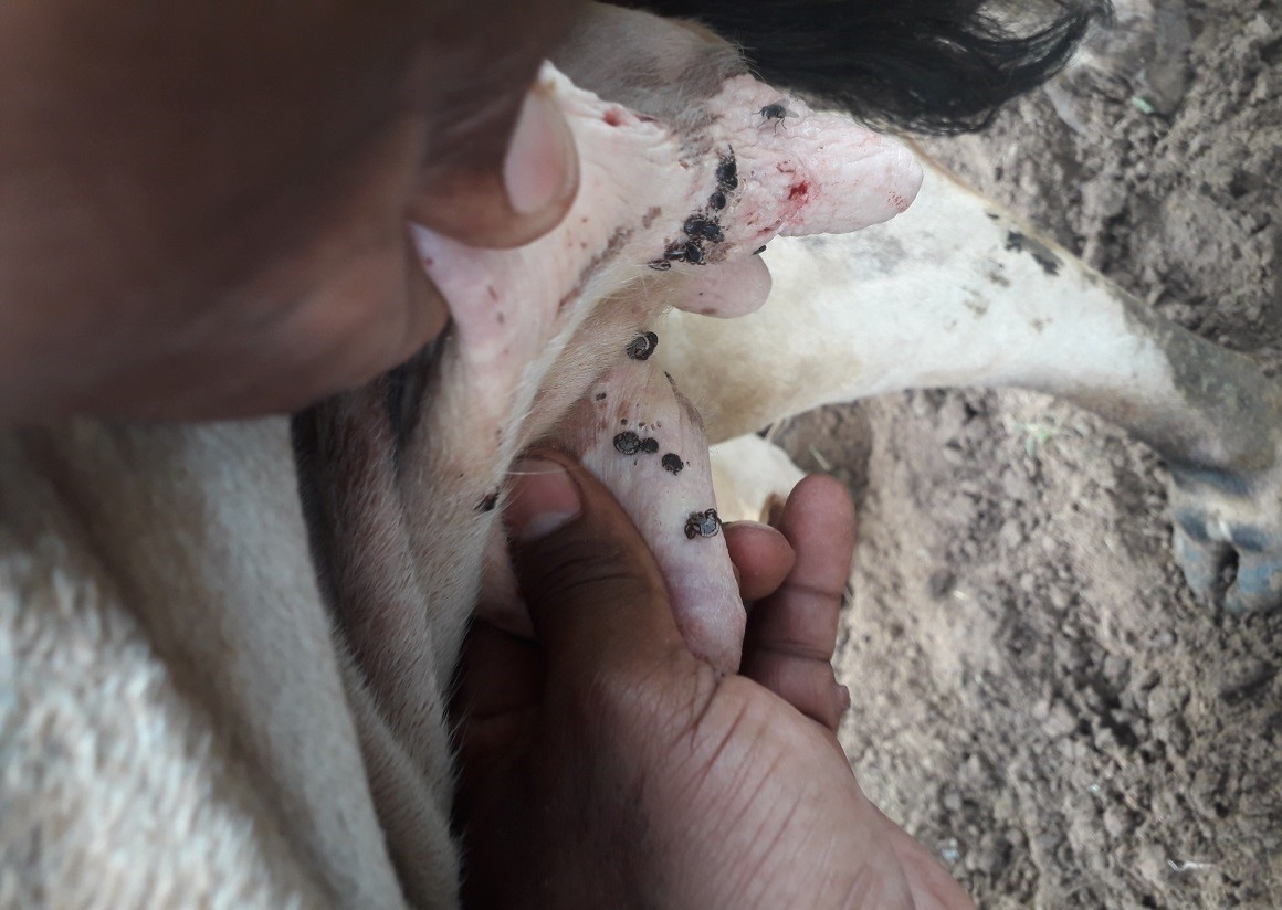
Ticks were hand-picked from under the tails, around the anus, on belly or groin regions and between hooves of randomly selected livestock. Ticks were carefully transferred into labeled 2 mL tubes and kept in a labeled ziploc bag at room temperature until refrigeration at the end of the sampling day. Ziploc bags were labeled with the sub-district, site coordinates, and date of collection. The ticks collected were morphologically identified using a Leica M205 C microscope (Leica Microsystems Ltd, Switzerland) which had a Leica MC190HD camera connected to it. Key features used for identification were adopted from guidelines provided by Walker et al (2003) [6].
Tick genomic DNA extraction
Ticks were incubated in 1 mL of Quick-DNA Tissue/Insect Miniprep Kit lysis buffer at 56 °C overnight. Tick tissue was dash frozen with liquid nitrogen and ground using a pestle and mortar. Tick tissue was then transferred to bashing tubes from the Quick-DNA Tissue/Insect Miniprep Kit (Zymo Research, USA). DNA extraction was then undertaken according to manufacturer’s instructions. Genomic DNA was eluted using 25 µL of nuclease-free water.
Polymerase Chain Reaction
A fragment of the mitochondrial marker cytochrome c oxidase subunit I (COI) was amplified using PCR using universal primers LCO 1490 (5ꞌ-GGTCAACAAATCATAAAGATATT GG-3ꞌ) and HCO 2198 (5′-TAAACTTCAGGGTGACCAAAAAATCA-3) described by Folmer [7] to yield a 710 bp amplicon. Twenty five µL PCR reaction samples were prepared in PCR vial tubes. Each reaction comprised of 12.5 µL of the Q5® High-Fidelity 2X Master Mix PCR Master Mix (Thermo Fisher Scientific, USA), 6.5 µL of nuclease free water (VWR International LLC, USA), 1 µL each of forward and reverse primers, and 2 µL of extracted genomic DNA. PCR reaction conditions were as follows; initial denaturation at 98 °C for 30 seconds, followed by 35 cycles of; denaturation at 98 °C for 10 seconds, annealing at 48 °C for 30 seconds and extension at 72 °C for 30 seconds. This was followed by a final extension at 72 °C for 7 minutes. The reaction was then stopped at 4 °C. The presence of amplicons in PCR products was verified through agarose gel electrophoresis.
Agarose gel electrophoresis
PCR products were electrophoresed on 1% agarose gel at 120 V for 90 minutes. The agarose gel was stained with ethidium bromide to visualize the amplicons.
Amplicon clean-up
PCR products were purified using the GeneJET PCR purification kit (Thermo Fisher Scientific, USA), according to manufacturer’s instructions.
Sequencing
Bidirectional, Sanger sequencing of the partial COI gene was performed at Inqaba Biotechnical Industries (Pretoria, South Africa).
Molecular identification of ticks
Contiguous sequences were analyzed using basic local alignment search tool (BLAST) to confirm matches with submissions made to the NCBI genbank.