Copper is an essential element used in metabolic processes for both humans and animals (Bhattacharya et al. 2016). It is needed for crosslinking connective tissue as well as in iron and lipid metabolism (Siddiqui et al. 2013). However, excessive copper is extremely toxic and can cause various pathological changes in tissues (Ozcelik et al. 2003). Besides, CuO-NPs was shown to catalyze the formation of free radical species capable of triggering lipid peroxidation (Rikans and Hornbrook, 1997; Paresh et al. 2010).
While the respiratory tract is the most common route of nanoparticle exposure, the gastrointestinal tract is also a potential route of entry. Copper nanoparticles can be exposed to the gastrointestinal tract either through direct ingestion of water and food or through the mucociliary clearance of the respiratory tract which is subsequently ingested after swallowing (Chen et al. 2006).
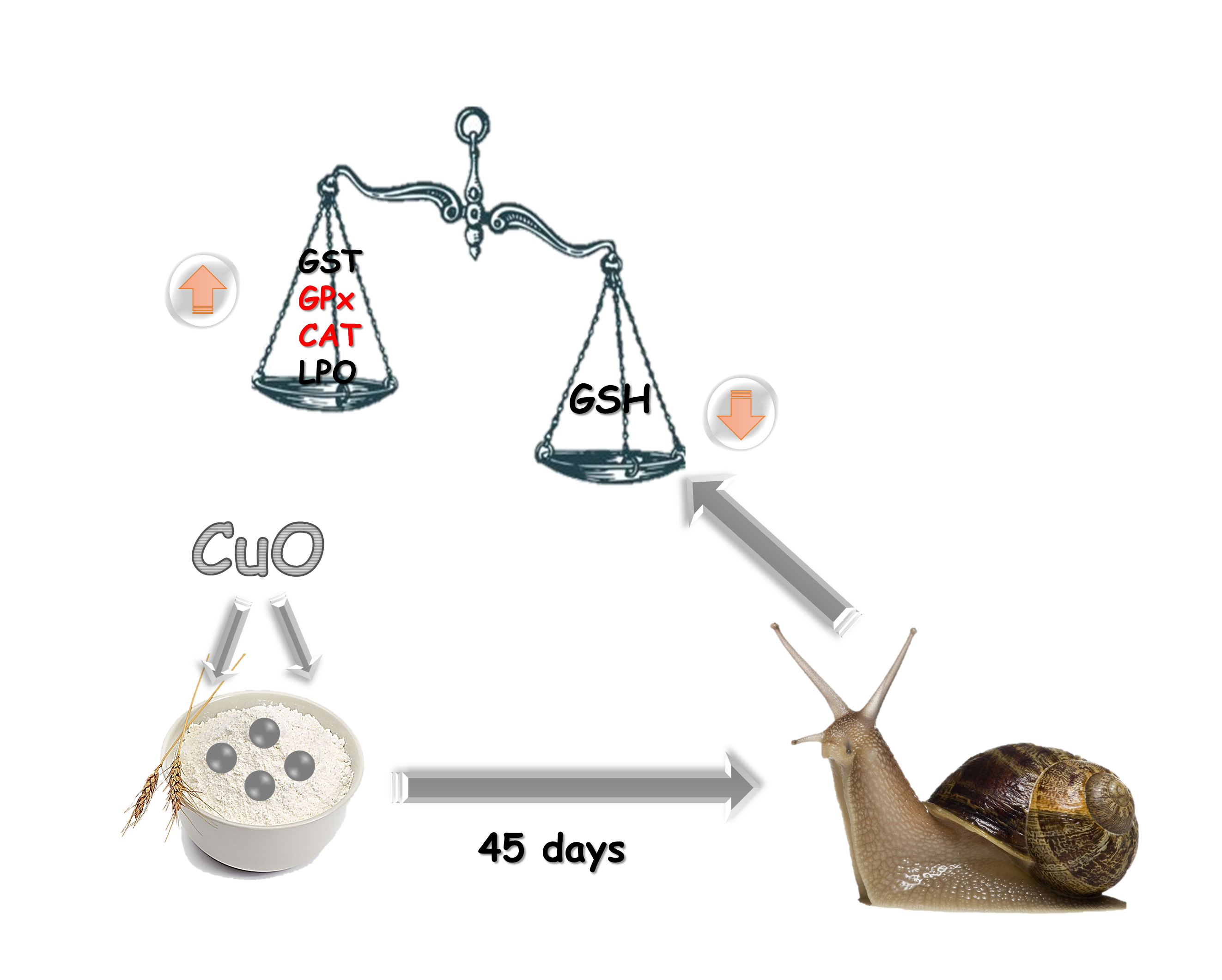
The main objective of this study is to evaluate the induction of oxidative stress by copper oxide on the land snail Helix aspersa exposed to mixing of the nanoparticles and the wheat flour; for 45 days (subchronic exposure), with a battery of stress biomarkers (GSH, GST, GPx, CAT and LPO)
Glutathione (GSH) is the most important water-soluble and low-molecular antioxidant found in cells (Deneke and Fanburg, 1989). It has multiples physiological roles; as hydrogen peroxide scavenging and preservation of SH groups in a reduced state in proteins, enzymes, and some other molecules (Meister,1985). It plays a key role in cellular defense and serves as a reservoir for the amino acid cysteine.
In the present study, GSH contents in the hepatopancreas and kidney tissues of Helix aspersa exposed to CuO-NPs showed a significant decrease, except those exposed to 50, 100, and 200 mg/kg in the case of kidney tissues, when compared with the control groups after 45 days. These results are similar to Xiong et al. (2011); who studied the impact of ZnO on zebrafish, Abdel-Khalek et al. (2015) who exposed Oreochromis niloticus to a series of CuO-NPs concentrations, and Anreddy (2018), who treated the Wistar rat by CuO-NPs, all teams constated that NPs suspension caused a decrease in GSH content in the hepatopancreas tissue. Redox-active metal ions such as Cu (II) readily catalyze the oxidation of GSH giving rise to thiol and hydroxyl radicals (Stohs and Bagchi, 1995). This depletion can be a result of increased binding of Cu (stabilization of Cu in the oxidative state), enhanced use of GSH’s oxidizing ability (conversion into GSSG, the oxidized form of glutathione), or an ineffective GSH regeneration (Pandey et al. 2001; Parvez et al. 2003; Ahmad et al. 2005; Parvez and Raisuddin, 2006). This depletion of GSH level reduces the cellular availability to scavenge free radicals and can lead to more oxidative stress-related damage (Elia et al. 2003).
GST is an enzyme that catalyzes in the cytosol the conjugation reaction of electrophilic xenobiotics and their metabolites with an endogenous polar ligand which is glutathione (Habig et al. 1974). It is a key detoxifying enzyme that also plays an important role in eliminating ROS and scavenging lipid peroxidation metabolite (Wang et al. 2016).
The reduction of GST activity is could be explained by the deficiency and absence of biotransformation of xenobiotics and androgenic substances. Therefore, the GST has an important role in the neutralization and detoxification of certain xenobiotics by increasing their water solubility and thus facilitating their elimination (Vander et al. 2003).
Induction of the GST activity has already been reported in the snail Helix aspersa, exposed to various pollutants (Belhaouchet et al. 2012; Radwan and Mohamed, 2013; Hamdeni et al. 2014; Larba and Soltani, 2014).
All living organisms have protective systems against the reactions of free radicals, including antioxidant and oxidative stress enzymes. The activity of these antioxidant enzymes depends on the concentration and duration of exposure to pollutants, and the susceptibility of the species studied (Ballesteros et al. 2009).
GPx has a crucial role in intracellular protection against toxic compounds such as Cu and Zn (Anderson, 1997; Anderson and Luo, 1998; Mosleh et al. 2005). GPx is responsible for enzymatic defense against hydrogen peroxide and is strictly linked with the concentration of GSH, it catalyzes the reaction between glutathione and hydrogen peroxide, resulting in the formation of glutathione disulfide (GSSG) (Alkaladi et al. 2013). In the present result, the GPx activity in the hepatopancreas tissues of Helix aspersa treated by copper oxide showed a significant increase when compared with control after 45 days. Witch is confirmed by Fahmy and Cormier (2009) who reported that CuO-NPs NPs were better able to increase the activity of GPx.
Catalase is a catalytic enzyme in the disproportionation of hydrogen peroxide (a SOD product generator) into the water and molecular oxygen (Delattre et al. 2005; Mohora et al. 2007). This hemoprotein protects tissues from hydroxyl radicals which are very reactive (Sathishsekar and Subramanian, 2005). In the present investigation, while CAT activities showed a significant increase in the case of hepatopancreas tissues of H. aspersa exposed to 04 concentrations of copper oxide, it showed a nonsignificant increase in kidney tissues, when compared with matched controls.
CAT activity in invertebrates depends on the mode of exposure, species, and even within the same organism. Regoli et al. (2006) reported that vehicles’ pollutants induced an increase in CAT activity in the digestive gland of Helix aspersa, however, it doesn’t change when the species exposed to electromagnetic fields (Regoli et al. 2005). Some authors proposed that seasonal variations can affect catalase activity (Ramos-Vasconcelos et al. 2005). In contrast, Chandran et al. (2005) showed that enzyme activity was inhibited in the presence of Cadmium and Zinc in the snail Achatina fulica, in both target organs "hepatopancreas and kidney".
Lipid peroxidation was the classic result of oxidative damage and its byproduct was often formed when free radicals attacked cellular membranes (Chen et al. 2010). In the present study, the excessive level of TBA, a stable metabolite of lipid peroxidation, would further confirm the generation of oxidative damage in hepatopancreas and kidneys. All the above data suggest that nano-CuO-NPs affects the oxidation–antioxidation homeostasis of both organs via increasing ROS and reducing antioxidant enzymes.
During the interpretation of our results, we observed the presence of a slight variation in the level of stress and defense biomarkers in the kidneys compared to hepatopancreas. A large number of studies have shown that excessive Cu results in inhibition of the levels of antioxidant enzymes while promoting the levels of lipid peroxidation in kidneys (Wang, 2017; Kumar, 2016).