Materials
Tetraethyl orthosilicate (TEOS), (3-aminopropyl)triethoxysilane (APTS), tetrakis(hydroxymethyl)phosphonium chloride (THPC), polyvinylpyrrolidone (PVP), gold (III) chloride trihydrate (HAuCl4), ascorbic acid (AA), Paraformaldehyde, 4-fluorobenzenethiol (4-FBT), 2-naphthalenethiol (2-NT), 3,5-dichlorobenzenethiol (3,5-DCT), 4-chlorobenzenethiol (4-CBT), 4-methylbenzenethiol (4-MBT), 4-mercaptophenol (4-MP), 4-bromobenzenethiol (4-BBT), 4-aminothiophenol (4-ATP), 4-mercaptobenzoic acid (4-MBA), 4-mercaptophenyl boronic acid (4-MPBA), benzenethiol (BT), 2-bromobenzenethiol (2-BBT), 3,4-dichlorobenzenethiol (3,4-DCT), and 2-fluorobenzenethiol (2-FBT) were purchased form Sigma-Aldrich (St. Louis, MO, USA). Ethanol (EtOH) and aqueous ammonium hydroxide (NH4OH) were purchased form Daejung (Sihung-si, Gyeonggi-do South Korea). Sodium hydroxide (NaOH) was purchased from Samchun (Pyeongtaek-si, Gyeonggi-do, South Korea). Deionized water (DW) was produced by a Millipore water purification system of Vivagen (Seongnam-si, Gyeonggi‐do, South Korea). HCT 116 cells were purchased from American Type Culture Collection (ATCC) (Manassas, VA, USA). RPMI 1640 was purchased from Biowest (Riverside, MO, USA). Fetal bovine serum (FBS) was purchased from JCBIO (Seoul, South Korea). Penicillin streptomycin was purchased from Welgene (Gyeongsan-si, Gyeongsangbuk-do, South Korea). Phosphate-buffered saline (PBS) was purchased from BYLABS (Hanam-si, Gyeonggi-do, South Korea). Sodium dodecyl sulphate (SDS) was purchased from LPS solution (Daejeon-si, South Korea). Eight-weeks-old female Balb/c athymic nude mice were purchased from Orient Bio Inc. (Seongnam-si, Gyeonggi-do, Korea).
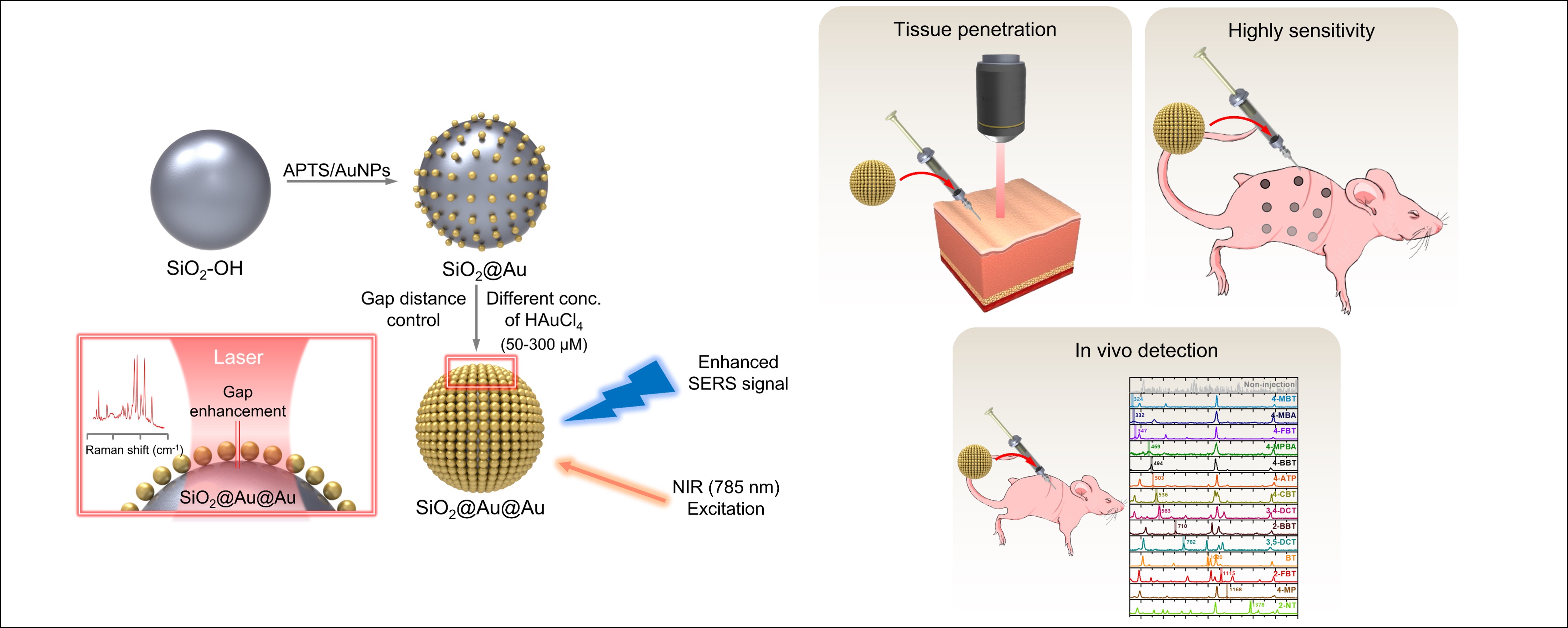
Preparation of SiO2@Au@Au
SiO2@Au was synthesized using a previously reported method [42]. Briefly, Au NPs (3 nm) were prepared using the Turkevich method. To obtain 3 nm Au NPs, 1.5 mL of sodium hydroxide (NaOH; 0.2 M), 12 μL of THPC, and 1.5 mL of chloroauric acid (HAuCl4) solution (50 mM) were added to 47.5 mL of distilled water (DW). The mixture was vigorously stirred for 1 h and stored in a refrigerator for at least 2 days. In addition, 62 μL of APTS and 40 μL of ammonium hydroxide (NH4OH) were added to 1 mL of SiO2 NPs (50 mg/mL), and the mixture was stirred at 700 rpm for overnight to produce aminated SiO2 NPs. Then, the SiO2-NH2 NPs were washed several times with ethanol via centrifugation, and 10 mL of Au NPs and 200 μL of SiO2-NH2 NPs (10 mg/mL) were mixed and stirred for overnight. SiO2@Au NPs were obtained after washing several times with ethanol via centrifugation. These SiO2@Au NPs were dispersed in 2 mL of DW containing 2 mg of PVP.
SiO2@Au@Au NPs were prepared as per the method described by our group [42] with some modifications. Briefly, SiO2@Au@Au NPs were synthesized according to the seed-mediated growth method using SiO2@Au NP seed and Au precursor. To grow Au into the SiO2@Au seed, 200 μL of SiO2@Au NPs (1 mg/mL) were dispersed in 9.8 mL DW containing 10 mg of PVP. This suspension was stirred with 20 μL of HAuCl4 (10 mM), and treated with 40 μL of AA (10 mM) every 5 min until the desired concentration of Au3+ was achieved (50, 100, 200, 300, 400, and 500 μM). Then, SiO2@Au@Au was obtained after washing several times with ethanol via centrifugation.
Labelling SiO2@Au@Au with Raman chemical
An RLC solution (2 mM) was prepared and added to 1 mL of SiO2@Au@Au NPs (1 mg/mL). The mixture was vigorously shaken for 1 h at 25 ℃, and the RLC-conjugated SiO2@Au@Au obtained was washed several times with ethanol via centrifugation. Subsequently, Raman-labeled SiO2@Au@Au (SiO2@Au@AuRLC) NPs were redispersed in 1 mL of ethanol.
SERS measurement for SiO2@Au@AuRLC
SiO2@Au@AuRLC suspensions (1 mg/mL) were injected into a capillary tube. The SERS spectra of each NP were measured thrice using a microscopic Raman system. Measurement was carried out using 532-nm photoexcitation at 1 mW, 660-nm photoexcitation at 1.2 mW, and 785-nm photoexcitation at 2.1 mW laser power, and a ×10 objective lens with a 5-s acquisition time.
Calculation of SERS enhancement factor (EF)
The SERS EF of SiO2@Au@Au4-FBT (with 500 μM of Au precursor) at 785 nm photoexcitation was estimated using the following equation: EF = (ISERS × Nnormal)/(Inormal × NSERS) where ISERS and Inormal indicate the intensity of the Raman band from SERS and normal Raman, respectively, and Nnormal and NSERS are the number of 4-FBT molecules in the pure form and assembled form, respectively, on the surface of SiO2@Au@Au4-FBT NPs. Raman signal intensity was measured for both pure 4-FBT and single particles using an identical ×100 objective lens under the following conditions: 0.3 mW laser power and 5 s acquisition time. The 4-FBT peak at 1075 cm−1 was used to estimate EF. Isers were obtained by averaging the peak intensities of 20 individual particles. The probing volume (18.84 μm2) for the normal Raman measurement was approximated by a cylindrical form with a diameter of 2 μm and height of 6 μm. Assuming that 4-FBT molecules form a monolayer on the surface of the NP, NSERS was calculated from the surface area of the NPs (assuming that SiO2@Au@Au4-FBT has a spherical shape, r = 115 nm) and the molecular footprint of 4-FBT (0.383 nm2/molecule) [43].
Cytotoxicity of SiO2@Au@Au4-FBT to HCT 116 cells
HCT 116 cells (human colon cancer cell line) were cultured in Roswell Park Memorial Institute (RPMI)-1640 medium with 10% heat-inactivated fetal bovine serum (FBS) and 1% penicillin/streptomycin at 37°C in humidified air with 5% CO2. Cytotoxicity tests of NPs were conducted using a crystal violet assay. Cells were seeded in 96-well plates and incubated with different concentrations (0, 1.95, 3.90, 7.81, 15.63, 31.25, and 62.50 mg/mL) of SiO2@Au@Au4-FBT NPs at 37°C for 24 h. After incubation, the culture medium was removed and the cells were fixed with 4% paraformaldehyde for 1 h. Then, the cells were washed with DW and air-dried. The cells in each well were treated with 100 μL of a 0.5% crystal violet solution. After 10 min, the solution was removed, and the plates were washed with DW and air-dried. Subsequently, the cells were lysed with 1% sodium dodecyl sulfate (SDS), and the absorbance was measured using a VICTOR X3 multilabel plate reader (PerkinElmer, Waltham, MA, USA) at 570 nm.
SERS imaging of HCT 116 cells
Cells were seeded in a 60-mm dish and incubated with 50 μg/mL SiO2@Au@Au4-FBT at 37°C for 24 h. After incubation, the culture medium was removed, and the cells were washed thrice with 1× phosphate-buffer saline (PBS). The cells were fixed with 4% paraformaldehyde for 1 h, washed with PBS, and dried at room temperature. Then, the SERS mapping images were obtained by point-by-point mapping with a 1-μm step size using a ×100 objective lens with a 785-nm excitation source, 0.3-mW laser power, and 1-s acquisition time.
Depth profile evaluation of SiO2@Au@Au SERS signal
To investigate the depth profile of SiO2@Au@Au SERS signal, NPs were injected into a porcine tissue, and Raman spectra were measured. First, 15 μL of SiO2@Au@Au4-FBT (1 mg/mL) was dispersed in DW and injected into the porcine tissue with a 26-gauge syringe at different depths (1, 3, 5, 7, and 9 mm). SERS signals of NPs inside the tissue were measured immediately after injection under ×10 objective lens. A 785-nm excitation source with 2.1-mW laser power and 10-s acquisition time was used for measurement.
In vivo multiplexing SERS imaging
To conduct multiplexing SERS imaging in nude mice, 14 types of RLCs (4-MBT, 4-MBA, 4-FBT, 4-MPBA, 4-BBT, 4-ATP, 4-CBT, 3,4-DCT, 2-BBT, 3,5-DCT, BT, 2-FBT, 4-MP, and 2-NT) were conjugated to SiO2@Au@Au. After adaptation for 1 week, the mice were euthanized and subcutaneously injected with 15 μL of SiO2@Au@AuRLC. Diluted SiO2@Au@Au4-FBT (1000, 500, 250, 125, 63, 31, 16, 8, and 4 μg/mL) were injected into another mouse. Each measurement was performed under ×10 objective lens using a 785 nm excitation source with 2.1 laser power and 10 s acquisition time. The mice were maintained in accordance with the guidelines approved by the Konkuk University Animal Care and Use Committee (IACUC).