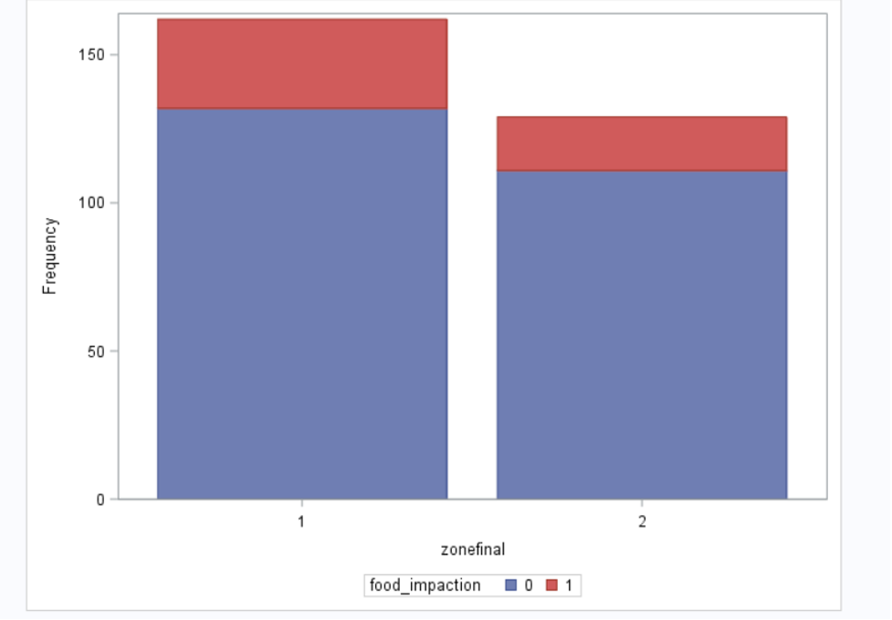
In our eosinophilic esophagitis (EoE) patient cohort, 48 cases (16%) had one or more esophageal food impactions (EFIs); male gender and the presence of classic EoE findings on endoscopy (longitudinal furrows and/or concentric rings in the esophagus) were associated with EFI. EFI is an indicator of severe fibrostenosing EoE or untreated disease, and can lead to serious life-threatening perforation.11 Our results are consistent with a large cohort study showing that males are more likely to have esophageal stenosis and food impaction events.12 Our results indicate that the presence of furrows and/or rings in EoE patients is a strong predictor of esophageal food impaction and this population should be treated more aggressively to prevent complications. Our findings are consistent with a pediatric study showing that the presence of furrows is associated with food impaction,13 no similar study was found in adult population.
Socioeconomic status (SES) of patients affects access to health care, and environmental exposures such as housing and pollution, in addition to food cost and accessibility. Asthma patients with lower SES have increased disease severity and hospitalizations.14,15 There are limited data on the role of SES in EoE patient outcomes. A study by Jensen et.al16 reviewed the role of different environmental factors in EoE and concluded that EoE might be higher in rural areas. Corder et al described association between EoE diagnosis and houses of brick exterior, forced air or gas heating.17 We examined the role of medical insurance and socioeconomic status using patients’ neighborhood adjusted gross income as a surrogate of household income. There was no difference in EFI risk across five different levels of household income. Furthermore, neighborhood income was grouped into two categories (high income versus low income) and there was still no difference in the risk of EFI (Supplementary Figure 1). We are the first study (to our knowledge) to examine the role of socioeconomic status in EoE outcomes.
All endoscopy images and patient pathology results of a cohort of 291 patients were reviewed in the current study. Most of the patients in our cohort (66%) had the classic EoE findings of esophageal furrows and/or concentric rings, which is higher than what was reported in a large systematic review and meta-analysis study of endoscopic findings in EoE patients that included 4678 patients.18 In the systematic review by Kim et al., the prevalence of linear furrows and concentric rings were 48% and 44%, respectively.18 17% of EoE patients in the Kim et. al study had normal endoscopic exam compared to 15% of patients in our cohort, this emphasizes the importance of obtaining esophageal biopsies in suspected EoE cases, despite normal endoscopic appearance of the esophagus.
It is known that GERD is a leading cause for EFI.19 About half of our patients had endoscopic findings suggestive of gastroesophageal reflux disease (GERD), including the presence of a hiatal hernia, erosive esophagitis or a Schatzki’s ring. Data on the presence of hiatal hernia or Schatzki’s ring in EoE patients is limited in the medical literature. A previous study showed that 10% of patients with Schatzki’s rings had EoE, 20 and a radiology study showed an association between EoE with GERD and Schatzki’s rings. 21 In our cohort, 35% of patients had hiatal hernia and 24% had Schatzki’s ring. The large systematic review and meta-analysis study of endoscopic findings in EoE patients by Kim et.al18 reported that 17% of EoE patients had evidence of erosive esophagitis compared to 11% of patients in our study that had LA Class B erosive esophagitis or greater. The lower prevalence of erosive esophagitis in our study may be attributed to the exclusion of patients with LA Class A esophagitis.
GERD is a very common disease with a prevalence of 18 to 28% in North America.22 The fact that GERD can lead to esophageal eosinophilia has impacted the diagnostic criteria of EoE and evolved the way EoE is diagnosed over time.8 A subset of EoE patients may have complete resolution of esophageal inflammation after treatment with acid suppression medications (proton pump inhibitors (PPIs)).9 These patients were previously labelled PPI-responsive esophageal eosinophilia (PPI-REE). Older guidelines (from 2011) recommended treatment with PPIs for 8 weeks to rule out PPI-REE before making the diagnosis of EoE.9 However, more recent 2018 guidelines confirmed that PPI-REE is a subtype of EoE and is not a subset of GERD.8,10,23 In our study, we used the updated EoE guidelines and did not exclude PPI-REE cases.
The differentiation between GERD and EoE in patients with esophageal eosinophilia is challenging. It is currently believed that EoE and GERD can coexist in many patients and possibly one condition can trigger the other.9 We showed that GERD and EoE coexistence may be more common than previously thought, given that half of our cohort had endoscopic findings suggestive of GERD. The mosaic pattern (Figure 2) clearly shows the different combinations of EoE and GERD endoscopic findings and the difficulty in drawing a line separating these conditions. Furthermore, Figure 2B shows that patients without EoE classic endoscopic findings (the left side of Figure 2B) are slightly more likely to have GERD-related findings than patients who have EoE classic endoscopic findings (the right side of Figure 2B); suggesting that some of our patients might have GERD with increased esophageal eosinophils [esophageal eosinophilia (EE)]; in some cases it is impossible to differentiate GERD with EE from EoE with coexisting GERD.
This study has a number of strengths; we included a relatively large number of patients, and all endoscopic images were reviewed by an expert gastroenterologist. In addition, the availability of subjects’ mailing addresses allowed us to investigate socioeconomic status. Our study also has a number of limitations. It is a retrospective study, and as such is subject the inherent limitations of a retrospective design. Some relevant information such as tobacco and ethanol use could not be reliably retrieved. Additionally, patients did not receive standard therapy for EoE, as EoE treatment was evolving over the relatively long timeframe represented in this study, and because patients were treated by multiple gastroenterology specialists. 49% of our patient underwent esophageal dilation, however, the presence edema, strictures and severity of strictures was not consistently documented in our cohort and the Eosinophilic Esophagitis Reference Score (EREFs) couldn’t not be calculated.