The pericellular matrix stiffness is strongly associated with its biochemical and structural changes During the aging and osteoarthritis progresses of articular cartilage. However, how substrate stiffness modulates the chondrocyte regulatory volume decrease (RVD) and calcium signaling remains unknown. This study aims to investigate the effects of substrate stiffness on the chondrocyte RVD and calcium signaling by recapitulating the physiologically relevant substrate stiffness. Our results showed that substrate stiffness induced completely different dynamical deformation between the cell swelling and recovering progresses. Chondrocytes swelled faster on the soft substrate but recovered slower than the stiff substrate during the RVD response induced by the hypo-osmotic challenge. We found that stiff substrate enhanced the cytosolic Ca2+ oscillation of chondrocytes in the iso-osmotic medium. More importantly, chondrocytes exhibited a distinctive cytosolic Ca2+ oscillation during the RVD response. Soft substrate significantly improved the Ca2+ oscillation during the cell swelling whereas stiff substrate enhanced the cytosolic Ca2+ oscillation during the cell recovering. Our work also suggests that TRPV4 channel are involved in the chondrocyte sensing substrate stiffness and RVD response by mediating Ca2+ signaling in a stiffness-dependent manner. It helps to understand a previously unidentified relationship between substrate stiffness and RVD response under the hypo-osmotic challenge.
Research Article
Substrate Stiffness-Dependent Regulatory Volume Decrease and Calcium Signaling in Chondrocytes
https://doi.org/10.21203/rs.3.rs-114377/v1
This work is licensed under a CC BY 4.0 License
Journal Publication
published 01 Nov, 2021
Read the published version in Acta Biochimica et Biophysica Sinica →
Version 1
posted
You are reading this latest preprint version
substrate stiffness
regulatory volume decrease
viscoelasticity
calcium signaling
Chondrocytes are the solitary cell type of articular cartilage and responsible for the slow turnover of matrix [1]. The pericellular matrix (PCM) with defined physical properties surrounds each chondrocyte [2] and plays a critical role in regulating the mechanical microenvironment of chondrocytes [3, 4]. During the aging and osteoarthritis (OA) progresses of articular cartilage, the mechanical properties (i.e. stiffness) of the PCM can markedly changes between 1 kPa and 205 kPa [5–8]. One study has shown that the PCM stiffness is reduced by 30–50% in the development of OA [9]. The reduction of PCM stiffness induced by OA modulates chondrocyte function-related spatial organization and biosynthetic activity [10, 11]. Furthermore, the loss of PCM stiffness corresponds to the increased extent of cell swelling and calcium signaling mediated by transient receptor potential vanilloid channel 4 (TRPV4) in response to osmotic stress [12, 13]. Cell volume regulation is important to chondrocyte physiology. Under the hypo-osmotic stimulus, chondrocyte volume rapidly increases, then followed by active regulatory volume decrease (RVD) which exhibit an active volume recovery process [14]. However, how changes in PCM stiffness influence the RVD and TRPV4-mediated calcium signaling in chondrocytes remains unknown.
As a key physical factor of cell microenvironment, cell-supporting substrate stiffness is well recognized to regulate diverse cell functions [15]. Substrate stiffness has been shown to modulate the chondrocyte morphology, phenotype and mechanical behavior [16, 17]. The RVD is crucial for many biophysical and biological responses of chondrocytes. A loss of chondrocyte RVD is associated with cell death and progression of OA [18, 19]. Changes to volume affect the phenotypic plasticity and matrix metabolism of chondrocytes [20, 21]. For example, RVD-induced changes in cell shape can affect membrane transporter activity potentially mediated via Ca2+ signaling. TRPV4, the primary regulator of Ca2+ signaling, has been proposed as a mechanosensitive channel that is involved in regulating the mechanotransduction and RVD response of chondrocytes [22–27]. It is assumed to involve in cell sensing substrate stiffness by locally altering the Ca2+ permeability [28, 29]. Since calcium signaling plays an important role in multiple signaling pathways and mechanotransduction in chondrocytes [30, 31], substrate stiffness may modulate the chondrocyte mechanotransduction via the TRPV4-mediated Ca2+ signaling, which eventually affects cell function [15]. Collectively, these findings highlight the potential role of substrate stiffness in regulating the RVD response and TRPV4-mediated calcium signaling in chondrocytes. Previous studies have suggested that PCM stiffness affects the chondrocyte volume swelling and TRPV4-mediated Ca2+ signaling [13]. However, these studies normally have been focused on the swelling process but neglected the dynamic recovering process of chondrocytes. To date, neither theoretical nor experimental studies have been focused on the role of substrate stiffness in the RVD response and TRPV4-mediated Ca2+ oscillations during the chondrocyte swelling and recovering processes.
Taken together, in this study, we hypothesize (I) that substrate stiffness modulates the mechanical properties of chondrocytes, resulting in the substrate stiffness-dependent RVD response, and (II) that TRPV4 channels are involved in chondrocytes sensing substrate stiffness by mediating cytosolic Ca2+ signaling. To test these hypotheses, Polydimethylsiloxane (PDMS) was utilized to engineer cell-supporting substrate with physiologically-relevant stiffness. The mechanical properties of chondrocytes were evaluated by using atomic force microscopy (AFM). Ca2+ oscillation of chondrocytes cultured on variable stiffness substrates was measured by using fluorescence confocal laser scanning microscopy and Fluo-4 AM ratiometric method.
2.1 Preparation of substrates with different stiffness
PDMS substrate stiffness was formulated to mimic the physiologically-related PCM elasticity that was either stiff (2.2 ± 0.3 MPa), medium (46.5 ± 5.8 kPa) and soft (2.1 ± 0.2 kPa), respectively. Briefly, PDMS curing agent (Sylgard184, Dow corning Corp) was mixed with a base agent in a mass ratio of 1:10 (stiff), 1:50 (medium) and 1:70 (soft), respectively. The mixture was poured into 35-mm petri dishes to create 1-mm thick films and then cured at 70 °C for 6 hours. After curing, these PDMS substrates were placed in an oxygen plasma cleaner (SBC-12, KYKY) for surface oxidation. The stiffness of PDMS substrate (elastic modulus) was determined using the indentation method and ElectroForceH3100 test instrument (Bose, Shanghai, China) as previously described [32]. Before seeding cells, substrates with varying stiffness were coated by 0.02mg/ml rat type I collagen (Shengyou Biotechnology) at 4°C overnight and sterilized by UV radiation for 45 minutes, as previously described [17].
2.2 Isolation of primary chondrocytes
A total of 24 animals of 5~6 day old mice (C57BL/6) were used in this study. All animal procedures were approved by the Animal Ethics committee of Taiyuan University of Technology and conducted in accordance with international standards on animal welfare. Mice were killed under general anesthesia. Femoral condyles and tibial plateau were isolated from the hind legs using scissors and pincer. The pieces of cartilage were incubated in the collagenase D digestion solution (3 mg/ml) for 45 min at 37 °C in a thermal incubator under 5% CO2. Then the cartilage pieces were placed in a new Petri dish with collagenase D solution (0.5 mg/ml) overnight at 37 °C, as previously described [33]. Cells were isolated by overnight incubation and filtered through a 40 mm cell strainer (BD-Falcon). The resulting cell suspension was washed twice in fresh Dulbecco’s modified eagle’s medium (DMEM) (1 g glucose per ml; no L-glutamine pH 7.0-7.6, Osmolality 304-336 mos/kg, Sigma, USA). Cells were re-suspended in DMEM supplemented with 10% fetal calf serum (Sigma) and 1% penicillin/streptomycin solution at the density of 8´103 cells/cm2 (the final osmolarity is 320 mOsm). Cells were grown on substrates of varying stiffness at 37°C in 5% CO2. In AFM experiments, chondrocytes were rinsed with phosphate buffered saline (PBS) solution to avoid proteins and cellular debris adhesive to tip prior to single-cell stress relaxation tests. In the RVD experiments, chondrocytes cultured on varying substrate stiffness were exposed to hypo-osmotic medium (180 mOsm). All experiments were performed at 37°C and controlled humidity (custom-built equipment) within 1.5h unless otherwise stated.
2.3 Cell diameter change during hypo-osmotic (180 mOsm) induced RVD response
The chondrocytes were cultured for 24 hours in the iso-osmotic medium (320 mOsm) before exposure to the 180 mOsm medium. The complete cell RVD response suggests that following hypo-osmotic challenge, cells initially swell passively, and then volume recovery. Cells were considered to show RVD response following hypo-osmotic (180 mOsm) if the cell diameter reduced by at least 1 µm during the imaging period [34]. The whole process of RVD response was tracked. Bright-field sequential time-lapse images of chondrocytes on substrates of varying stiffness were obtained every 10 s for 45 min by microscopy (FV1000, Olympus) with a 40´ oil immersion objective lens. The diameters d of single cells were quantitatively calculated from the mean of the two orthogonal diameters measured using ImageJ software. Then, we analyzed the following parameters. The swelling time, TS, is the time the cell takes to swell until it reaches its maximum cell diameter. In contrast, the recovering time, TR, is the time the cell takes to recover from its maximum cell diameter. The responding time, TRes, is the entire time during the whole RVD process which equals the sum of TS and TR. The cell diameter rate, Vd, is the change of diameter over time during the RVD response.
2.4 AFM testing
In AFM experiments, we only focused on the changes in the mechanical properties of chondrocytes on substrates of varying stiffness during the cell swelling. The mechanical properties of single chondrocyte were measured by an AFM (NTEGRA Solaris, NT-MDT, Russia) combined with a phase contrast microscopy (IX-70, Olympus). Chondrocytes were tested at a density of 2´104 cells/cm2, so that individual cells would not be connected with others. A gold-coated, 5-mm diameter, spherical tip cantilevers (spring constant is 0.05 N/m, Novascan Technologies, Inc.) was used for indentation and stress-relaxation experiments. Indentations were applied with a force trigger of 2.5 nN. An approach velocity of 6.53 μm/s was used and followed by a 100-s relaxation period. A constant displacement of about 2μm indentation was hold for all the stress-relaxation experiments. The elastic modulus and viscoelastic parameters of chondrocyte were determined according to a modified Hertz equation (Eq.1) and a stress relaxation model of viscoelastic solid (Eq.2), respectively. They are given by [35],

Where F is the applied force; R is the relative radius, n is the Poisson’s ratio which is assumed to be 0.5, C is a constant depending only on the indentation during stress relaxation. The elastic modulus, Eelastic, is determined by fitting the indentation approach curves with Hertz equation (Eq.1). d is the indentation depth. Data from the relaxation phase of the test were fit with the viscoelastic equation (Eq. 2) to determine the ER, τσ, and τε. E0 and m were calculated using Eq. 2. ER and E0 are instantaneous modulus and relaxed modulus, respectively. τσ represents the time of relaxation of deformation under constant load, τε represents the time of relaxation of load under constant deformation. m is the apparent viscosity. All AFM experiments were carried out at 37°C and controlled humidity, as previously described [17].
2.5 Calcium imaging
Chondrocytes on substrates of varying stiffness were loaded with Fluo-4 AM (Life Technology) in pure DMEM buffer for 35 min at 37°C before imaging. To modulate the TRPV4 channel activity, we applied 10 μM GSK205 or 1 µM 4αPDD (Merck Millipore) to the bath solution. GSK205 is a TRPV4 selective antagonist while 4αPDD is a TRPV4 selective agonist. Ca2+ signaling imaging of chondrocytes was recorded in response to substrates of varying stiffness and hypo-osmotic (180 mOsm) challenge by using fluorescence confocal laser scanning microscopy (FV1000, Olympus). Time-series images of baseline Ca2+ oscillations were recorded every 3 s for a total of 15 min and fluorescence was analyzed by using the ImageJ software. The difference (ΔF) between the mean fluorescence measured in a given region of interest (ROI) and the corresponding control value for each ROI (F0) was expressed as the fraction of the control (ΔF/F0) and then plotted as a function of time. In the RVD experiments, we simultaneously monitored the cell diameter change and chondrocyte Ca2+ oscillations. A cell was defined as responsive if it showed a calcium peak with a magnitude four times higher than its maximum fluctuation along the baseline [36].
2.6 Statistical Analysis
Statistical significance between the groups was estimated using one-way analysis of variance (ANOVA) followed by a Tukey post hoc test using Origin (OriginLab, Northampton, MA). P values less than 0.05 are considered to be significant. All data are presented as mean ± standard deviation.
3.1 Substrate stiffness affects the RVD response of chondrocytes
Our results showed that the RVD response of chondrocytes was intimately related to substrate stiffness. The soft substrate markedly increased the percentage of RVD response in chondrocytes (p<0.001) (Fig.1A). Substrate stiffness caused distinctive swelling and recovering behaviors of chondrocytes (Fig. 1B). The change of cell diameter d depended on the substrate stiffness during the RVD response (Fig.1C). The trend of non-dimensional diameter d/d0 (d0 is the initial cell diameter) over time remained the same with Fig.1C, and it was independent of the cell’s initial diameter (Fig.1D). We found that stiff substrate significantly prolonged the swelling time TS of chondrocytes (p<0.001) (Supplementary Table I and Fig.1E), and soft substrate significantly increased the percentage increase of diameter in chondrocytes during the cell swelling (p<0.001) (Fig.1F). In contrast, during the cell recovery, the recovery time TR in chondrocytes on the stiff substrate was significantly shorter than that on the soft substrate (p<0.05) (Fig.1G). Furthermore, stiff substrate can obviously enhance the capacity of cell diameter recovery during cell recovering (p<0.01) (Fig.1H). Interestingly, the trend of the swelling and recovering time was completely opposite to that of the percentage increase of diameter in chondrocytes during cell RVD responding. Lastly, the soft substrate significantly increased the mean RVD responding time TRes of chondrocytes (p<0.01) (Fig.1I).
The cell diameter rate was obtained by fitting the cell diameter vs. time curves from substrates of varying stiffness. The cell diameter vs. time curves from all substrates during the cell swelling exhibited an approximately linear relationships (Fig.2A-C). Moreover, the soft substrate markedly increased the cell diameter rate during the cell swelling caused by hypo-osmotic (180 mOsm) medium (p<0.001) (Fig.2D). Then, we fitted the approximately linear part of cell diameter vs. time curves during the cell recovering (Fig. 2E-G). However, stiff substrate significantly increased the recovering cell diameter rate (p<0.001) (Fig.2H). For the first time, our results indicated that hypo-osmotic (180 mOsm) challenge-induced chondrocyte deformation is substrate stiffness-dependent.
3.2 Stiff substrate enhances the mechanical properties of chondrocytes during the cell swelling
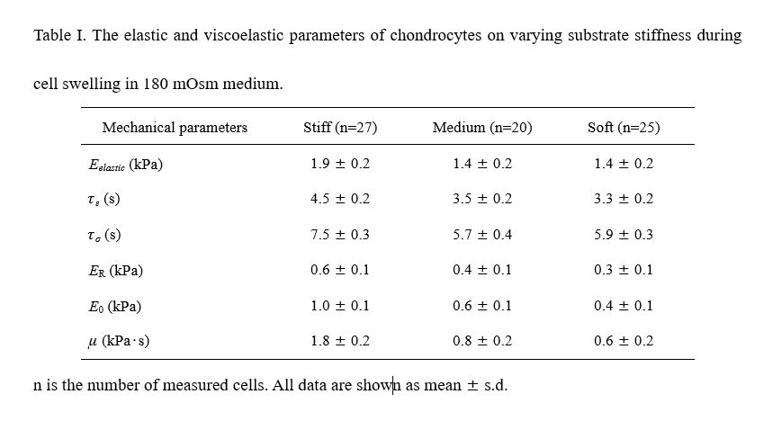
AFM experiments were used to measure the mechanical properties of chondrocytes on substrates of varying stiffness in the iso-osmotic (320 mOsm) and hypo-osmotic (180 mOsm) medium (Fig. 3A). When the constant indentation displacement 2 μm was held on chondrocytes, chondrocytes first exhibited typical elastic response consistent with the Hertz model and then exhibited stress relaxation consistent with the viscoelastic theoretical model (Fig.3B). Theoretical model fitting of all experimental data exhibited excellent consistency for elastic and viscoelastic equations respectively (Fig. 3C). Our results indicated that stiff substrate enhances the elastic (Eelastic) and viscoelastic parameters (ER, E0 and m) of chondrocytes during the cell swelling in hypo-osmotic (180 mOsm) medium (p<0.001) (Table I and Fig. 3D-G).
Then we compared mechanical parameters of chondrocytes on variable stiffness substrates between the iso-osmotic (Supplementary Table II) and hypo-osmotic (180 mOsm) medium. Our results showed compared to iso-osmotic medium, chondrocytes on the stiff substrate during the cell swelling in the hypo-osmotic (180 mOsm) showed higher elastic and viscoelastic parameters including Eelastic, ER, E0 and m (p<0.001) (Fig. 3H-K). Moreover, the mean percentage increases in elastic modulus Eelastic of chondrocytes were higher on the soft substrate than that on the stiff substrate during the cell swelling. However, in response to the hypo-osmotic medium (180 mOsm), the mean percentage increases in viscoelastic parameters (ER, E0 and μ) of chondrocytes were higher on the stiff substrate than those on the soft substrate (Supplementary Table III). Our results suggested that substrate stiffness determines regulates the dynamic elastic and viscoelastic mechanical properties of chondrocytes during the hypo-osmotic (180 mOsm) challenge.
3.3 Stiff substrate enhances cytosolic Ca2+ oscillation of chondrocytes in iso-osmotic medium
Chondrocytes on variable stiffness substrates showed cytosolic Ca2+ oscillations in the iso-osmotic medium (320 mOsm) (Fig. 4A). Stiff substrate showed the greatest percentage of Ca2+ oscillations of chondrocytes (p<0.001) (Fig. 4B). We recorded the amplitude and frequency of Ca2+ oscillation in chondrocyte on variable stiffness substrates (Fig. 4C, D). Our results showed that stiff substrate markedly enhanced both the amplitude and frequency of Ca2+ oscillations in chondrocytes (p<0.001) (Fig. 4E, I). To test if TRPV4 channels are involved in substrate stiffness mediating Ca2+ signaling in chondrocytes, we applied TRPV4 channel activator 4aPDD and inhibitor GSK205 when recording Ca2+ oscillations, respectively. TRPV4 activator 4aPDD significantly enhanced the calcium responsive rate, amplitude and frequency of Ca2+ oscillations in chondrocytes on all substrates of varying stiffness while TRPV4 inhibitor GSK205 weakened those Ca2+ effects. (p<0.01) (Fig. 4F-H, J-L, Supplementary Table IV). In addition, when treated with TRPV4 activator 4aPDD, chondrocytes on the stiff substrate showed the highest amplitude and frequency of Ca2+ oscillations (p<0.001) (Fig. 4E, I). They also showed a bigger percentage increase of amplitude and frequency of Ca2+ oscillations than chondrocytes on the soft substrate (Supplementary Table V). On the other hand, when treated with the TRPV4 inhibitor GSK205, chondrocytes on varying substrates showed no significant difference in the amplitude of Ca2+ oscillations (p>0.25) (Fig. 4G), though chondrocytes on the stiff substrate exhibited a higher frequency of Ca2+ oscillations (p<0.05) (Fig. 4J). Chondrocytes showed a bigger percentage decrease of amplitude and frequency of Ca2+ oscillations on the stiff substrate than the soft substrate with the application of GSK205 (Supplementary Table V). Taken together, these results suggested that mechanosensitive TRPV4 channel is involved in chondrocytes sensing substrate stiffness and mediating the calcium signaling.
3.4 Substrate stiffness affects the hypo-osmotic (180 mOsm) challenge-induced Ca2+ oscillations in chondrocytes
Only chondrocytes that exhibited both RVD response and Ca2+ oscillations were used in this analysis. Our results show that soft and medium substrates significantly increased the percentage of chondrocytes exhibited both RVD response and Ca2+ oscillations (42 ± 5% and 39 ± 4%, respectively) than the stiff substrate (31 ± 5%) (p<0.01). We then quantified the amplitude and frequency of Ca2+ oscillation in chondrocytes cultured on variable stiffness substrates during the cell swelling and recovering (Fig. 5A, B). The results showed that soft substrate induced a significantly higher percentage of Ca2+ oscillations (58 ± 7%) in chondrocytes than medium (77 ± 8%) and stiff substrates (79 ± 8%) during the hypo-osmotic (180 mOsm) challenge (p<0.001). Application of TRPV4 activator 4aPDD or the TRPV4 inhibitor GSK205 significantly enhanced or weakened the calcium responsive rate of Ca2+ oscillations in chondrocytes on variable stiffness substrates, respectively (p<0.01) (Supplementary Table VI). During the cell swelling, the soft substrate enhanced both the amplitude and frequency of Ca2+ oscillations (p<0.01) (Fig. 5C, F). With the treatment of TRPV4 activator 4aPDD, both the amplitude and frequency of Ca2+ oscillations in chondrocytes were significantly higher on soft substrate (p<0.005) (Fig. 5D, G). With the treatment of TRPV4 inhibitor GSK205, the amplitude and frequency of Ca2+ oscillations were significantly higher on the stiff substrate (p<0.05) (Fig. 5E, H). Moreover, the relative percentage increase of Ca2+ oscillations (amplitude and frequency) in chondrocytes treated with TRPV4 activator 4αPDD was higher on the soft substrate than the stiff substrate during the cell swelling. On the other hand, the decrease of Ca2+ oscillations induced by GSK205 was also higher on the soft substrate than the stiff substrate. (Supplementary Table VII).
During the cell recovering process, the stiff substrate enhanced both the amplitude and frequency of Ca2+ oscillations in chondrocytes (p<0.001) (Fig. 5I, L). With the treatment of TRPV4 activator 4αPDD, both the amplitude and frequency of Ca2+ oscillations in chondrocytes were significantly higher on the stiff substrate (p<0.01) (Fig. 5J, M). With the treatment of TRPV4 inhibitor GSK205, the amplitude of Ca2+ oscillations in chondrocytes showed no significant difference between varying substrates (p>0.05) (Fig. 5K), but the frequency of Ca2+ oscillations in chondrocytes was significantly higher on the stiff substrate (p<0.05) (Fig. 5N). Moreover, with the treatment of TRPV4 activator 4αPDD or the TRPV4 inhibitor GSK205, stiff substrate induced a higher percentage increase or decrease in Ca2+ oscillation in chondrocytes than the soft substrate (Supplementary Table VIII), respectively. Our results, therefore, suggested that the TRPV4 channel is involved in chondrocyte RVD response, exhibiting a completely different substrate stiffness-dependent mode of Ca2+ oscillations between the cell swelling and recovering progress.
In this study, we only focused on the effects of the microenvironmental physical factor, substrate stiffness, on the RVD and calcium signaling in chondrocytes. Our results showed that substrate stiffness induced deformational discrepancies between the cell swelling and recovering under hypo-osmotic (180 mOsm) stimulus. Soft substrate induced robust swelling behaviors of chondrocytes while stiff substrate contributed to a faster recovery behavior of chondrocytes during RVD response (Fig. 2D, H). This is not surprising because chondrocytes on the stiff substrate have been shown to exhibit higher stiffness and lower cell height before the RVD response [16], which might make chondrocytes more difficult to swell in response to the 180 mOsm osmolarity. In contrast, the lower mechanical properties of chondrocytes on the soft substrate might make them easy to rapidly swell. But it is unclear yet why chondrocytes on the stiff substrates recover faster than ones on soft substrates. It is well known that stiff substrate induces cytoskeletal stiffening and hypo-osmotic (180 mOsm) challenge induces chromatin condensation, both of which contribute to the cell stiffening [34, 37, 38]. Therefore, the higher increase in mechanical properties of chondrocytes on the stiff substrate may make them store higher cytoskeletal tension and more recovery energy. This would prevent chondrocytes from swelling but instead provide them with greater motility to recover. It has been suggested that cell stiffness decreases with increasing volume as cells are grown on a substrate with fixed stiffness [38], this is contrary to our finding that the mechanical parameters of chondrocytes on substrate of varying stiffness are all increased in varying degrees during hydro-osmotic (180 mOsm) challenge.
Previous studies have been focused on the inactive mechanical response of chondrocytes to external osmolarity or biochemical stimulus [34, 39-41]. Under the two-dimensional (2D) substrate test conditions, the observed RVD response on substrates of varying stiffness is directly related to not only the substrate stiffness-induced adhesive force but also to applied hypo-osmotic (180 mOsm) challenge. Comparing non-dimensional mechanical parameters, Eelastic, ER, E0 and m with cell diameter rate Vd, we found that the Vd was negatively correlated with those substrate stiffness-dependent cell moduli during the cell swelling (Fig. 6A). We also found that both the amplitude and frequency of Ca2+ oscillations were negatively correlated with those parameters during the cell swelling (Fig. 6B).
Here, we show that stiff substrate enhanced Ca2+ oscillations of chondrocytes. Moreover, the Ca2+ oscillations of chondrocytes in the hypo-osmotic medium (180 mOsm) were more robust compared with those in the iso-osmotic medium. The phenomena may be due to the large amounts of membrane area are involved in membrane stretching during the hypo-osmotic (180 mOsm) challenge. However, this explanation is only suitable for the cell swelling process but not for the cell recovering process. Unfortunately, no mechanism underlying this phenomenon is available.
We found that the amplitude and frequency of Ca2+ oscillations were consistently correlated with the change in Vd during the cell swelling process (Fig. 6C, D). During cell recovering process, the amplitude of Ca2+ oscillations were not significantly consistently correlated with the change in Vd (Fig. 6E), but the frequency of Ca2+ oscillations were consistently correlated with the change in Vd (Fig. 6F). That suggests the relation between cell diameter rate Vd and Ca2+ oscillations during the RVD response contributes to elucidating the mechanism underlying the RVD response. The observed difference in Ca2+ oscillations of chondrocytes on variable stiffness substrates may have important implications for regulating cell function through the cell-instructive substrate or scaffold. For example, inappropriate increases in chondrocyte volume lead to the progression of OA [18]. The robust Ca2+ oscillations can enhance anabolic gene expression and matrix synthesis that are required to produce functional cartilage [27].
In vivo PCM may involve other mechanical and biochemical factors to co-regulate Ca2+ oscillations. Here, we only focus on the effects of 2D substrate stiffness on hypo-osmotic challenge-induced Ca2+ oscillations of chondrocytes. However, our recent study indicated that the more physically-relevant 3D matrix induces Ca2+ oscillations different from the 2D substrate [42]. Therefore, 3D cell-instructive substrate is necessary to better understand the nature of mechanical microenvironment of chondrocytes in the future studies [43]. Our study further confirms the hypothesis that Ca2+ signaling is mediated by the TRPV4 channel, in a stiffness-dependently manner, in chondrocytes sensing substrate stiffness in the iso-osmotic (320 mOsm) and hypo-osmotic (180 mOsm) conditions. However, TRPV4-mediated Ca2+ oscillations of chondrocytes exhibited a more complex form during the RVD responding induced by hypo-osmotic challenge. For example, soft substrate significantly improved TRPV4-mediated Ca2+ oscillations during the cell swelling, which is consistent with a previous study that soft PCM increases the TRPV4-mediated Ca2+ response to hypo-osmotic (180 mOsm) challenge [15]. On the other hand, stiff substrate greatly enhanced the TRPV4-mediated Ca2+ oscillations during the cell recovering.
Note that substrate stiffness-induced cell stiffness may determine the capacity for cell volume regulation and TRPV4-mediated Ca2+ oscillation. TRPV4 functions as a volume sensor and is activated by increased cell volume irrespective of the molecular mechanism underlying the cell swelling [44]. However, the detailed regulatory mechanisms remain unknown. Therefore, it is needed to further examine the relationship between the rate of membrane deformation and calcium signaling during the cell swelling and recovering progress. Additionally, the Ca2+ oscillation characteristics in native tissue remain to be determined in that it is quite difficult to determine whether the impact of substrate stiffness on Ca2+ signaling is chondroprotective or disease-promoting.
Our work shows that the substrate stiffness plays a critical role in regulating the RVD of chondrocyte and TRPV4-mediated mechanotransduction, though here we do not prove that substrate stiffness directly regulates the TRPV4 function. TRPV4 can be activated by a variety of other stimuli [45]. For example, the cell swelling increases the membrane stretch and leads to increases in contacts between the adhesive molecular and TRPV4, which could activate TRPV4 channels by conformational change-associated ‘‘gate’’ opening through actin reorganization [46]. But it has been proven challenging to demonstrate, in vitro, that the TRPV4 channel is directly gated by mechanical inputs [47].
In summary, our work highlights the importance of substrate stiffness as a physical cue of the microenvironment in modulating the RVD behavior and TRPV4-mediated Ca2+ signaling of chondrocyte during the hypo-osmotic challenge. Thus, RVD response and TRPV4-mediated Ca2+ signaling, which are critical features of chondrocytes, may change in pathological conditions such as matrix stiffness variance in OA, therefore significantly affecting many cellular functions. For future directions, it is necessary to explore the relationship between calcium signaling and deformation rate in the cell swelling and recovering by direct membrane stretching experiments with different frequency and magnitude. It is well known that a three-dimensional (3D) cell matrix that mimics key factors of tissue is much more representative of the in vivo microenvironment than two-dimensional (2D) substrate. Thus, with an increasing list of available three-dimensional cell-supporting biomaterials, investigating the roles of TRPV4 in regulating chondrocyte RVD and calcium signal based on in vivo animal models would be clinically significant. Our study coupled with a recent progress of ion channels mechanism of cell sensing matrix stiffness is beneficial for the design of cell-instructive scaffolds, which would be crucial for chondrocytes to obtain appropriate mechanobiological behavior. Furthermore, modulation of substrate stiffness-related mechanotransduction pathways, possibly by altering the TRPV4 function, could provide a basis for therapeutic interventions for OA.
Acknowledgements
This work was supported by research grants (No.11872263 and No.11632013) from the National Natural Science Foundation of China.
Author contributions
All authors aided in revising this manuscript for intellectual content and approved the final version to be published. Conceived and designed the experiments: QYZ, WYC. Data acquisition: QYZ, MZ, XAW, GLD. Data analysis and interpretation: QYZ, GLD, MZ, XAW, WYC. Wrote and Revised Manuscript: QYZ, MZ, WYC. Critical revision and final approval of the manuscript: All authors.
Competing interest statement
The authors have no competing interests.
- R.F. Loeser, S.R. Goldring, C.R. Scanzello, M.B. Goldring, Osteoarthritis: a disease of the joint as an organ, Arthritis Rheum. 64(2012)1697–707.
- C.A. Poole, Articular cartilage chondrons: form, function and failure, J Anat. 191 (1997)1–13.
- L.G. Alexopoulos, L.A. Setton, F. Guilak, The biomechanical role of the chondrocyte pericellular matrix in articular cartilage, Acta Biomater. 1(2005)317–25.
- M. Khoshgoftar, P.A. Torzilli, S.A. Maher, Influence of the pericellular and extracellular matrix structural properties on chondrocyte mechanics, J. Orthop. Res. 36(2018)721–9.
- M.A. McLeod, R.E. Wilusz, F. Guilak, Depth-dependent anisotropy of the micromechanical properties of the extracellular and pericellular matrices of articular cartilage evaluated via atomic force microscopy, J Biomech. 46(2013)586–92.
- L.G. Alexopoulos, M.A. Haider, T.P. Vail, F.Guilak, Alterations in the mechanical properties of the human chondrocyte pericellular matrix with osteoarthritis, J. Biomech. Eng. 125(2003)323–33.
- E.M. Darling, R.E. Wilusz, M.P. Bolognesi, S. Zauscher, F. Guilak, Spatial mapping of the biomechanical properties of the pericellular matrix of articular cartilage measured in situ via atomic force microscopy, Biophys. J. 98(2010)2848–56.
- R.E. Wilusz, J. Sanchez-Adams, F. Guilak, The structure and function of the pericellular matrix of articular cartilage, Matrix Biol. 39(2014)25–32.
- F. Guilak, R.J. Nims, A. Dicks, C.L. Wu, I. Meulenbelt. Osteoarthritis as a disease of the cartilage pericellular matrix, Matrix Biol. 71–72(2018):40–50.
- T.L. Vincent, Targeting mechanotransduction pathways in osteoarthritis: a focus on the pericellular matrix, Curr Opin Pharmacol. 13(2013)449 – 54.
- M. Danalache, R. Kleinert, J. Schneider, A.L Erler, M. Schwitalle, R. Riester, F. Traub, U.K. Hofmann, Changes in stiffness and biochemical composition of the pericellular matrix as a function of spatial chondrocyte organisation in osteoarthritic cartilage, Osteoarthritis Cartilage. 27(2019)823 – 32.
- P. Tanska, S.M. Turunen, S.K. Han, P. Julkunen, W. Herzog, R.K. Korhonen, Superficial collagen fibril modulus and pericellular fixed charge density modulate chondrocyte volumetric behavior in early osteoarthritis, Comput Math Methods Med. (2013)164146.
- N.A. Zelenski, H.A. Leddy, J. Sanchez-Adams, J. Zhang, P. Bonaldo, W. Liedtke, F. Guilak, Type VI collagen regulates pericellular matrix properties, chondrocyte swelling, and mechanotransduction in mouse articular cartilage, Arthritis Rheum. 67(2015)1286–94.
- P.G. Bush, A.C. Hall, Regulatory volume decrease (RVD) by isolated and in situ bovine articular chondrocytes. J Cell Physiol.187(2001)304–14.
- V. Vogel, Unraveling the Mechanobiology of Extracellular Matrix, Annu Rev Physiol. 80(2018)353 – 87.
- B. Cao, R. Peng, Z. Li, J. Ding, Effects of spreading areas and aspect ratios of single cells on dedifferentiation of chondrocytes, Biomaterials. 35(2014) 6871–81.
- Q.Y. Zhang, Y. Yu, H.C. Zhao, The effect of matrix stiffness on biomechanical properties of chondrocytes, Acta Biochim Biophys Sin. 48(2016)958–65.
- P.G. Bush, A.C. Hall, The volume and morphology of chondrocytes within non-degenerate and degenerate human articular cartilage, Osteoarthritis Cartilage. 11(2003)242–51.
- P.G. Bush, A.C. Hall, Passive osmotic properties of in situ human articular chondrocytes within non-degenerate and degenerate cartilage, J Cell Physiol. 204(2005)309–19.
- A.C. Hall, The role of chondrocyte morphology and volume in controlling phenotype-implications for osteoarthritis, cartilage repair, and cartilage engineering, Curr Rheumatol Rep. 21(2019) 38.
- R. Lewis, C.H. Feetham, R. Barrett-Jolley, Cell volume regulation in chondrocytes, Cell Physiol Biochem. 28(2011)1111–22.
- F. Guilak, H.A. Leddy, W. Liedtke, Transient receptor potential vanilloid 4: the sixth sense of the musculoskeletal system?, Ann N Y Acad Sci. 1192(2010)404–9.
- M.N. Phan, H.A. Leddy, B.J. Votta, S. Kumar, D.S. Levy, D.B. Lipshutz, S.H. Lee, W. Liedtke, F. Guilak, Functional characterization of TRPV4 as an osmotically sensitive ion channel in porcine articular chondrocytes, Arthritis Rheum. 60(2009)3028–37.
- M.R. Servin-Vences, J. Richardson, G.R. Lewin, K. Poole, Mechanoelectrical transduction in chondrocytes, Clin Exp Pharmacol Physiol. 45(2018) 481–8.
- C. Goswami, J. Kuhn, P.A. Heppenstall, T. Hucho, Importance of non-selective cation channel TRPV4 interaction with cytoskeleton and their reciprocal regulations in cultured cells, PLoS One. 5(2010) e11654.
- B.D. Matthews, C.K. Thodeti, J.D. Tytell, A. Mammoto, D.R. Overby, D.E. Ingber, Ultra-rapid activation of TRPV4 ion channels by mechanical forces applied to cell surface beta1 integrins, Integr Biol. 2(2010)435–42.
- C.J. O’Conor, H.A. Leddy, H.C. Benefield, W.B. Liedtke, F. Guilak, TRPV4-mediated mechanotransduction regulates the metabolic response of chondrocytes to dynamic loading, Proc Natl Acad Sci U S A. 111(2014)1316–21.
- T. Kobayashi, M. Sokabe, Sensing substrate rigidity by mechanosensitive ion channels with stress fibers and focal adhesions, Curr Opin Cell Biol. 22(2010)669–76.
- C.L. Jablonski, S. Ferguson, A. Pozzi, A.L. Clark, Integrin α1β1 participates in chondrocyte transduction of osmotic stress, Biochem Biophys Res Commun. 445(2014)184–90.
- D.E. Clapham, Calcium signaling, Cell. 131(2007)1047–58.
- X. Gong, W. Xie, B. Wang, L. Gu, F. Wang, X. Ren, C. Chen, L. Yang, Altered spontaneous calcium signaling of in situ chondrocytes in human osteoarthritic cartilage, Sci Rep. 7(2017)17093.
- Q.Y. Zhang, Y.Y. Zhang, J. Xie, C.X. Li, W.Y. Chen, B.L. Liu, et al, Stiff substrates enhance cultured neuronal network activity, Scientific Reports. 4(2014)6215.
- M. Gosset, F. Berenbaum, S. Thirion, C. Jacques, Primary culture and phenotyping of murine chondrocytes, Nat Protoc. 3(2008)1253–60.
- Z. Wang, J. Irianto, S. Kazun, W. Wang, M.M. Knight, The rate of hypo-osmotic challenge influences regulatory volume decrease (RVD) and mechanical properties of articular chondrocytes, Osteoarthritis Cartilage. 23(2015)289–99.
- E.M. Darling, S. Zauscher, J.A. Block, F. Guilak, A thin-layer model for viscoelastic, stress-relaxation testing of cells using atomic force microscopy: do cell properties reflect metastatic potential?, Biophys. J. 92(2007)1784–91.
- S.W Donahue, H.J Donahue, C.R Jacobs, Osteoblastic cells have refractory periods for fluid-flow-induced intracellular calcium oscillations for short bouts of flow and display multiple low-magnitude oscillations during long-term flow, Biomech. J. 36(2003)35–43.
- J. Irianto, J. Swift, R.P. Martins, G.D. McPhail, M.M. Knight, D.E. Discher, D.A. Lee, Osmotic challenge drives rapid and reversible chromatin condensation in chondrocytes, Biophys. J. 104(2013)759–69.
- P.A. Janmey, D.A. Fletcher, C.A. Reinhart-King, Stiffness sensing by cells, Physiol. Rev. 100(2020)695–724.
- F. Guilak, G.R. Erickson, H.P. Ting-Beall, The effects of osmotic stress on the viscoelastic and physical properties of articular chondrocytes, Biophys. J. 82(2002)720–7.
- M. Guo, A.F. Pegoraro, A. Mao, E.H. Zhou, P.R. Arany, Y. Han, et al, Cell volume change through water efflux impacts cell stiffness and stem cell fate. Proc Natl Acad Sci U S A. 114(2017) E8618-27.
- Y. Zhou, M. Park, E. Cheung, L. Wang, X.L. Lu, The effect of chemically defined medium on spontaneous calcium signaling of in situ chondrocytes during long-term culture, Biomech. J. 48(2015) 990–6.
- Q.Y. Zhang, J.D. Bai, X.A. Wu, X.N. Liu, M. Zhang, W.Y. Chen, Microniche geometry modulates the mechanical properties and calcium signaling of chondrocytes, Biomech.J. 104 (2020) 109729.
- K.E. Scott, K. Rychel, S. Ranamukhaarachchi, P. Rangamani, S.I. Fraley, Emerging themes and unifying concepts underlying cell behavior regulation by the pericellular space, Acta Biomater. 96(2019)81–98.
- T.L. Toft-Bertelsen, O. Yarishkin, S. Redmon, T.T.T. Phuong, D. Križaj, N. MacAulay, Volume sensing in the transient receptor potential vanilloid 4 ion channel is cell type-specific and mediated by an N-terminal volume-sensing domain, J. Biol. Chem. 294(2019)18421–34.
- W.G. Darby, M.S. Grace, S. Baratchi, P. McIntyre, Modulation of TRPV4 by diverse mechanisms, Int J Biochem Cell Biol. 78(2016)217–28.
- M. Jin, J. Berrout, R.G. O'Neil, Regulation of TRP Channels by Osmomechanical Stress. In: M.X. Zhu, editor. TRP Channels. Boca Raton (FL): CRC Press/Taylor & Francis. Chapter 16(2011).
- A.P. Christensen, D.P. Corey, TRP channels in mechanosensation: direct or indirect activation?, Nat Rev Neurosci. 8(2007)510–21.
Due to technical limitations, table 1 is only available as a download in the Supplemental Files section.