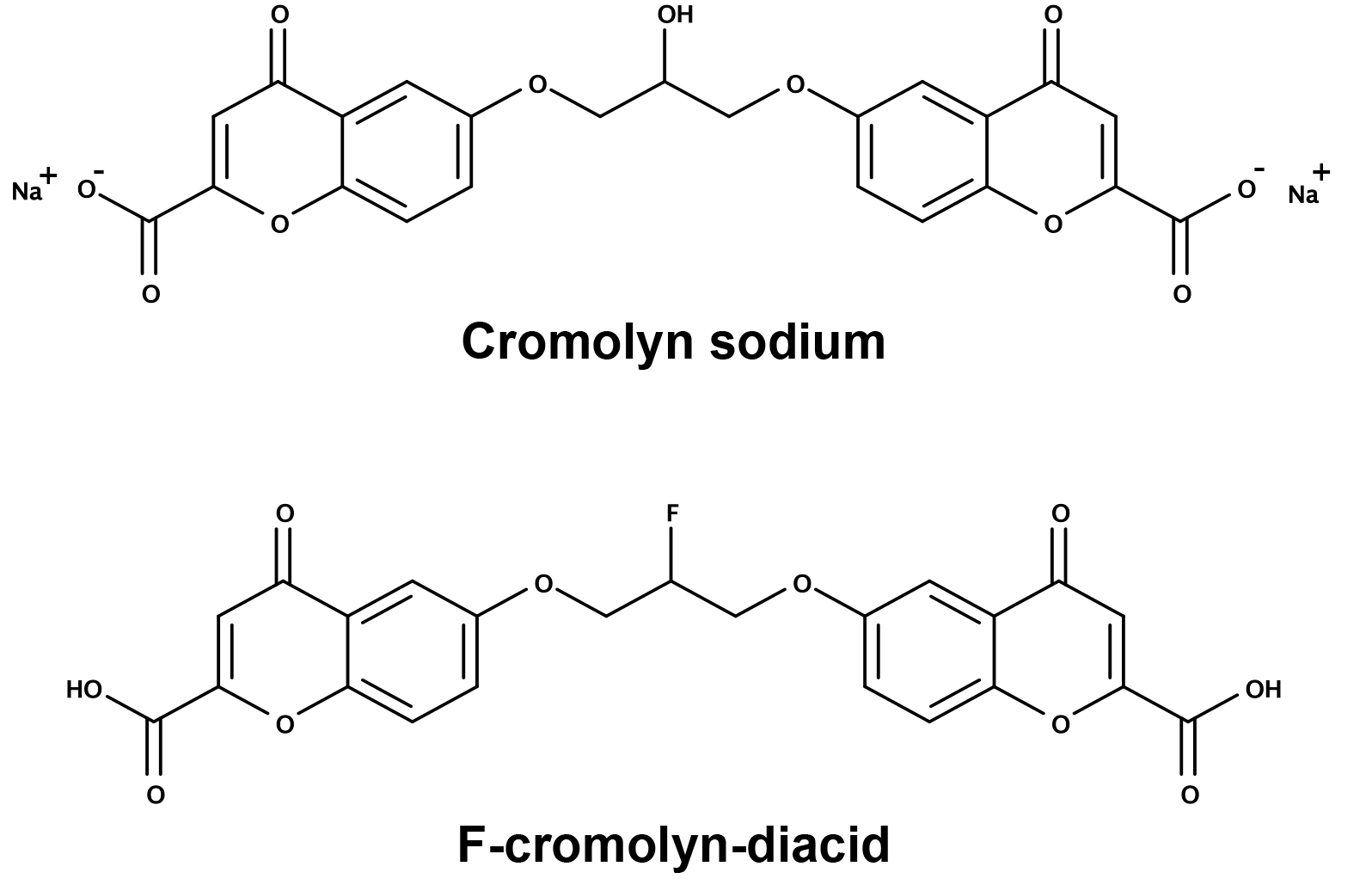
Chemicals and reagents
DMEM (Dulbecco’s Modified Eagle Medium) (Cat#1995065), DMEM without phenol red (Cat#21063029), PBS-pH7.2 (Cat#20012050), L-glutamine (Cat#25030081), Trypsin-EDTA (Cat#25200056), and penicillin-streptomycin (Cat#15140122) were products of Gibco, Thermo Fisher Scientific. FBS (fetal bovine serum) (Cat#F4135), DMSO (dimethyl sulfoxide) (Cat#D2438), and TWEEN-20 (Cat#P1379) were purchased from Sigma-Aldrich. 3 mL Syringe/Needle Combination with Luer-Lok™ Tip (Cat#8936G82), 13 mm syringe filter (PVDF, 0.22 µm) (Cat#1159T77) were purchased from Thomas Scientific. Recombinant human TNF-α (Cat#300-01A), recombinant human IFN-γ (Cat#300-02) were purchased from PeproTech. Cromolyn and F-cromolyn were provided by AZTherapies and DMEM was used as diluent to achieve final concentrations as indicated.
Cell line and cell culture
Human microglial cell line HMC3 (CRL-3304) was purchased from ATCC (American Type Culture Collection). HMC3 cell line was cultured in DMEM medium with 10% FBS, 1% L-glutamine, and 1% penicillin/streptomycin. Cells were maintained in 37°C incubator at 5% CO2.
Flow cytometry
HMC3 cells were resuspended, counted using the LUNA-II Automated Cell Counter, seeded in the 6-well plate (300 K cells/2 mL medium/well), and incubated overnight to allow cells to attach. Cells were treated with TNF-α or IFN-γ at five different concentrations (0, 10, 30, 100, 300 ng/mL) for 24 hr to induce cell activation. The supernatant medium was completely aspirated and 1 mL of fresh DMEM was added to wash the cell layer to remove detached cells and debris. The attached cells were resuspended and washed with PBS. Cells were washed with FACS buffer (2% FBS in PBS) prior to incubation with Zombie Violet Fixable Viability Dye (#423114, Biolegend, 1:500 dilution) and Human Fc Receptor Blocking Solution (#422302, Biolegend, 1:20) diluted in FACS buffer at RT for 10 min in the dark. Cells were then washed with FACS buffer and incubated at RT for 20 min with the following cell-surface antibodies: FITC anti-MHC II (#361706, Biolegend), PerCP/Cy5.5 anti-CD11b (#301328, Biolegend), BV605 anti-CD40 (#334336, Biolegend), PE anti-CD86 (#374206, Biolegend), PE/Cy7 anti-CD163 (#326514, Biolegend), APC anti-CD206 (#321110, Biolegend), and PerCP/Cy5.5 anti-CD14 (#367110, Biolegend). All of the cell-surface antibodies were diluted 1:20 in FACS buffer. Cells were permeabilized using the Transcription Factor Fixation/Permeabilization Buffer Set (#424401, Biolegend). Then cells were incubated with the following intracellular antibodies: APC/Cy7 anti-CD68 (#333822, Biolegend), purified anti-IBA1 (#PA5-27436, Invitrogen), Goat anti-Rabbit-Alexa Fluor 647 (#A21245, Invitrogen), purified anti-GFAP (#644702, Biolegend), and Goat anti-Mouse-Alexa Fluor 488 (#11029, Invitrogen). All of the intracellular antibodies were diluted 1:100 in the Permeabilization buffer (1x). Cells were washed with Permeabilization buffer and fixed in Fixation Buffer (#420801, Biolegend). Flow cytometry was performed using the Attune NxT Acoustic Focusing Cytometer (Thermo Fisher Scientific). Analysis of flow cytometry data was performed via FCS Express 7 (DeNovo Software).
Cell viability assay by AO/PI stain
Cell viability was assessed with a LUNA-FL Automated fluorescence cell counter. Viable nucleated cells show green fluorescence and dying nucleated cells show red fluorescence. 18 μL of sample was mixed with 2 μL Acridine Orange/Propidium Iodide (AO/PI) Stain.
MSD U-PLEX assay platform – cytokine and chemokine secretions
HMC3 cells were resuspended, counted using the LUNA-II Automated Cell Counter, seeded in the 6-well plate (400 K cells/2 mL medium/well), and incubated overnight to allow cells to attach. The media and detached cells were removed next day. The cell layer was washed three times in PBS and once in serum- and phenolred-free DMEM (SPFM). Cells were incubated in SPFM for 4 hr prior to treatment with TNF-α (0.3 µg/mL) and/or Cromolyn (0.3 µM, 3 µM) or F-cromolyn-diacid (0.3 µM, 3 µM) for 24 hr. The conditioned medium was collected and centrifuged at 1,000 x RCF (g) for 5 min to pellet detached cells and large debris, which was subsequently passed through a 0.22 µm filter with PVDF membrane to remove smaller debris. Samples of the supernatant medium were put in a CoolRack (#07210041, Fisher Scientific) on dry ice for Snap-freezing and kept at -80ºC until use.
Meso-scale U-PLEX plates that detect a cytokine panel including IL-1β, IL-2, IL-4, IL-6, IL-8, IL-10, IFN-γ, TNF-α, and a chemokine panel including IP-10/CXCL10, MCP-1/CCL2, MIP-1α/CCL3, MIP-1β/CCL4, Eotaxin/CCL11, were used as per manufacture’s protocol. 25 µL of the conditioned medium was used in each well of the MSD plates to detect the analytes. The plates were then analyzed on an MSD QuickPlex SQ120 instrument. All standard concentration curves for cytokines and chemokines tested in this study are provided in the Supplemental Figure S1.
Quantification and statistical Analysis
All the data were presented as mean ± standard error from at least three times, each done in triplicate. The statistical significance between two groups was determined by Student’s t test, whereas the comparisons of multiple groups were carried out by one-way ANOVA, followed by Bonferroni’s post-test using GraphPad Prism 7 (GraphPad Software, Inc.). A probability value of * p < 0.05 was considered to be significant.