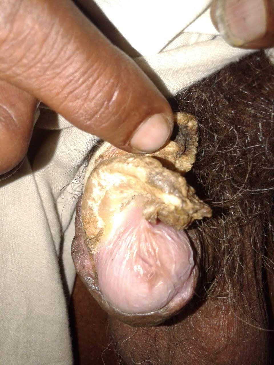
Sebaceous horns also known as cutaneous horns [1, 2] commonly occurring on sun-exposed areas of the skin, rarely seen on the penis. Clinically diagnosis is by horn-like projections of compacted keratin seen above the surface of the skin having flat, nodular, or crateriform base. The aetiology is uncertain, but may be formed by drying up of secretions from the sebaceous gland or cysts forming compacted keratin over a period of time. Lesions like squamous cell carcinoma, actinic keratosis, keratoacanthoma, Bowen’s disease, seborrheic keratosis, basal cell carcinoma, haemangioma, keratotic and micaceous pseudo papillomatous balanitis, Kaposi’s sarcoma, and sebaceous adenoma [1] may be seen in the base of the horn. Described first in 1854, and since then, only about 100 cases of cutaneous horn have been reported. Only one case of multiple cutaneous horns is reported [3]. Malignant change can be seen at the base of the horn. Microscopically, hyperkeratosis, acanthosis, dyskeratosis, papillomatosis, and chronic inflammatory infiltration of the adjacent dermis may be seen. Excision of the lesion with a rim of normal tissue at the base should be done. HPE should be done to rule out malignant transformation [4, 5]. Partial penectomy with or without inguinal block dissection is recommended if malignant change is found in histopathology. One-third of cases of penile horns are associated with underlying malignancies. [6]