Characterization of the Cell/IL/Ag-AgCl NPs
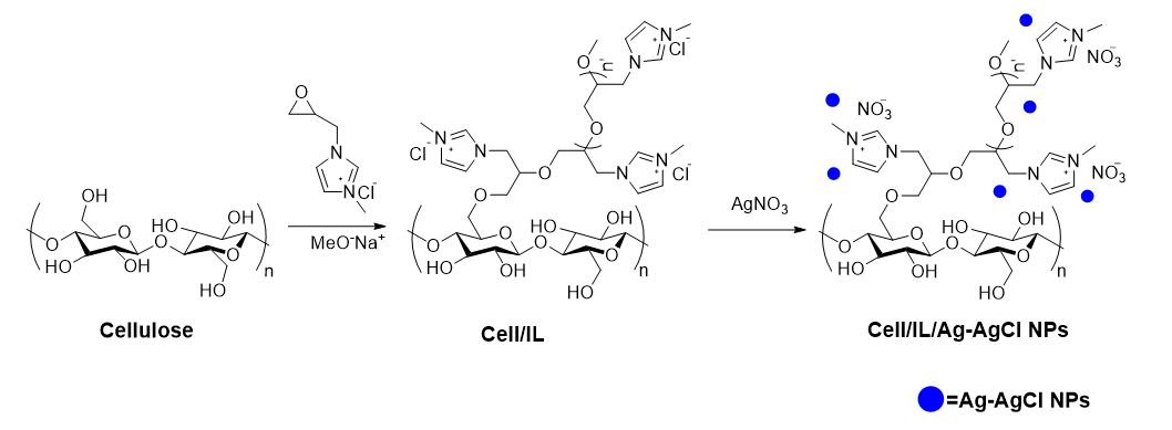
The synthesis of polyelectrolyte cellulose-based macromolecule was expertly designed using the following steps as presented in Scheme 1.
The synthesis process of Ag-AgCl NPs immobilized on Cell/IL was described as follows (a) At first, to a mixture of microcrystalline cellulose in dimethyl formamide (as a solvent), 1-Methyl-3-(oxirane-2-yl-methyl)-1H-imidazolium chloride was placed to give the ionic liquid anchored cellulose (Cell/IL). (b) Continuing, AgNO3 was added to an aqueous solution of Cell/IL and then the reaction was pursued 24 h at 30 °C to create the Cell/IL/Ag-AgCl NPs polyelectrolyte (Fig. 1). Flame atomic absorption spectrometry (FAAS) was applied to specify the percentages of Ag ions loaded on the polyelectrolyte to be 6.39 wt%.
The synthesized materials were characterized using FT-IR spectroscopy. As presented in Fig. 2(a), in the cellulose spectrum, the wide peak at 3344 cm-1 is affiliated to the stretching vibrations of the hydroxyl group in the biopolymer. The peak at 2895 cm-1 relates to stretching vibrations of the CH2 groups and the absorption peak at 1596 cm-1 is associated with the bending mode of the physically absorbed water. The peaks at 1434, 1378, and 1328 cm-1 are assigned to the bending vibrations of the C–H bond. The bonds at 1167, 1116, and 1058 cm-1 are ascribed to asymmetric bridges stretching the C–O bond, the crystal absorption bond at cellulose, and pyranoid ring skeletal vibrations of C–O–C respectively. The band at 897 cm-1 corresponds to the b-glycosidic linkages. FT‐IR spectrum of ionic liquid anchored cellulose (Cell/IL) was shown the bond at 1648 and 1427 cm-1 can be assigned to the C═N and C═C due to stretching vibrations in the imidazolium rings, respectively (Shojaei et al. 2017b, a). After the adsorption of Ag-AgCl NPs, a slight decrease in peaks intensity corresponding to the O-H, C-O, C-H, and C-N groups is observed that indicate binding interaction of Ag-AgCl NPs with functional groups on support. Also, the band at 3344 cm-1 was transferred to 3349 cm-1 by the adsorption of Ag-AgCl NPs.
The crystalline structure of the polyelectrolyte (Cell/IL/Ag-AgCl NPs) was inspected by an XRD pattern. As seen in Fig. 2(b), a comparison of Ag-AgCl NPs (dark line) with Cell/IL (red line), shows new peaks (2θ) at 38.27 (226), 44.45 (200), 77.39 (201) which assigned to the cubic Ag (JCPDS file: 65-2871). Also peaks at 27.97(111), 32.40(200), 46.39(220), 55.00(311), 57.64(222), 64.66(400) related to the cubic AgCl (JCPDS file: 31-1238). The particle size of the Ag-AgCl nanoparticles was estimated by the Scherrer formula to be 8 nm at 2θ = 38.27 for Ag and 15 nm at 2θ = 32.40 for Ag-AgCl.
The information of BET surface area, pore size, and pore volume of the Cell/IL and Cell/IL/Ag-AgCl NPs were also investigated by nitrogen adsorption-desorption isotherms Fig. 2(c). The pore volume and BET surface area of Cell/IL were determined as 5.92 m2 g-1 and 0.025 cm3g-1, whereas after introducing Ag-AgCl nanoparticles, Cell/IL/Ag-AgCl NPs, it was reduced to 4.22 m2 g-1 and 0.016 cm3g-1 respectively(Table 1). Additionally, the accommodating of the Ag-AgCl nanoparticles might lead to reducing in pore volume and surface area for the Cell/IL/Ag-AgCl NPs.
Table 1. BET surface area and porosity data of the Cell/IL and Cell/IL/Ag-AgCl NPs.
|
Salmple
|
BET surface area (m2g-1)
|
BJH Analysis
|
|
Pore Volume (cm3g-1)
|
Pore diameter (nm)
|
|
Cell/IL
|
5.92
|
0.025
|
1.64
|
|
Cell/IL/Ag-AgCl NPs
|
4.22
|
0.016
|
1.85
|
Thermogravimetric analysis (TGA) in an N2 atmosphere of Cell/IL/Ag-AgCl NPs sample is represented in Fig. 2(d). Curve analysis showed that two weight loss steps were beheld for composite. The first, around 50-120 °C, is related to the deprivation of adsorbed water and solvent with ∼6% weight loss. The next step around 250–450 °C with ∼41.8% weight loss is attributed to the breakdown of the organic and inorganic moieties (cellulosic, ionic liquid parts and, Ag-AgCl NPs) (Shojaei et al. 2017b). Additionally, no residue was observed after burning cellulose, while the Cell/IL/Ag-AgCl NPs had weight residue, which indicated the existence of silver in the macromolecule. Meanwhile, due to the presence of silver nanoparticles, Cell/IL/Ag-AgCl NPs had higher residuals which showed that the thermal stability of the cellulose after adding Ag nanoparticles was ameliorated.
In the process of Ag-AgCl NPs synthesis, cellulose acted as a successfully protecting support to resist the agglomeration Ag-AgCl NPs and also as a reducing agent for Ag+ to metallic silver. The microstructures of synthesized composites were analyzed by SEM and TEM studies (Fig. 3). For Cell/IL/Ag-AgCl NPs composite, dark Ag nanoparticles were anchored on cellulose and a light black AgCl layer surrounded the around of Ag NPs (Fig. 3d). The TEM analysis confirmed that the Ag-AgCl NPs are spherical in shape and also revealed that the particles are well separated, not aggregated with an average particles size at approximately ∼3 nm (Fig. 3e).
To identify elements in the Cell/IL/Ag-AgCl NPs, EDX analysis was also conducted (Fig. 3f). The presence of C, O, N, Ag, and Cl elements in the nanocomposite indicates the incorporation of silver into the polyelectrolyte surface.
The Ag nanoparticles distribution on the polyelectrolyte was appraised by EDX mapping. As shown in Fig. 4, the homogeneous density of Ag nanoparticles has been diffused on the surface of the cellulosic sample. It can be determined that the combining XRD analysis with these supportive results, indicated the successful synthesis of Ag-AgCl NPs.
Antibacterial activity of Cell/IL/Ag-AgCl NPs
In this research, the in-vitro antibacterial activity of Cell/IL/Ag-AgCl NPs was evaluated against methicillin-resistant staphylococcus aureus (ATCC 43300) and multidrug-resistant Pseudomonas aeruginosa (ATCC 27853). The antibacterial effect (MICs and MBCs values) of the compound against the bacteria evaluated was demonstrated in Fig. 5.
Our studies displayed that the antimicrobial effect of Cell/IL/Ag-AgCl NPs against Pseudomonas aeruginosa was higher than that of Staphylococcus aureus. Previous studies have shown that cellulose nanocomposites are able to inhibit the growth of infectious agents(Morones et al. 2005; Yan et al. 2016). Numerous reports on cellulosic substances and similar compounds confirm our findings(Konwarh et al. 2013; da Silva Dannenberg et al. 2017; Burdușel et al. 2018). It seems that Cell/IL/Ag-AgCl NPs (due to their biocompatible nature, good toughness, relatively low cost, and large surface area) could be used as a substitute for common antibiotics in the future, killing multidrug-resistant bacteria(Kalwar and Shen 2019b). On the other hand, using the microtiter plate method, the antibiofilm effect of this compound against bacteria was shown. This effect against Pseudomonas aeruginosa was remarkable (Fig. 6). It is possible that the use of these compounds can inhibit the biofilm formation of bacteria on surfaces and equipment, which in turn can reduce nosocomial infections, especially in the intensive care unit, surgery unit/room in the hospital(Marambio-Jones and Hoek 2010; Hasanzadeh et al. 2021).
Toxicity of the Cell/IL/Ag-AgCl NPs
To assess the toxic effects of the Cell/IL/Ag-AgCl NPs, human intestinal Caco-2 cells were acted with multifold concentrations of the synthesized material (0, 100, 200, 300, 400 ppm) for 24h. According to our results Fig. 7, different doses of Cell/IL/Ag-AgCl NPs up to 400 ppm did not show a significant decrease in the on cell viability compared with the control. These results are in line with previous studies that low concentrations of silver nanoparticles are nontoxic(Nguyen et al. 2017).
Our results also showed that cellulosic materials with excellent physical and biological properties are favorable candidates for biomedical products due to their low cytotoxicity, biodegradability, and biocompatibility(Annamalai and Nallamuthu 2016).