Participants
One hundred and ninety-nine Aβ- CU participants were included in this study. Participants had a stable clinical classification for an average of 5.3 years (± 4.1) prior to, and at the time of their tau PET scan. Figure 1 shows the distribution of Me SUVR and entorhinal SUVR across the entire cohort.
Tau burden in a mesial temporal composite (Me) and the entorhinal cortex as measured by tau PET SUVR. A) The red dashed line separates the cohort by the 95% percentile Me SUVR (top 5% vs lower 95%), while the black dashed line separates the cohort by the 90% percentile Me SUVR (top 10% vs lower 90%); B) the black dashed line represents a visually-derived threshold used to discriminate higher (EC+) from lower entorhinal SUVR (EC-).
Demographics and characteristics of the cohort are shown in Table 1, split by the 90% percentile Me SUVR (lower 90% and top 10%). Participants with higher MTL tau were significantly older than participants with lower MTL tau. Participants with higher MTL tau also had lower hippocampal volumes, which remained significant after correction for age. The results were similar using the 95%ile threshold and the visually derived entorhinal cortex threshold (see Supplementary Table 1 and 2, Additional Files 1 and 2).
Table 1. Demographics and characteristics of the Aβ- cognitively unimpaired cohort split by the 90%ile Me SUVR
|
|
90%ile Me SUVR
|
|
|
Lower 90%
(n = 179)
|
Top 10%
(n = 20)
|
|
Age (y)
|
74.3±5.0
|
78.3±5.7**
|
|
Sex, F n (%)
|
99 (55.3%)
|
14 (70.0%)
|
|
APOE ε4+, n (%)a
|
40 (22.3%)
|
6 (30.0%)
|
|
Education (y)
|
14.3±3.1
|
13.5±3.2
|
|
HV (cm3)b
|
2.97±0.3
|
2.82±0.2**
|
|
Centiloid
|
2.02±7.0
|
3.36±11.1
|
|
SMC, n (%)
|
102 (57.0%)
|
11 (55.0%)
|
Abbreviations: Me = mesial temporal composite; SUVR = standardized uptake value ratio; APOE = Apolipoprotein E; HV = hippocampal volume; SMC = subjective memory complaint.
Mean (SD), unless otherwise specified. *p≤0.05, **p≤0.01 compared to the lower 90% group.
aAPOE data was not available for 3 participants in the lower 90% group.
b HV was only available for 17/20 participants in the top 10% group and 153/179 participants in the lower 90% group. Results remain significant after correction for age (p=0.04); effect size, Cohen’s d = 0.34.
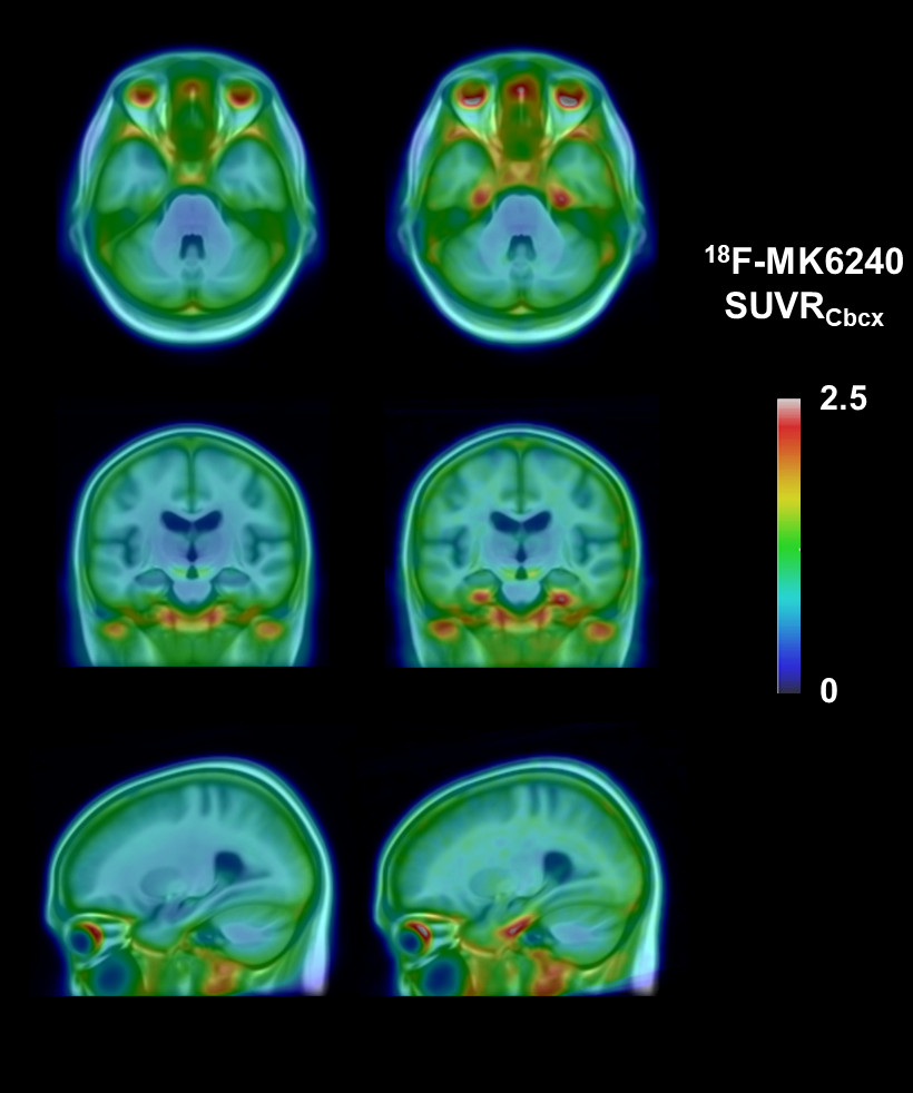
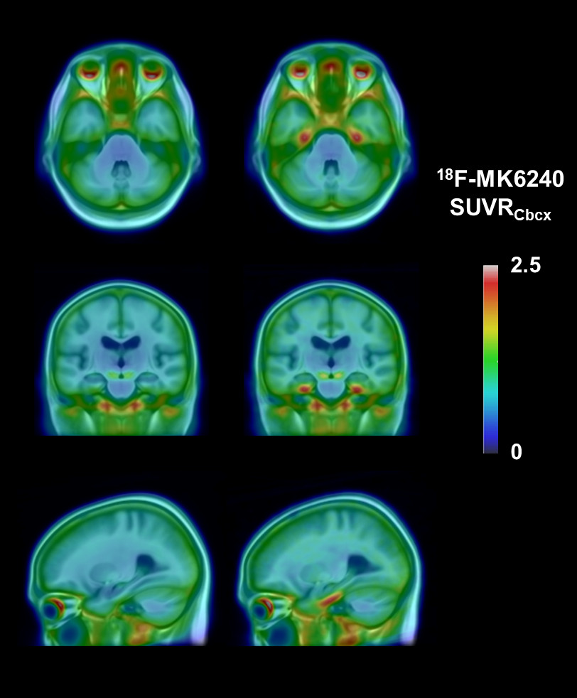
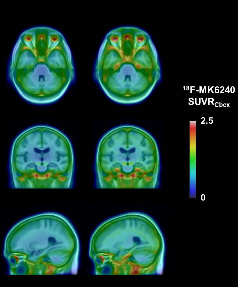
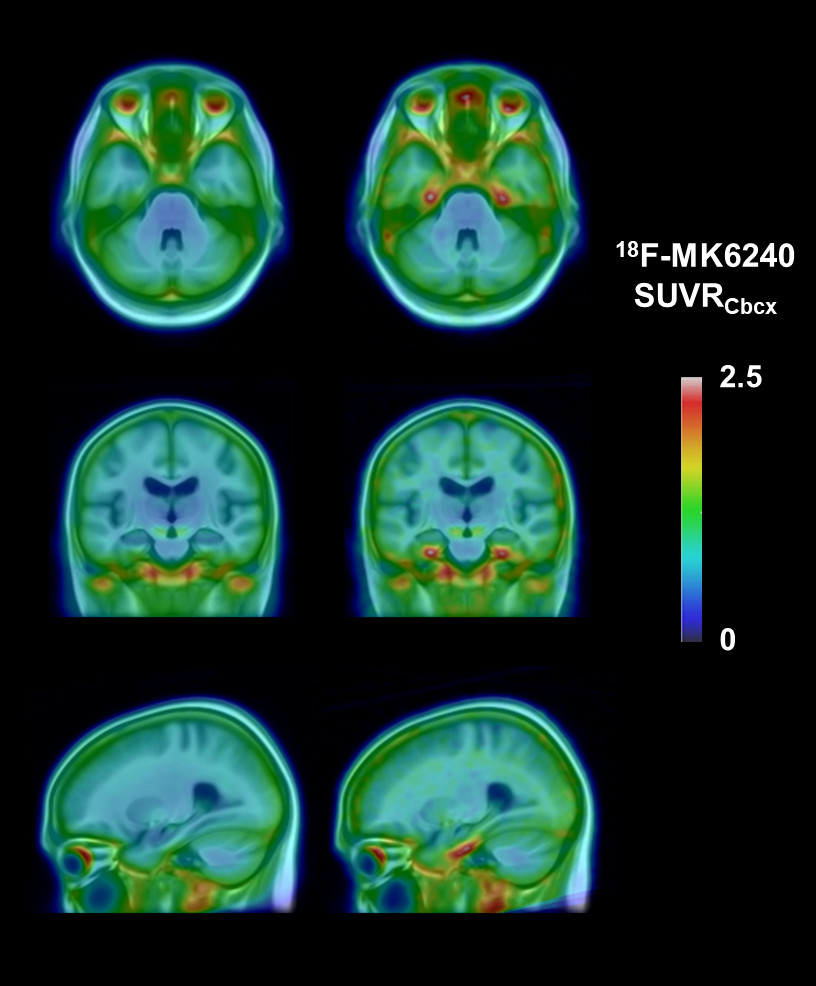
Using the thresholds specified, participants with higher MTL/higher entorhinal tau were visually observed to have a focal increase in tau tracer retention in a distribution consistent with Braak stage I-II when compared to participants with lower MTL/entorhinal tau (Figure 2; Supplementary Figures 1 and 2, Additional files 3 and 4). Additionally, these participants were visually observed to have a subtle increase in signal across the brain and extracerebral structures, though much less than the focal increase seen in the MTL.
Mean tau 18F-MK6240 SUVR images overlaid on a T1 MRI template for the cohort, lower 90% (left) and top 10% Me SUVR (right) showing tau tracer retention confined to Braak stage I-II.
Participants with high MTL tau also had a higher mean neocortical tau tracer retention (Te and R), which was significantly different compared to participants with lower MTL tau (Table 2). However, the mean images in Figure 2 show that this increase is seen across the entire image including white matter and extracerebral structures suggesting a reference region problem.
In a sub-analysis to determine whether differences in the reference region were driving these results, the groups were compared on their cerebellar cortex SUV. Participants in the top 10% Me SUVR were found to have significantly lower mean cerebellar cortex SUV values than participants in the lower 90% (t=3.09, p=0.001). Six alternative reference regions were evaluated. The alternative reference region with the lowest variance and for which the SUV did not differ between the top 10% and lower 90% groups (subcortical white matter) was selected, and composite ROI values were recalculated using this reference region, leaving the same participants classified as top 10% and lower 90%. The results showed that participants in the top 10% still had higher Me SUVR than the lower 90% (as expected), but participants in the top 10% no longer had higher neocortical (Te, R) SUVR values (Table 3). Table 3 shows that the lower 90% had significantly higher Te and R SUVR than the top 10%.
Table 2. Mesial temporal tau and neocortical tau (cerebellar cortex reference region)
|
|
90%ile Me SUVR
|
|
|
Lower 90%
(n=179)
|
Top 10%
(n=20)
|
p-value
|
Effect size
|
|
Composite ROI (SUVRcbcx)
|
|
Me SUVR
|
0.79±0.11
|
1.12±0.08
|
p<0.001
|
d = +2.97
|
|
Te SUVR
|
0.99±0.12
|
1.14±0.09
|
p<0.001
|
d = +1.24
|
|
R SUVR
|
0.86±0.12
|
0.96±0.11
|
p<0.001
|
d = +0.85
|
Mean (SD). T-test (two-tailed). Effect size = Cohen’s d. Abbreviations: Me = mesial temporal composite; Te = temporoparietal composite; R = rest of neocortex composite.
Table 3. Mesial temporal tau and neocortical tau (subcortical white matter reference region)
|
|
90%ile Me SUVR
|
|
|
Lower 90%
(n=179)
|
Top 10%
(n=20)
|
p-value
|
Effect size
|
|
Composite ROI (SUVRswm)
|
|
Me
|
1.11±0.13
|
1.30±0.23
|
p=0.001
|
d= +1.36
|
|
Te
|
1.41±0.18
|
1.32±0.16
|
p=0.029
|
d= -0.52
|
|
R
|
1.22±0.16
|
1.11±0.17
|
p=0.005
|
d= -0.68
|
Mean (SD). T-test (two-tailed). Effect size = Cohen’s d. Abbreviations: Me = mesial temporal composite; Te = temporoparietal composite; R = rest of neocortex composite.
Mesial temporal tau was associated with age
There was a significant association between age and Me SUVR (r=0.24, p<0.001) (Figure 3A), but no association between age and neocortical SUVR (Te, r=-0.03, p=0.33; R, r=-0.09, p=0.10). For mesial temporal sub-regions, there was an association between age and SUVR generated for the entorhinal cortex (r=0.29, p<0.001) (Figure 3B), amygdala (r=0.21, p=0.002), hippocampus (r=0.21, p=0.002), and parahippocampal gyrus (r=0.14, p=0.02).
Scatterplots showing A) the correlation between age and mesial temporal (Me) SUVR; and B) the correlation between age and entorhinal SUVR.
Mesial temporal tau was not associated with Aβ
There was no association between Aβ burden (measured in Centiloids) and Me SUVR (r=0.08, p=0.13). There was also no association between Centiloids and tau SUVR generated for Me sub-regions (entorhinal cortex, r=0.05, p=0.23; amygdala, r=0.10, p=0.07; hippocampus, r=0.09, p=0.11; and parahippocampal gyrus, r=0.07, p=0.17). Re-defining Aβ negative as less than 10 Centiloids did not affect the Me SUVR 90th or 95th percentile thresholds (see Supplementary Figures 3 and 4, Additional File 5).
Mesial temporal tau burden did not differ for individuals with and without subjective memory complaint
Across the cohort, 113/199 (56.8%) of participants had a subjective memory complaint (SMC). There was no significant difference in Me SUVR or entorhinal SUVR for individuals who had SMC compared to those who did not.
Age but not tau burden is associated with worse cognition
Participants who were in the top 10% for Me SUVR and those who were EC+ did not have significantly different MMSE and composite memory scores compared to the other participants. EC+ also did not have significantly different AIBL-PACC scores than EC-. The top 10% had worse CNMS (t=1.83, p=0.03) and AIBL-PACC scores (t=1.79, p=0.04) than the lower 90%. Using the 95%ile threshold, the top 5% Me SUVR did not differ significantly than the lower 95% on any of the cognitive scores. However, as noted earlier, the higher MTL tau groups were significantly older than the lower MTL tau groups.
There was no correlation between cognitive performance (MMSE, CMS, CNMS and AIBL-PACC) and SUVR in Me or the four Me sub-regions, both with and without the covariate of age.
Models combining age and Me SUVR were overall significant in accounting for the variance in MMSE, CNMS and AIBL-PACC scores. However, these models accounted for only 4% of the variance in the MMSE (R2 = 0.04), 1.8% of the variance in CMS (R2 = 0.018), 10% of the variance in CNMS (R2 = 0.10) and 7.5% of the variance in the AIBL-PACC (R2 = 0.075). Of the variance explained, age was a significant contributor, while Me SUVR did not contribute significantly (Table 4). The results were similar with entorhinal cortex SUVR and age as independent variables and cognitive score as dependent variables (See Supplementary Table 3, Additional file 6).
Table 4. Multiple linear regression models of the relationship between Me SUVR, age and cognitive composite scores
|
|
|
Dependent variable = MMSE
|
|
|
β
|
SE
|
t
|
P value
|
|
Age
|
-0.20
|
0.02
|
-2.75
|
0.007
|
|
Me SUVR
|
-0.001
|
0.60
|
-0.008
|
0.99
|
|
|
|
Dependent variable = CMS
|
|
Age
|
-0.12
|
0.01
|
-1.66
|
0.10
|
|
Me SUVR
|
-0.04
|
0.43
|
-0.49
|
0.62
|
|
|
|
Dependent variable = CNMS
|
|
Age
|
-0.33
|
0.01
|
-4.69
|
<0.001
|
|
Me SUVR
|
0.03
|
0.30
|
0.38
|
0.71
|
|
|
|
Dependent variable = AIBL-PACC
|
|
Age
|
-0.27
|
0.01
|
-3.77
|
<0.001
|
|
Me SUVR
|
-0.03
|
0.40
|
-0.40
|
0.69
|
Abbreviations: β = standardized beta coefficient; SE = standard error; Me = mesial temporal; SUVR = standardized uptake value ratio; MMSE = mini-mental state examination; CMS = composite memory score; CNMS = composite non-memory score; PACC = pre-clinical Alzheimer cognitive composite.