Patients and tissue samples
A total of 113 peripheral blood samples were collected, including 42 from cervical cancer patients, 35 high-grade squamous intraepithelial lesion (HSIL) patients, and 36 from subjects with normal cervix. In addition, we collected 11 pairs of cervical cancer and adjacent tissues. All the included cervical cancer patients underwent primary surgery. All samples were collected from the Qilu Hospital of Shandong University. The study was approved by Qilu Hospital's ethics committee.
Cell Isolation
Isolation of peripheral blood mononuclear cells (PBMC) from whole blood using Ficoll (TBD science, Tianjin, China) density gradient centrifugation. We obtained tumor single cell suspension from fresh tumor tissue. According to the manufacturer’s instructions, a gentle MACS C tube (Milteny Biotec, Bergisch Gladbach, Germany) was used for mechanical dissociation and a tumor dissociation kit (Milteny Biotec) Enzymatic hydrolysis. The digested cells were filtered through a 70µm mesh, centrifuged with Ficoll (Solarbio, Beijing), and the monocytes were resuspended in RPMI 1640.
Cell Culture
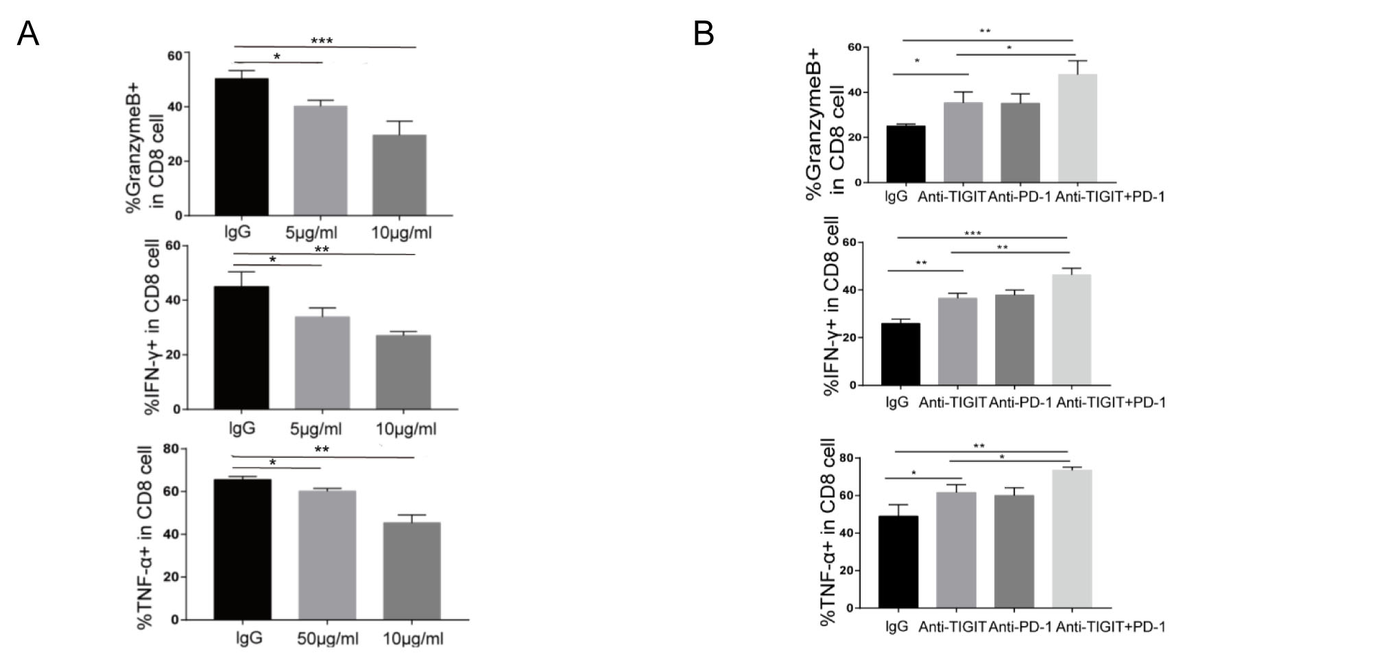
Mouse cervical cancer cell line U14 was obtained from the National Biomedical experimental cell resource bank (Beijing, China). U14 was cultured in DMEM supplemented with 10% fetal bovine serum (all from Gibco, Grand Island, NY, USA), 50 U/mL penicillin and 50 mg/mL streptomycin (all from Solarbio Science & Technology, Beijing, China). CD8+T cells were purified from PBMC positive selection by using a kit (Milteny Biotec, Bergisch Gladbach, Germany). CD8+T cells were stimulated with anti-CD3/CD28(Stemcell, Canada) in T cell expansion medium (Stemcell, Canada). Activated CD8+T cells were treated with 5µg/mL CD155-Fc, 10µg/mLCD155-Fc. Activated CD8+T were cocultured with tumor cells at a 10:1 ratio.
Add 10µg/ml anti-PD-1 mAb or 5µg/ml anti-TIGIT mAb (R&D Systems), respectively. As isotype control, we used α-human IgG1 (R&D Systems). After 48 hours, CD8+T cells were collected to determine cytokine production using the T cell function assay.
Flow Cytometry
PBMCs isolated from cervical cancer patients or normal people were stained with fuorochrome conjugated PE-conjugated-anti-CD8 (Elabscence, Wuhan, China), PE-conjugated-anti-TIGIT-FITC (eBioscience) antibodies for 30 min in 4°C. After washing 3 times, collect samples and use flow cytometry for detection. For intracellular staining, cell surface marker-stained cells were fixed and permeabilized with a fixation and permeabilization kit (BD Bioscience) for 20 minutes, and then treated with the fluorochrome-conjugated antibody APC-conjugated-anti-TNF-α (eBioscience), APC-conjugated-anti-IFN-γ (eBioscience), APC-conjugated-anti-GramB (eBioscience) for intracellular staining at 4°C for 30 minutes. Finally, the stained cells were analyzed using a FACS Calibur flow cytometer (Becton Dickinson, USA), and the data were analyzed using Flow Jo software.
Immuno histochemistry (IHC)
For immunohistochemical analysis, the sections are deparaffinized, and then citric acid buffer is used for heat-mediated antigen retrieval. For testing, follow the manufacturer's instructions and use an immunohistochemistry detection kit (Zhongshan Jinqiao, Beijing, China). Sections were incubated at 4°C overnight with primary antibodies in PBS (anti-human TIGIT, 1:100, Cell Signaling Technology, Danvers, MA; anti-human CD155, 1:100, Cell Signaling Technology Danvers, MA, Abcam; anti-human CD8, Abcam, Cambridge, UK; anti-mouse CD8, 1:200, Cell Signaling Technology Danvers, MA). The slides were then incubated for 10 minutes at 37°C with biotin-labeled goat anti-rabbit IgG secondary antibody. Incubate the streptavidin peroxidase with the slides for 15 minutes at 37°C before staining with DAB (Zhongshan Jinqiao, Beijing, China). Meyer's hematoxylin (Solarbio Science & Technology, Beijing, China) was used to stain the slides for 5 minutes. Seal the slices with neutral glue after they have been dehydrated.
Multiplex Immunohistochemistry Staining (Mihc)
For immunofluorescence analysis, we use multiple fluorescence immunohistochemical staining kits (Absin, Shanghai, China). Heat-mediated antigen retrieval and primary antibody incubation are the same as immunohistochemistry. After incubating the secondary antibody for 10 minutes, incubate the slides with the fluorescent staining amplification solution for 10 minutes at 37°C. After washing three times with TBST, incubate the slides with 4′,6-diamidino-2-phenylindole (DAPI) for 5 minutes. Finally, an anti-fluorescence quenching agent was used to seal the slides.
Real-time Quantitative Rt-pcr (Qrt-pcr)
Trizol reagent (Invitrogen) was used to extract total RNA from cells. After total RNA was quantified by spectrophotometry, reverse transcription was performed using the PrimeScript RT kit (Accurate biology, Hunan, China). Real-time PCR was performed using SYBR Premix Ex Taq (Accurate biology, Hunan, China) and 7900HT fast real-time PCR system (Applied Biosystems, Waltham, MA, USA). The primer sequences of genes including TIGIT, PD-1, LAG3, Tim3 and β-actin are shown in the supplementary information. The mRNA level of a specific gene were normalized with β-actin.
Western Blot
After washing the cells three times with PBS, they were lysed on ice in radioimmunoprecipitation analysis buffer (RIPA; Beyotime, China Institute of Biotechnology, 1% phenylmethylsulfonyl fluoride (PMSF); 1% NaF) for 30 minutes. Centrifuge at 12,000 rpm for 10 minutes at 4°C, and collect the supernatant. Next, the proteins were separated by SDS-PAGE and transferred to a PVDF membrane (Merck Millipore, Burlington, Massachusetts, USA). Incubate the membrane with the primary antibody β-actin (1:1000, Cell Signaling Technology), TIGIT (1:1000, Cell Signaling Technology), SHIP-1 (1:1000, Cell Signaling Technology), ERK (1:1000, Cell Signaling Technology), p-ERK (1:1000, Cell Signaling Technology), p-IκBα (1:1000, Cell Signaling Technology), p-NF-κBP65 (1:1000, Cell Signaling Technology) overnight at 4°C, and then incubate with the appropriate secondary antibody. ImageJ software (National Institutes of Health) was used to analyze relative protein levels, and β-actin was used as an endogenous control.
Immunoprecipitation
The cells were placed in lysis buffer (Beyotime Biotechnology, China), lysed on ice for 30 minutes, and centrifuged at 15000 rpm for 15 minutes at 4°C. At 4°C overnight, incubate 10 mg of antibody with 1000 mg of protein supernatant. Collect the supernatant and incubate it with Protein A/G Sepharose beads (Santa Cruz, USA) for 6 hours. The beads were washed three times and boiled before the immunoprecipitated protein was detected using western blotting.
Cas9-sgrna Knockout
Cas9-sgRNA knockout
Cas9 and single-guide RNA (sgRNAs) lentiviruses were designed and constructed by OBiO Technology Company (Shanghai, China). A lentivirus containing Cas9 and sgRNA (sg-scramble, sg-CD155) was introduced into U14 cells. The sgRNA sequence used is ATTCGACAGGCGTCTTGGGAGGG. After 48 hours, the transfected cells were selected in a medium containing puromycin. Compared with the control group, the silencing efficiency of U14 cells is nearly 99%.
In Vivo Treatments
Female C57BL6 mice (18-22g, 4–6 weeks old) were purchased from Beijing Vital River Laboratory Animal Technology Co., Ltd. for this study. U14 cells were trypsinized, resuspended in PBS, and inject 200ml (1×10⁷ cells) of the cell suspension into the subcutaneous area of the right armpit of each mice. For in vivo blockade, 3 days after injection of cell suspension, mice were randomly allocated to the anti-PD-1 mAb (100µg, clone BE0188, BioXcell, West Lebanon, USA), anti-TIGIT mAb (100µg, clone 1G9, BioXcell, West Lebanon, USA), anti-TIGIT mAb + anti-PD-1 mAb and IgG (Mouse IgG1, clone MOPC-21, BioXcell) group. Three times a week intraperitoneal injection of blocking antibody and isotype control group. To investigate the antitumor effects by target CD8+T cells, mice were subcutaneously inoculated with 1×10⁶ U14-NC-CD155 or U14-KO-CD155.
Statistical Analysis And Bioinformatics
GraphPad Prism 7.0 (GraphPad Software, San Diego, CA) was used for statistical significance analysis. The results are expressed as mean ± standard deviation. The two-tailed Student’s t-test was used for statistical comparison between two independent groups; a non-parametric test was used for data that does not conform to the normal distribution. Gene expression and clinical annotation data were downloaded from GEO (Gene Expression Omnibus) and TCGA (The Cancer Genome Atlas). The “limma” package was used to analyze diferentially expressed genes between cervical cancer and normal. For all experiments, a p-value less than 0.05 is the key to a significant difference (P value is expressed as: *P < 0.05, **P value < 0.01, ***P < 0.001).