Synthesis and characterization of the commercial dye-loaded PSMA NPs
We prepared PSMA nanoparticles with a diameter of 73.4 ± 11.9 nm (Fig. 1a). The single void (~ 17.1 ± 6.6 nm) in the central region of the PSMA nanoparticle was created by removing the metallic Cu core from the interior of the Cu@PSMA nanostructure (Fig. 1b) via the HCl etching process (Figure S1). Then, we loaded organic dyes with low molar mass (479.1-753.9 g/mol) into these nanosponges. The dye loading efficiency was ~ 3.7% for sodium iron chlorophyllin (FeChl), 50% for Cy5, and 26.5% for rhodamine 6 G (R6G). This phenomenon can be explained by the positively charged Cy5 and R6G dyes, which prefer to load into/onto the negatively charged PSMA NPs (-22.3 mV). These dye-loaded PSMA NPs exhibited fluorescence peak positions similar to those of their free-form molecules in the solution (Fig. 1c). Although the FeChl molecules were less attached to the PSMA NPs, the FeChl-loaded PSMA NPs could produce a prominent deep red to NIR emission. Without the loading of emitters, the PSMA NPs showed no significant emission signals upon excitation at 365–600 nm (Fig. 1d).
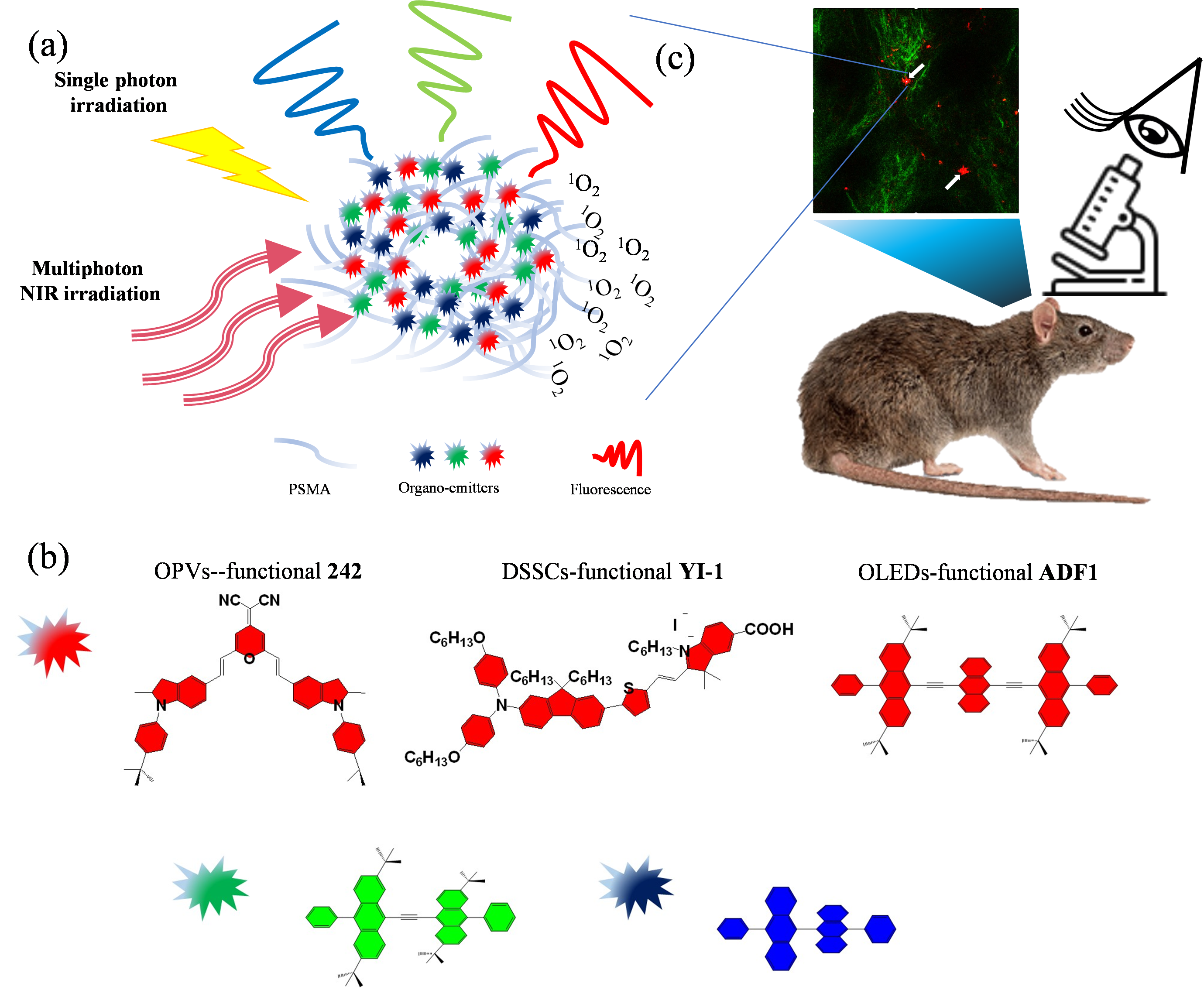
Photophysical properties of the PSMA NPs loaded with OLED-, OPV-, and DSSC-functional molecules
Next, we examined the loading abilities of molecules with larger sizes and molar masses to demonstrate the versatility of the developed nanoformulation platform. We loaded PSMA NPs with an OPV-functional molecule, i.e., 242 (~ 2.5 nm in length; 772.4 g/mol), with a 4-dicyanomethylidenepyran-based core and 2-methylindoline-based donor moieties, and a DSSC-functional molecule, i.e., YI-1 (~ 3 nm in length; 1081.7 g/mol), with a donor-π bridge-acceptor structure using bis-(4‐alkoxyphenyl)amine as the electron donor and indolium as the electron acceptor. In our previous report, we found that 242 was highly emissive in the deep red and NIR regions and that 242-based OLEDs exhibited high external quantum efficiency [43]. On the other hand, YI-1 exhibited both strong π-π transitions and intramolecular charge transfer (ICT) absorption bands ranging from the visible to NIR regions. Further details of the synthesis of 242 and YI-1 were described in the Supporting Information, including NMR and mass spectra results (Scheme S1 and Figure S2 ~ S15). The loading efficiencies of PSMA NPs for 242 and YI-1 were both larger than 85% (Figure S16). As shown in Fig. 2a, 242 exhibited a characteristic absorption band at 498 nm, which is redshifted by 12 nm (J-aggregation) [44] after loading into the PSMA NPs. On the other hand, a redshifted emission band for the PSMA-242 NPs was also observed when compared with that of 242 alone (Fig. 2b). A similar redshifting trend was found in the absorption and emission spectra for YI-1 and PSMA-YI-1 NPs (Fig. 2a and 2b).
In addition, we loaded three OLED-functional ADF-based molecules (Scheme 1b, Fig. 2c, and Figure S17) into PSMA NPs. Loading efficiencies of 85–88% were achieved for ADF1 (~ 2.8 nm in length; 954.5 g/mol), ADF2 (~ 2.1 nm in length; 754.5 g/mol), and ADF3 (~ 1.8 nm in length; 506.2 g/mol) (Figure S16). In terms of their fluorescence properties, ADF1-, ADF2-, and ADF3-loaded PSMA NPs exhibited a red emission band at 660 nm, green emission band at 515 nm, and blue emission band at 435 nm, respectively.
Analysis of multiphoton fluorescence spectra of dye-loaded nanoparticles
For deep-tissue imaging applications, we further investigated the multiphoton spectra of PSMA-242, PSMA-YI-1, and PSMA-ADF1 NPs (Fig. 2d-2f). The PSMA-242 NPs exhibited two-photon fluorescence peaks at approximately 700 nm under varying excitation wavelengths from 1000 nm to 1200 nm (Fig. 2d). Interestingly, the PSMA-YI-1 NPs have two fluorescence peaks at 600 and 800 nm (Fig. 2e). Both PSMA-242 and PSMA-YI-1 exhibited NIR-excited multiphoton NIR-fluorescence emission features. In contrast, PSMA-ADF1 NPs exhibit weak luminescence at ~ 605 nm when excited at 920 nm (Fig. 2f).
Surface characterization of dye-loaded PSMA NPs
Based on the FT-IR spectrum analysis (Figure S18), the typical absorption bands of 242, YI-1, and ADF1 overlapped with those of the PSMA NPs at 1720–1760 cm-1 resulting from C = O groups, 1601 − 1584 cm-1 resulting from C = C stretching within the ring of styrene, 1450 cm-1 resulting from νasymCOO- mode and 1340 cm-1 resulting from νsymCOO- mode. The IR spectra of the dye-loaded PSMA NPs and the bare PSMA NPs are almost the same, indicating that most organic molecules were loaded in the cores of PSMA NPs. The zeta potential of each of the dye-loaded PSMA NPs was approximately − 20 mV (Figure S19), indicating a negatively charged surface with carboxylate terminals exposed to water. This surface charge property is beneficial for the good dispersion of PSMA NPs in aqueous solutions.
The transmission electron microscopy (TEM) images revealed that PSMA NPs showed increased contrast after loading with photosensitive molecules with larger size and higher hydrophobicity (Figure S20). The increased contrast can be attributed to the high loading efficiencies of PSMA NPs for the photosensitive molecules (85% for ADF1, 99% for 242, and 87% for YI-1). The resulting particle size was slightly increased by 1.5–7.8 nm, and some particles had enlarged void sizes in the interior (Table S1), suggesting that the sponge-like swelling nature of the PSMA NPs allows the penetration of these photosensitive molecules into the cores of NPs (Scheme 1a) and not just the adsorption of the photosensitive molecules on the surfaces of NPs. Moreover, the potential leakage of the dyes was below 6.25% after two days of aging (Figure S21a).
Without help from carriers, hydrophobic 242, YI-1, and ADF1 photosensitive molecules will aggregate in the blood [45] and undergo undesired rapid clearance by the endothelial reticular system. One common solution for this is to carry these chromophores in a polymer through a phospholipid packaging method to form encapsulated nanoparticles (NPs) [46, 47]. The disadvantage of this method is a low loading rate. In addition, there are concerns about the undesired release of the dyes. Although synthetic methods, such as polymer grafting, AIE self-assembly, and coprecipitation methods, can be used to assemble light-emitting dye nanoparticles [9, 12–14, 16], the synthesis parameters of these approaches still require optimization. In our current work, the assembly of PSMA NPs is achieved by a straightforward strategy (Scheme 1a) to load small commercially available dyes and newly synthesized large dyes. It should be a more appropriate method to prepare organic nanoemitters without tedious synthetic procedures.
Cell viability of dye-loaded PSMA NPs
The cytotoxicity of these 242-, YI-1-, and ADF1-loaded PSMA NPs was further evaluated by MTT assay. The cell viability was over 80.2% throughout the concentration range of 0 ppm[dye]-50 ppm[dye] for all samples incubated with MB49 cancer cells (1 day), demonstrating the reduced cytotoxicity of these molecules (Fig. 3a) compared to that of free dye molecules (Figure S21b). Their low cytotoxicity implies the low leakage of dyes (Figure S21a). Direct treatment of these dyes at the same released concentration did not cause cell toxicity (Figure S21b). Using JC-1 staining, we further confirmed the unchanged bioactivity of the mitochondria in MB49 cells after treatment with dye-loaded PSMA NPs (Figure S22).
Single-photon cellular imaging of dye-loaded PSMA NPs
After observing the red-NIR emission characteristics of the dye-loaded PSMA NPs (Fig. 2b and 2c), we treated MB49 cells with PSMA NPs loaded with 242, YI-1, and ADF1 and explored their potential applications as biological probes. Fluorescence images (Fig. 3b) showed that all three nanoemitters appeared red within MB49 cells after 24 h of coculture. The 530–550 nm excited red fluorescence was detected by employing a longpass filter (Olympus, UFGW) with an edge wavelength of 575 nm. Based on the confocal microscopy image, these red fluorescent dots of PSMA-ADF1 (as a modeling nanoemitter) in cells were overlaid with the green fluorescence of the lysosome tracker, which is represented as yellow (S23a).
The wavelength tunability of the labeling can be easily achieved by choosing the emitter color of fluorescence. As a demonstration, we prepared ADF1-, ADF-2-, and ADF3-loaded PSMA NPs that exhibited individual red, green, and blue fluorescence properties to stain MB49 cancer cells after 24 h of incubation (Figure S23b). Then, we codelivered both ADF1- and ADF2-loaded PSMA NPs to MB49 cancer cells. A fluorescence microscope could separately visualize the emission signals from the green and red emitters in the MB49 cells (Fig. 3c). In the merged image, most of the emission color manifested as yellow due to the colocalization of the ADF1- and ADF2-loaded PSMA NPs, and combined with the confocal microscopy images of PSMA-ADF1 and lysosomes, it showed a colocalized position (Figure S23a), suggesting the internalization of ADF1- and ADF2-loaded PSMA NPs in the endolysosomes. We believe that these nanoemitters, combining different colors of dye molecules with strong fluorescent properties, can replace small molecular dyes, thereby allowing the development of biological immunofluorescence staining technology with low concentrations of biomolecules.
Multiphoton bioimaging in vitro and in vivo
To demonstrate the multiphoton contrasts of dye-loaded PSMA NPs in biomedical imaging, we treated MDA-MB-231 cells with PSMA-242 and PSMA-YI-1 NPs for 16 h. The cells were observed using an inverted multiphoton microscope (Nikon, A1 MP + with Ti2 E). The multiphoton fluorescence signals from PSMA-242 (Fig. 3d) and PSMA-YI-1 NPs (Fig. 3e) were mainly distributed in the cytoplasm, demonstrating that the nanoparticles were biocompatible and able to enter cells through endocytosis. To demonstrate in vivo deep-tissue imaging, we tail-vein injected the PSMA-242 and PSMA-YI-1 NPs to circulate them in the blood of mice and successfully observed their presence in the blood vessels of mouse ears (Fig. 3f and 3g). These nanoparticles were able to circulate to the ear (white arrows) after tail-vein injection (see supplementary movies 1 and 2). These features validated that dye-loaded PSMA NPs can serve as NIR-active multiphoton contrast agents in deep-tissue biomedical imaging. We also loaded DTDPTID (Scheme S2) into PSMA NPs. At a threefold concentration of PSMA-242, a sixfold two-photon fluorescence intensity was obtained (Figure S26). PSMA-DTDPTID exhibited a better two-photon quantum yield due to its D-A-D-configured molecular structure, which can generate efficient charge transfer interactions and has a more promising detection ability for highly sensitive biolabeling in vivo.
NIR-based PDT
Finally, we examined the photodriven toxicity of the PMSA-YI-1 NPs by using a RNO/imidazole indicator [48]. The stability test revealed that the loading of YI-1 into PSMA NPs increased their optical stability in a physiological buffer (PBS, pH = 4 and 7) environment (Figure S27) compared to that of the dye molecules alone under the same conditions. Considering the broadened absorption range of the PMSA-YI-1 NPs (Fig. 2a), we utilized a 660 nm laser and 808 nm laser (Fig. 4a) to access the energy transfer from the triplet state of the PMSA-YI-1 NPs to 3O2, leading to the production of 1O2 species by the intersystem crossing pathway. We observed a decrease in the absorbance at 440 nm by the RNO molecule as a function of irradiation at 660 nm (75 mW/cm2) and 808 nm (0.9 W/cm2), indicating the successful production of 1O2 molecules after the light excitation of the PMSA-YI-1 NPs. Approximately 40.5% at 660 nm and 54.2% at 808 nm of PDT activity decay were measured for the PMSA-YI-1 NPs compared to the performance of 1O2 species generation from the free YI-1 photosensitive molecules (Figure S28). This decreased efficiency could be attributed to the tremendous adsorption of the YI-1 molecules and thus caused energy compulsion via intermolecular interactions [45]. The PSMA NPs did not contribute to the production of 1O2 molecules when irradiated with 808 nm laser light.
On the other hand, we found that the NIR-response of 1O2 produced by the PMSA-YI-1 NPs was larger than that of the other DSSC-based analog molecules (Figure S28). This result can be attributed to the strong absorbance of YI-1 in the NIR region (ε = 23,000 M-1•cm-1 at 808 nm), which is advantageous for the generation of singlet oxygen through electron transfer in the excited states. Such an extended π-conjugated system also results in a small energy gap of YI-1 (1.29 eV) that could facilitate electron transfer from the sensitizer to oxygen. In addition, the efficient generation of 1O2 by YI-1 can also be explained by its low fluorescence quantum yield, suggesting a decreased rate of fluorescence decay from the excited state and therefore increasing the chance of the generation of triplet excitons through intersystem crossing.
Because of the exposure of the carboxylate groups in the dye-loaded PSMA NPs, 4-carboxyphenylboronic acid (CPBA) was conjugated to the nanoemitters to enhance targeted delivery to the glycoprotein receptors of the MB49 cells. Figure 4b shows an ~ 50% decrease in cell viability in PSAM-YI-1 NP-treated MB49 cells by 808 nm laser irradiation (0.9 W/cm2, 10 minutes). On the other hand, we found that the culture medium was maintained below 38 ℃ during the same light incident (Figure S29), indicating successful PDT-elicited phototoxicity and low side effects by heating. By screening with the other dye-loaded PSMA NPs, they presented NIR-photoactive ROS (e.g., PDT) that were negligible with regard to cells (Figure S30).
To suggest the concept that NIR-PDT treatment is promising for the depletion of solid tumors, we further investigated the in vivo PDT efficacy of PSMA-YI-1 NPs in an orthotopic bladder cancer mouse model[39]. Mice were first intrabladder inoculated with tumors of 1×106 MB49 cells at 10 days before starting treatment and received irradiation every 5 days (Fig. 4c). The mice receiving 120 ppm CPBA-conjugated PSMA-YI-1 NPs combined with 30 minutes of 660 nm laser irradiation (75 mW/cm2) showed a significant tumor volume decrease after 2 rounds of PDT treatment according to ultrasound imaging observations, while the tumor size in the PBS-treated group continuously increased and eventually occupied almost the entire bladder cavity by Day 20 (Fig. 4d and 4e). The subsequent pathology assessment of mouse organs through H&E stain revealed that no obvious damage or inflammation in heart, liver, spleen, lung and kidney was observed in PSMA-YI-1 NPs-treated mice (Figure S31), demonstrated no systemic toxicity of PSMA-YI-1 NPs on mice model. Our preliminary results suggested that the PSMA-YI-1 NPs not only possess in vitro PDT properties but also exhibit successful in vivo PDT efficacy.