Chemicals and Reagents
Interleukin 4 (IL-4) and interferon gamma (IFN-γ) assay kits were purchased from Sunbio Biotech Co. Ltd. (Beijing, China). AH was purchased from the National Institutes for Food and Drug Control (Beijing, China) with more than 99% purity. AH was dissolved in saline for treatment.
Experimental Procedures and Treatment
Twenty-four male Beijing white pigs (12–14 months old, 30 ± 2 kg) were purchased from the Institute of Zoology, Chinese Academy of Sciences (Beijing, China). All animals were housed in the specific pathogen-free environment. Pigs had free access to food and water during the experimental period. Pigs were fasted overnight but allowed free access to water before the experiment. All animal experiments were performed following the Chinese legislation on the use and care of laboratory animals and approved by the Institutional Animal Care and Use Committee of Capital Medical University (IACUC protocol No. 2019–2256, Beijing, 9/23/2019). We have reported our study according to the relevant Equator network guideline and followed the Animal pre-clinical research guideline (ARRIVE). Effects were made to minimize the number of animals utilized and to decrease their suffering.
After an intramuscular injection of midazolam (0.5 mg/kg, Sinopharm Chemical Reagent Co. Ltd., Shanghai, China), anesthesia was induced by the intravenous injection of propofol (1.0 mg/kg, Sinopharm Chemical Reagent Co. Ltd., Shanghai, China) and maintained with intravenous infusion of pentobarbital (8 mg/kg/h, Sinopharm Chemical Reagent Co. Ltd., Shanghai, China). Heart rate and electrocardiogram measurements were monitored using a four-channel physical recorder (BL-420F Data Acquisition& Analysis System; TME Technology Co. Ltd, Chengdu, China). A cuffed 6.5 mm cannula was advanced into the trachea. Pigs were ventilated with a volume-controlled ventilator (Servo 900C; Siemens, Germany) with a fraction of inspiration O2 (FiO2) at 0.35 and a respiratory frequency of 12 breaths/min using a tidal volume of 15 ml/kg. The aortic pressure was measured by an angiographic catheter inserting from the femoral artery into the aortic arch. All hemodynamic parameters were monitored by the M1165 system (Hewlett-Packard, Palo Alto, CA, USA).
Prior to the induction of cardiac arrest, pigs were allowed to equilibrate for 30 mins to reach the stable level after anesthesia. The conductor of temporary pacemaker was inserted into the right ventricle through the right sheath and connected to an electrical stimulator (GY-600A; Kaifeng Huanan Equipment Co., Ltd., China) with the S1S2 mode (300/200 ms, 40 V), which provided the continuous electrical stimulation with a proportion of 8:1 and a step length of 10 ms, until ventricular fibrillation (VF) occurred [17] and the mean aortic pressure (MAP) suddenly dropped to zero.
Ventilation was withheld for 8 mins after the onset of VF. Manual CPR was then conducted at a frequency of 100 compressions/min with ventilation at FiO2 of 100% and a compression-to-ventilation ratio of 30:2. The quality of chest compressions was controlled by a HeartStart MRx Monitor/Defibrillator with Q-CPR (Philips Medical Systems, the Netherlands). If the spontaneous circulation was not restored, defibrillation was attempted with the mode of 150 J.
ROSC was defined by the systolic blood pressure above 50 mmHg for more than 10 min. If spontaneous circulation was not restored within 30 min, the pig was considered dead [18]. Immediately after successful CPR, pigs were randomly divided into 2 groups (n = 8): Saline and AH. Saline or AH (4 mg/kg) was administered via central venous injection. The same procedures without VF initiation were conducted in the Sham group. At the end of the study, pigs were euthanized with an overdose of pentobarbital (150 mg/kg) via the femoral artery.
Outcome Measurement
Both the real-time mean arterial blood pressure (ABP) and central venous pressure (CVP) were measured. Continuous cardiac output (CO) and global ejection fraction (GEF) were checked through a pulmonary artery catheter.
Arterial Blood Gas
Arterial blood was collected at baseline and 0, 1, 2, 4, and 6 hr after ROSC and arterial blood gas was measure using the blood gas analyzer (GEM Premier 3000, Instrumentation Laboratory, Lexington, MAs).
Measurement of Cytokines
Serum IL-4 and IFN-γ were measured using enzyme-linked immunosorbent assay (ELISA) in accordance with the manufacturer’s instructions (Sunbio Biotech Co. Ltd., China).
Hematoxylin and Eosin (H&E) Staining
After sacrifice, hearts were harvested and fixed with 4% formalin for 24 h and embedded in paraffin. 5-µm serial sections were stained with H&E. Histological analysis was performed by an experienced pathologist blinded to the experimental groups using an Olympus BX60 microscope (Tokyo, Japan).
Terminal Deoxynucleotidyl Transferase dUTP Nick-end Labeling (TUNEL) Staining
TUNEL staining was performed with an in situ cell death detection kit according to the manufacturer’s instructions. Slides were mounted in DAPI containing Vectashield (Vector) and visualized with an Olympus BX60 microscope (Tokyo, Japan).
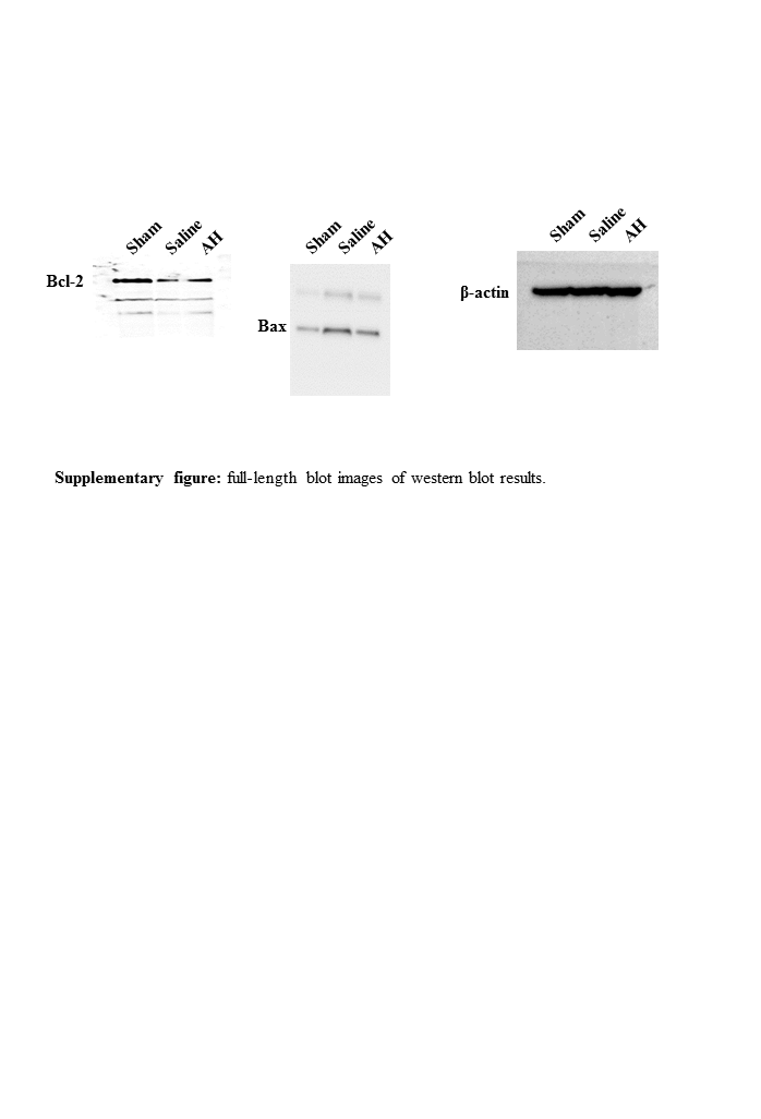
Western Blot
The heart tissues were harvested, washed in PBS three times and then homogenized in lysis buffer with protease inhibitors. Protein concentration was determined by bicinchoninic acid assay assay. Samples with 50 µg protein were run on a 10% sodium dodecyl sulfate (SDS)-polyacrylamide gel, and proteins were electrotransferred to PVDF membranes. The membranes were blocked with 5% non-fat milk in PBS for 1 hr and then incubated with primary antibodies at 4 oC overnight. After washing, blots were washed with PBS, and then incubated with the horseradish peroxidase-conjugated secondary antibody for 1 hr. Signals were detected using an ECL kit (Bio-Rad, Hercules, CA, USA).
Microcirculation
A video microscope, namely the sidestream dark field imaging (MicroScan, Microvision Medical, Amsterdam, Netherlands) was used for the observation of the microcirculation. Shortly, a handheld video microscope emits stroboscopic green light, which is absorbed by the erythrocytic hemoglobin and released back immediately. The images of the erythrocytes moving in the micro-vessels are transmitted to the camera through the microscope, thereby obtaining a non-invasive real-time image of the microcirculation. The microcirculation of the experimental animals at 0, 1, 2, 4, and 6 hr after ROSC was recorded by manual operation. In order to observe the intestinal microcirculation, 2–3 cm segment of jejunum has been isolated. Additionally, it has been coated with gauze soaked in warm saline. The mesentery on the serosal side was used as an observing region to evaluate intestinal microcirculation. After the observation, the intestine was back to the peritoneal cavity. The abdominal wall was sutured, and the skin incision was closed through the mode of a wound clip. The images were stored and analyzed offline using AVA-Automated Vascular Analysis 3.1 (Microvision Medical) to obtain Perfused vessel density (PVD) and microvascular flow index (MFI) parameters. These parameters were calculated for microvessels (≤ 20 µm in diameter). For PVD, microvessels were classified as either perfused (hyperdynamic, continuous or slow blood flow in the vessels) or non-perfused (intermittent or no blood flow) based on the microcirculation. For the quantification of MFI, the images were put into four parts. In addition, the microcirculatory condition was shown in a scheduled scale.
Statistical Analysis
Data were presented as mean ± SD and analyzed by SPSS 17.0 (SPSS Inc, Chicago, IL, USA). Data between two groups were compared using the Student’s t-test; data among multiple groups were compared using one-way analysis of variance (ANOVA). A value of p < 0.05 was considered significant difference.