The past decade in the LUAD research has been characterised by a greater understanding of cancer biology and management, with targeted therapy and immunotherapy providing significant survival benefits and manageable toxicity profiles in selected patients. However, major challenges still remain, including low response rate and drug resistance(Rotow and Bivona, 2017; Hegde and Chen, 2020). Thus, there is a clear urgent need to identify new driver gene alterations to expand the population that benefit from targeted therapy or immunotherapy, predict treatment responses and prevent or overcome the drug resistance. In this study, through employing open-access databases for comprehensive analyses, we found that TEDC2 could be involved in the tumorigenesis and progression of LUAD, and might contribute to the formation of immunosuppressive microenvironment in LUAD patients. These results suggested that TEDC2 could be regarded as a novel potential target for the treatment of LUAD.
The role of TEDC2 has been explored in few studies. Lim, D. H. et al. reported that TEDC2 could be predominantly expressed in primary central nervous system (CNS) diffuse large B-cell lymphoma (DLBCL) compared to non-CNS DLBCL(Lim et al., 2015). Hsu, M. K. et al. suggested that TEDC2 might be one of the potential genes for the tumorigenesis of LUAD and the construction of accurate classification systems distinguishing tumor from normal tissues(Hsu et al., 2015). TEDC2 was also seemed to act as a potential marker for treatment effect in male schizophrenia patients(Rukova et al., 2014). However, TEDC2 is not yet thoroughly studied and currently its function remains unclear. Our study preliminarily demonstrated a part of functions of TEDC2 in LUAD.
According to the analyses of data from TCGA and GEO databases, the mRNA expression of TEDC2 was significantly upregulated in LUAD compared to normal tissues. The protein level of TEDC2 was also confirmed to be higher in LUAD by immunohistochemistry analysis in Human Protein Atlas. In addition, ROC curve analysis found that TEDC2 could distinguish patients with LUAD from the normal population regardless of tumor stage. These evidences indicated that TEDC2 might play an important role in the tumorigenesis of LUAD and could serve as a new diagnostic marker for LUAD patients. To determine whether TEDC2 could be used as a prognostic marker in LUAD, we investigated the prognosis of LUAD patients with different TEDC2 expression levels. Kaplan-Meier curve analysis revealed that high TEDC2 expression was associated with inferior survival outcomes including OS, DSS and PFS. Cox regression analysis proved that high TEDC2 expression was an independent risk factor for poor prognosis, suggesting the potential prognostic value of TEDC2 in LUAD.
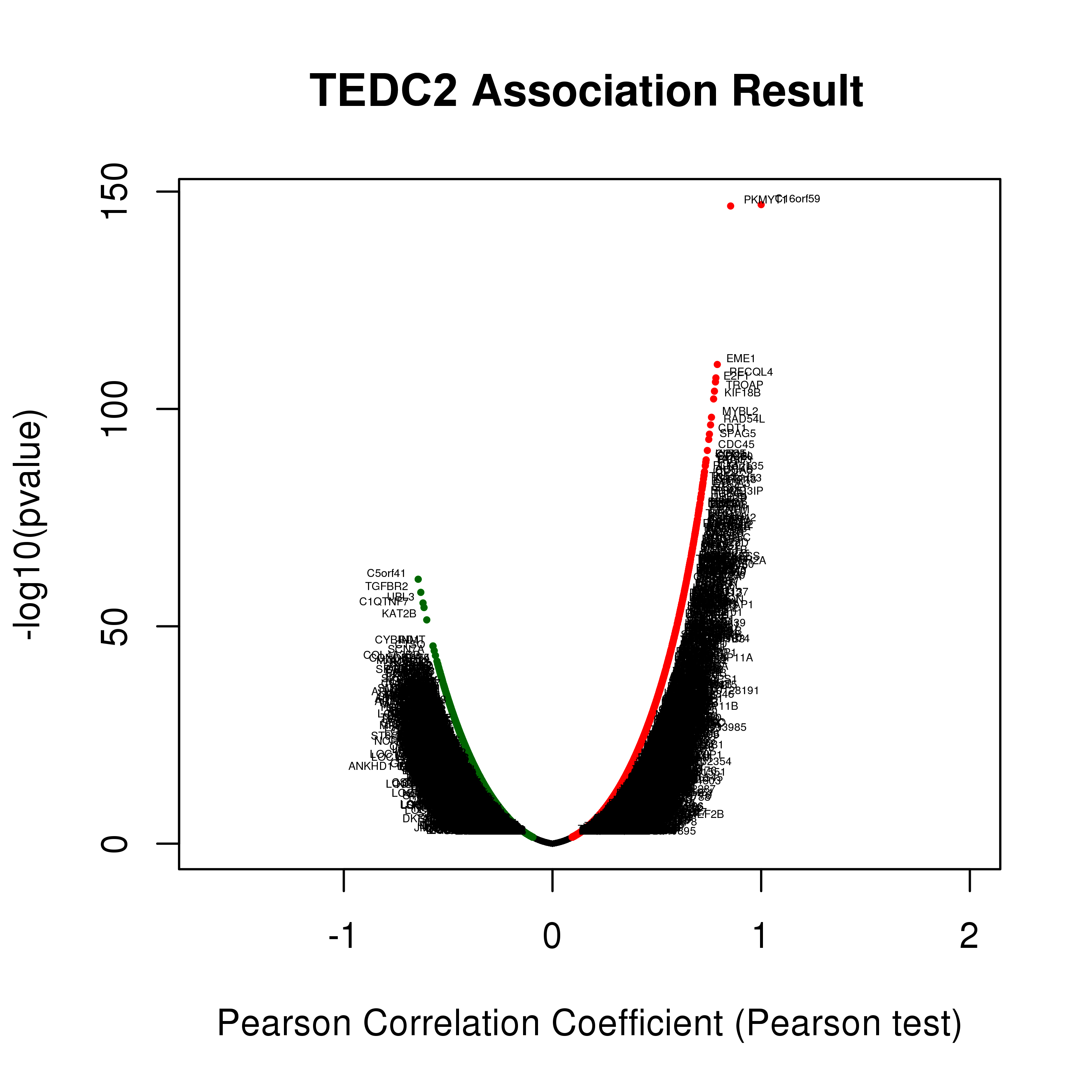
To unravel the biological functions of TEDC2, co-expression analysis and functional enrichment analysis were performed. LinkedOmics database analysis pointed out that most co-expressed genes with TEDC2 were mainly enriched in mitotic cell cycle processes, including chromosome segregation, DNA replication and cell cycle phase transition, suggesting that these genes could act as oncogenes to promote LUAD by accelerating cell cycle phase.
Another vital aspect of this study was that TEDC2 might be involved in regulating immune microenvironment in LUAD. There is increasing evidence proving the important role of tumor immune microenvironment in cancers(Remark et al., 2015; Hiam-Galvez et al., 2021). With the application of ssGSEA and ESTIMATE algorithms, we identified that the expression of TEDC2 was significantly negatively associated with immune infiltrates, which implied that TEDC2 might induce immunosuppressive context. The relationships between TEDC2 and immune infiltrates in LUAD were also analyzed by TIMER2.0 and GEPIA2 databases. The results demonstrated that TEDC2 expression showed negative correlation with DC and B cell. DC is one of the major regulators of immune response and can elicit T cell response, and previous studies have proved that DC could be associated with cytotoxic T cell infiltration and predict favorable outcome(Goc et al., 2014; Fucikova et al., 2016). B cell has emerged as a key player in immune microenvironment and correlates with better prognosis in NSCLC(Germain et al., 2014; Ghosh et al., 2021).
In addition, we conducted a systematic analysis of more than 40 common immune checkpoint genes and found that PDCD1, LAG3 and CD276 were highly correlated with TEDC2 expression, and the analysis of GEPIA2 database also proved these correlations. PDCD1 is an inhibitory receptor and negative regulator of T cell function, which can promote disease progression in patients with NSCLC(Altorki et al., 2019). LAG3 is able to function in coordination with other checkpoints such as PDCD1 to inhibit the activity of effector T cells and promote the suppressive activity, but effects and signaling events after LAG3 activation have not been completely understood(Rotte et al., 2018). CD276 expression on lung cancer leads to a lower number of tumor infiltrating lymphocytes and promotes lymph node metastasis, suggesting a role for CD276 in immune evasion and tumor progression(Picarda et al., 2016). Besides, TEDC2 expression was also correlated with TNFRSF25, TNFRSF4 and TNFRSF18 in our analysis, which might be correlated with immune evasion and poor outcome in lung cancer, but the molecular mechanisms of these immune checkpoint molecules still remain elusive and need further investigation(Luo et al., 2021; Kim et al., 2022).
This study preliminarily demonstrated the prognostic and diagnostic values of TEDC2 in LUAD, as well as the immune characteristics. However, there were certain limitations in our study. First, data heterogeneity was inevitable due to all the data in this study obtained from online databases. Second, gene expression analysis based on open-source databases might not be sufficiently accurate, which required additional experiments to provide a better understanding of the underlying biological mechanisms of TEDC2. Finally, although the regulatory effect of TEDC2 on immune microenvironment was evaluated by various algorithms in this study, the actual status of specific immune processes should be further investigated by in vitro and vivo models.