3. Methods
3.1 Controlled ovarian stimulation (COH)
COH protocols are chosen on the evaluation of ovarian reserve including age, anti-Müllerian hormone (AMH), basal hormone levels and antral follicle count (AFC).The long-term protocol of ovulatory induction is most frequently used, and it is mainly applied to young women with good ovarian reserve. The short-term protocol is used in women less than 40 years old whose ovarian reserve is not satisfying enough. Women with poor response to the long-term protocol or with polycystic ovarian syndrome (PCOS) can be induced by the GnRH-antagonist protocol. The ultra-long protocol is mainly suitable for female patients with endometriosis.
3.2 Oocyte retrieval
Human chorionic gonadotropin (hCG) was injected when at least one follicle with a diameter over 18 mm was present. Oocytes were retrieved 36 hours later.
3.3 In vitro fertilization
IVF or IVF/ICSI was chosen according to the sperm parameters on the day of oocyte retrieval in the control group. PGT was done in IVF/ICSI cycles to eliminate the effect of sperm and guarantee the accuracy of biopsy. In our center, the indication of IVF/ICSI includes: 1) Sperm concentration < 5 × 10⁶ /ml; 2) Sperm motility < 10%; 3) Rate of sperm with normal morphology < 1%.
3.4 Biopsy
Firstly, blastocysts were graded according to the Gardner blastocyst morphologic scoring system [11]. Approximately 4–6 trophoblast cells from high quality blastocysts were biopsied 5–6 days after fertilization by an experienced embryologist. In our center, blastocysts were simply shrunk by proper energy at first, then minor laser energy was used to slot the zona pellucida at the position of biopsy. The trophoblast cells to be biopsied were sucked into the biopsy needle through negative pressure, and then were cut by laser along the interface between zona pellucida and biopsy needle. Finally, trophoblast cells were completely segregated. Generally, the biopsied trophoblasts were lysed, and DNA was amplified by whole genome amplification (WGA, SurePlex, Illumina, Inc., San Diego, CA, USA) following the manufacturer’s protocol.
3.5 Blastocysts cryopreservation and thawing
We used vitrification technique for blastocyst cryopreservation, which was carried out using the Mukaida protocol with cryoloop [12]. To begin with, blastocysts were put into base medium containing 7.5% (v/v) DMSO and 7.5% (v/v) EG (vitrification solution I). After 2 min, the blastocysts were suspended in base medium containing 15% (v/v) DMSO and 15% (v/v) EG, 10 mg/ml Ficoll 70 (Pharmacia Biotech, Sweden, CB9248463) and 0.65 mol/l sucrose (vitrification solution II) for 30 s. Finally, they were put into liquid nitrogen as soon as possible.
Warming was performed in a four-well multidish using the Mukaida protocol. Briefly, blastocysts were incubated in base medium containing 0.33 mol/l sucrose (thawing solution I), base medium containing 0.2 mol/l sucrose (thawing solution II), and base medium at 37℃ for 2 min, 3 min, and 5 min.
Vitrified blastocysts were thawed on the morning of transfer day. After 30 min, hatching was assisted by laser with pulse length of 0.180 mS, applied for the thawed blastocysts. Only the expanded blastocysts were transferred after they were incubated for 4–5 h.
3.6 PGT technology
Before October 2016, PGT procedures were performed in the molecular laboratory of the center with a 24sure microarray chip (Illumina) according to the manufacturer’s protocol. Briefly, Cy3-labelled embryo WGA products were hybridized with Cy5-labelled SureRef (Illumina) reference male DNA WGA products. A microarray scanner (InnoScan 900) was used to scan microarray slides. And the images were analyzed using BlueFuse Multi software (BFM, Illumina). Aneuploid embryos that were unbalanced for the translocated chromosomes or/and abnormal for aneuploidy of chromosomes unrelated to the translocation were ascertained when those with a ratio greater than + 0.3 log 2 ratio were considered to be trisomic, and those with a ratio less than − 0.3 log 2 ratio were considered to be monosomic. Segmental abnormality in chromosomes unrelated to the parental rearrangement was indicated when chromosomal segmental aneuploidy of more than 10 Mb was involved.
After October 2016, NGS was implemented. After DNA fragmentation and library construction, the VeriSeq PGS kit—MiSeq (Illumina) was used for parallel sequencing and alignment, which were performed using the manufacturer’s protocol. The bioinformatic analysis was completed using BlueFuse Multi Software V4.4 (Illumina). Data quality, read depth, dynamic range/noise, the presence of ramping artifacts or step changes, and any recorded issues with biopsy or cell-to-tube transfer (e.g. the presence of lysed cells) were also taken into account in the final diagnosis.
As far as we know, NGS is expanding its applications in PGT given its capability to detect items of wide applications and detect unknown fragments. However, NGS also has limitations such as lower number of available bins of each chromosome compared to the a-CGH platform. Therefore, chromosome rearrangements that are predicted to be small and can be tested by a-CGH may not be detectable using NGS. By these means, we screen or diagnose numerical and structural chromosomal abnormalities from cells of trophectoderm. The embryos will not be thawed until the screening results come out.
3.7 Endometrial preparation and luteal phase support
In the control group, nearly all patients underwent fresh embryo transfer unless they had a tendency for ovarian hyperstimulation syndrome (OHSS) after controlled ovarian hyperstimulation (COH). Generally, fresh embryo transfer cases did not need extra endometrial preparation because the endometrium was able to reach its maximal thickness after COH. In our center, we usually use oral dydrogesterone (40 mg/d) and vaginal progesterone (200 mg/d) for each patient. Luteal support was initiated from the day of oocytes retrieval to the 12th week of pregnancy in most circumstances.
In the PGT group, all cases underwent frozen embryo transfer and the luteal phase support varied with the endometrial preparation protocol. At the third (or later) menstrual cycle after oocyte retrieval, natural ovulation was monitored by ultrasonography. Thirty milligrams of oral dydrogesterone were administered daily from the day of ovulation until the 12th week of pregnancy. If the natural ovulation cycle was cancelled due to anovulation or poor endometrial development, an artificial cycle was used for endometrial preparation. Luteal phase support was started when the endometrial thickness reached at least 7 mm and continued daily until 12 weeks of gestation with 40 mg daily of oral dydrogesterone and 200 mg daily of vaginal progesterone. Ovulation induction cycle was used if the endometrium was still responding poorly after natural ovulation and artificial cycles. Luteal phase support was initiated from the day of ovulation and the dosage was between the above two protocols.
4. Outcome Measures
In each cycle, pregnancy outcomes of the first transfer (including cryopreserved and fresh embryo transfers) were evaluated, including biochemical pregnancy, clinical pregnancy, ongoing pregnancy, live birth, miscarriage rates of the first transfer as well as the cumulative live birth rates. Biochemical pregnancy is defined as a hCG serum levels of more than 25 mIU per milliliter as measured at 12 days after embryo transfer. Clinical pregnancy refers to the signals of fetal heart beating in gestational sac assessed by ultrasonography during the 7th or 8th week. Ongoing pregnancy is regarded as the presence of a vital fetus with a positive heart beat at 11–12 weeks of gestation. Live birth is defined as the delivery of a live-born baby infant after at least 28 weeks of gestation. Miscarriage means termination of pregnancy before 28 weeks of gestation or a fetal weight lower than 1000 g [13].
We compared the differences of pregnancy outcomes between PGT and control group in order to find out if PGT could improve pregnancy outcomes of chromosomal inversion carriers. First, we compared factors which could potentially influence the pregnancy outcomes between the groups. These included maternal age, history of recurrent spontaneous abortion (RSA), duration of infertility, body mass index (BMI), hydrosalpinx, uterine leiomyoma, previous pelvic or intrauterine surgery and uterine abnormality. Since we found that the duration of infertility and the RSA rate in the PGT group were significantly different compared to the control group and maternal age has always been regarded as being of influence to pregnancy outcomes, logistic regression analysis was performed to determine if the factors besides the assisted reproduction method used would have any impact on pregnancy outcomes. According to the American Society for Reproductive Medicine’s criteria [14], RSA is defined as pregnancy loss for twice or more. BMI is calculated when kilograms of weight are divided by meters square of height. Hydrosalpinx is observed by ultrasonography of adnexa and/or salpingography. Uterine abnormalities included endometrial polyps, untreated intrauterine adhesions and uterine malformations.
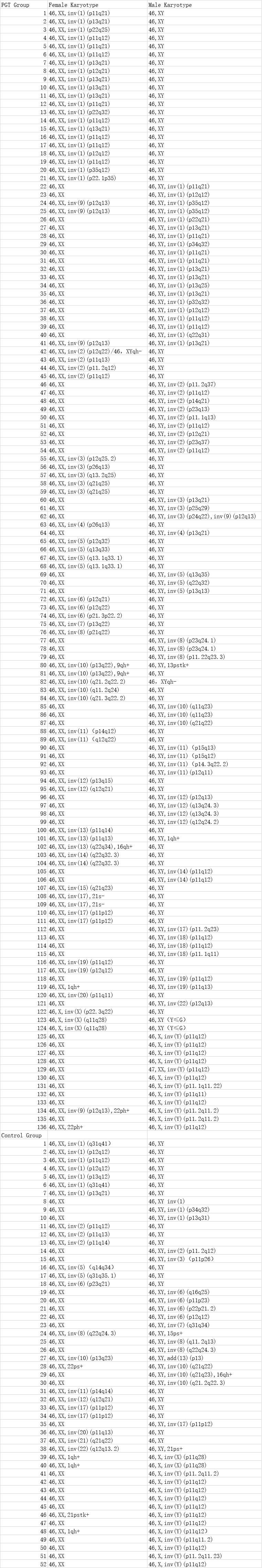
In the PGT group, the aneuploid rate was compared between the pericentric and paracentric inversion subgroups, and between the male and female carrier subgroups.