Survey of desmoid-type fibromatosis
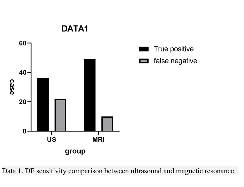
Desmoid-type fibromatosis is a rare fibrous tissue tumor. At the beginning of this century, the World Health Organization classified it as myofibroblastic tumor. Extra-abdominal desmoid-type fibromatosis commonly occurs in the musculoskeletal system. Although the disease has no tendency of distant metastasis, it has the characteristics of local recurrence, and its biological behavior is between benign and malignant. In this study, among 117 patients, 61 patients were followed up, and recurrence was found in 33 patients with a recurrence rate of 54.1%, which was consistent with previous studies. Many scholars believed that the occurrence of extra-abdominal desmoid-type fibromatosis is associated with genetic factors, history of trauma and history of surgery. In the ultrasound group and MRI group, among the 117 cases of extra-abdominal desmoid-type fibromatosis, 34 had a history of trauma, 27 had a history of surgery, and no one had a family history.
Ultrasound Imaging Performance
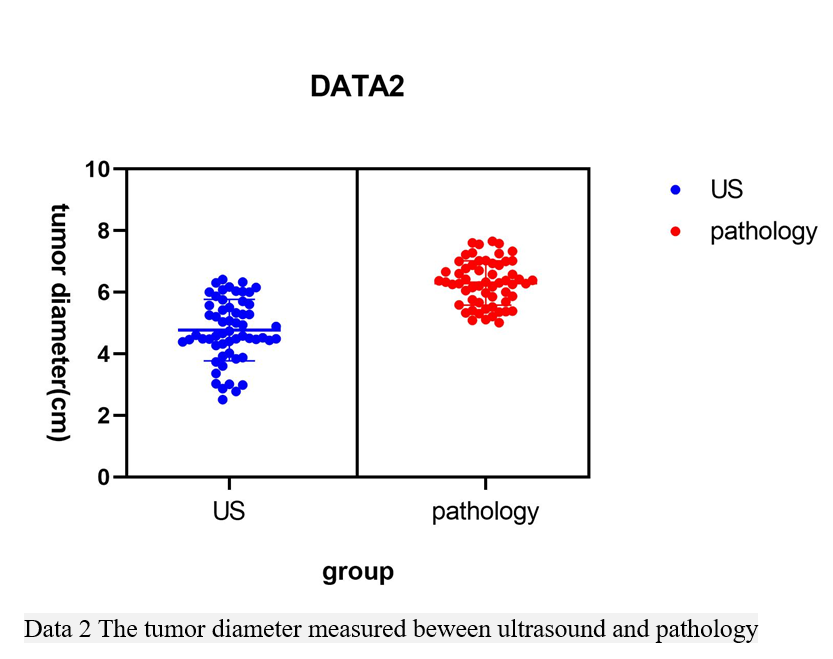
Ultrasound has a strong ability to display fibrous tissue. With the performance of ultrasound instrument greatly enhanced, the accuracy of color Doppler ultrasound alone in the diagnosis of soft tissue tumors has been significantly improved[3, 4]. The results from this study demonstrated that the preoperative diagnostic sensitivity of B-mode ultrasound was 62.07%. Ultrasound can significantly improve the identification of extra-abdominal desmoid-type fibromatosis by judging the blood flow signals and distribution in and around the tumor. In the present study, among 58 patients detected by ultrasound, the blood supply in 37 tumors was relatively abundant, and all experienced the first onset, which was not consistent with previous studies[2, 4]. The difference may be associated with our selection of extra-abdominal cases. In all ultrasound-detected cases, no obvious liquid anechoic area was detected. Fibrous echo could be obvious in 53 cases. These could be used as a diagnostic basis to distinguish other soft tissue tumors of musculoskeletal system. The disadvantages of ultrasonic diagnosis of desmoid-type fibromatosis are the subjective factors of diagnostic physicians and the limited ability of ultrasound itself to show the extent of invasion of surrounding tissues. In all 58 cases of ultrasound group, the tumor size was less than the pathological results in 30 cases(Data2). Because of its well-known advantages, ultrasound is still the first choice for patients with extra-abdominal desmoid-type fibromatosis.
MRI imaging performance
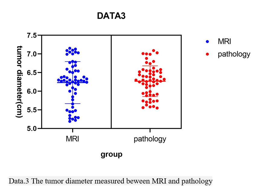
Since the advent of MRI, its superior soft tissue resolution has made it one of the indispensable inspection methods for imaging soft tissue lesions, especially soft tissue tumors. MRI diagnosis of extra-abdominal desmoid-type fibromatosis has certain advantages in all imaging examinations. In this study, the sensitivity of MRI reached 83.05%. On the MRI images of a typical extra-abdominal desmoid-type fibromatosis, T1WI showed equal or slightly lower signal intensity, and T2WI and STIR showed high signal intensity. Spot-like and cordlike low-signal fiber components could be seen in high signals. Most of the lesions showed more than medium homogeneous enhancement[5, 6, 7, 8]. In this study, 25 lesions showed obvious enhancement, accounting for 42.37%, which indicated that the blood supply of the tumor itself was relatively rich. The morphology of extra-abdominal desmoid-type fibromatosis is generally large, but there are few signs of liquefaction or necrosis in the lesions, which is one of the key points to distinguish it from other soft tissue tumors. MRI could perfectly reveal the invasion of the peritumoral tissue. Because of the high recurrence rate of extra-abdominal desmoid-type fibromatosis[9], the actual boundary of extra-abdominal desmoid-type fibromatosis has always been the focus of clinicians' attention(Data3). The determination of the resection range is the key factor to determine whether the tumor will recur after resection. MRI has become an important method for the diagnosis of extra-abdominal desmoid-type fibromatosis, the guidance of clinical tumor staging, the selection of surgical methods and the scope of resection.
Differential Diagnosis Of Extra-abdominal Desmoid-type Fibromatosis
The typical extra-abdominal desmoid-type fibromatosis has obvious imaging features on ultrasound and MRI, but it is difficult to distinguish it from some fibrous tumors, especially with some atypical fibrous tumors.
Fibroproliferative fibroma
The disease occurs frequently in the mandible, mainly at the age of 10–30 years old. The onset age coincides with that of desmoid-type fibromatosis. Fibroproliferative fibroma has the same characteristics as extra-abdominal desmoid-type fibromatosis tissues. It is extremely difficult to distinguish with imaging methods such as ultrasound and MRI. The final diagnosis relies on pathological results.
Fibrosarcoma
Fibrosarcoma is more common in the middle-aged and the elderly and the lesions grow faster. The center of the lesion easily suffers from necrosis and bleeding. The degree of enhancement is more obvious than that of desmoid-type fibromatosis. Different from desmoid-type fibromatosis’s compression of the surrounding bone, fibrosarcoma can invade the surrounding bone, which can cause many kinds of map-like, insect-like bone destruction. According to the general conditions such as medical history, it is not difficult to make the corresponding diagnosis by ultrasound and MRI, but attention should be paid to the identification of fibrosarcoma without early signs of liquefaction or necrosis.
Benign fibrous histiocytoma
The disease commonly occurs at the age of 20–60 years old in the pelvis and femur. The appearance of fat in the center of the lesion can be used as a characteristic diagnosis.
Malignant fibrous histiocytoma
A typical malignant fibrous histiocytoma has a large tumor mass; liquefaction and necrosis easily appear in the lesion. The boundary between the lesion and surrounding tissues is unclear. The onset age shows a bimodal distribution (20–40 years old and 60–80 years old). Ultrasound and MRI can make a correct diagnosis based on the above conditions.
In summary, ultrasound and MRI can make a qualitative diagnosis of extra-abdominal desmoid-type fibromatosis based on its inherent superiority and its good ability to display the composition of extra-abdominal desmoid-type fibromatosis fibers. The ability of MRI to show the true boundary of tumor mass and the extent of invasion to the surrounding area can provide clinical help as much as possible. However, it is still difficult for the imaging physicians to make a differential diagnosis between extra-abdominal desmoid-type fibromatosis and some atypical soft tissue tumors using ultrasound and MRI.