Although anion channel activities have been demonstrated in sarcoplasmic reticulum/endoplasmic reticulum (SR/ER), their molecular identities and functions remain unclear. Here, we link rare variants of CLCC1 (Chloride Channel CLIC Like 1) to ALS (amyotrophic lateral sclerosis)-like pathologies. We demonstrate that CLCC1 is a pore-forming component of an ER anion channel and that ALS-associated mutations impair the channel conductance. CLCC1 forms homomultimer and its channel activity is inhibited by luminal Ca2+ but facilitated by phosphatidylinositol 4,5-bisphosphate (PIP2). We identified conserved residues D25 and D181 in CLCC1 N-terminus responsible for Ca2+ binding and Ca2+ luminal inhibition on channel open probability and K298 in CLCC1 intraluminal loop as the critical PIP2-sensing residue. CLCC1 maintains steady-state [Cl-]ER and [K+]ER and morphology and regulates ER Ca2+ homeostasis including efficiency of internal Ca2+ release and steady-state [Ca2+]ER. ALS-associated mutant CLCC1 increase steady-state [Cl-]ER and impair ER Ca2+ homeostasis, and animals with the ALS-associated mutation sensitize to stress challenge-induced protein misfolding. Phenotypic comparisons of multiple Clcc1 loos-of-function alleles, including ALS-associated mutations, reveal a CLCC1 dosage-dependence in severity of disease phenotypes in vivo. As CLCC1 rare variants appear dominant in ALS, ~10% K298A heterozygous mice displayed ALS-like phenotypes, underscoring a mechanism underlying channelopathy dominant-negatively caused by a loss-of-function mutation. Conditional knockout of Clcc1 cell-autonomously causes motor neuron loss and ER stress, misfolded protein accumulation, and characteristic ALS pathologies in the spinal cord. Thus, we argue that disruption of ER ion homeostasis maintained by CLCC1 underlies etiology of neurodegenerative diseases.
Research Article
Disruption of ER ion homeostasis maintained by an ER anion channel contributes to ALS-like pathologies
https://doi.org/10.21203/rs.3.rs-1499219/v1
This work is licensed under a CC BY 4.0 License
Version 1
posted
You are reading this latest preprint version
Although Cl- is the most abundant anion in living cells, chloride currents and their functional significance had been understudied until the CLC family of chloride channels and CFTR (cystic fibrosis transmembrane conductance regulator) were cloned and their dysfunctions were linked to human diseases 1-4. In addition to those on the cell surface, Cl- channels have long been proposed to exist in the intracellular membrane-bound organelles 3,5. However, the previously postulated intracellular Cl- channels, like CLCAs (chloride channel Ca2+-activated) and CLICs (chloride intracellular channels), are now considered not likely to function as anion channels 6,7. Therefore, the molecular identities and functions of organellar anion channels, including those in the SR/ER, remain largely unknown.
As the major internal Ca2+ store, Ca2+ release from SR/ER is mediated by two cation channels, RyRs (ryanodine receptors) and IP3Rs (inositol 1,4,5-trisphosphate receptors) 8-10. During the release, SR/ER membrane is charged upon the Ca2+ efflux, that hinders the continued Ca2+ release. Previously reported TRICs (TRimeric Intracellular Cation channels) acts as counter-ion channel to balance the loss of positive charges from the SR/ER as a result of the release 11. In addition to cations, anions have also been proposed to function as counter-ion for the release, and various Cl- channel activities have been long demonstrated in microsome preparations 12-17. A previous study using mouse forward genetics revealed that loss of CLCC1 (Chloride Channel CLIC Like 1), an ER-resident protein 18,19, leads to ER stress and neurodegeneration 19. However, despite the name, CLCC1 has little sequence similarity with CLIC family members or any known ion channels. In addition, question remains whether the recorded chloride currents in microsome prepared from the CLCC1 overexpressing cells were actually mediated by CLCC1 20,21. Therefore, further evidence is needed to know if CLCC1 functions as an anion channel.
Here, we demonstrate that CLCC1 is a pore-forming component of an ER anion channel by incorporating purified CLCC1 into lipid bilayer. Depletion of CLCC1 reduces ER Ca2+ release, probably through a counter-ion mechanism, but increases steady-state [Cl-]ER and [K+]ER. We identified CLCC1 rare variants in a Chinese ALS cohort and the disease-associated non-synonymous mutations impair CLCC1 channel conductance and promote misfolded protein accumulation in the mutation knockin mouse brain and spinal cord. Conditional removal of Clcc1 in ChAT-positive motor neuron cell-autonomously leads to ubiquitin-positive and mislocalized TDP-43, a pathological hallmark of ALS, and motor neuron loss. Therefore, we argue that misregulation of ER ion homeostasis maintained by an ER anion channel underlies ER unfolded protein response (UPR) and etiology of neurodegenerative diseases.
CLCC1 forms homomultimer in the ER membrane
Based on its primary sequence, CLCC1 shares little sequence similarity with any known ion channel but is predicted to contain three transmembrane segments (TMs) and a N-terminal signal peptide (Fig. 1a). We generated antibodies against the N- and C-termini of CLCC1 (Extended Data Fig. 1a). Using the C-terminal antibody, we confirm that as suggested by a previous report 18,19 CLCC1 is predominantly ER-localized, as demonstrated by its co-localization with CALNEXIN, an ER-resident protein (Extended Data Fig. 1b).
To understand how CLCC1 functions in the ER, we treated human 293FT cells with disuccinimidyl suberate (DSS), a crosslinker with a spacer length of 11.4 Å. The C-terminal antibody detected high molecular weight complexes in a DSS dosage-dependent manner from whole cell lysate. From the complex sizes, we speculated that CLCC1 forms homomultimers (Fig. 1b), which was supported by co-immunoprecipitations of differentially tagged CLCC1 co-expressed in the same cells (Extended Data Fig. 2a) and of exogenous tagged CLCC1 with endogenous CLCC1 (Extended Data Fig. 2b). Consistent with our cell culture data (Fig. 1b) and a previous report 20, our native gel experiments suggested complex formations (~180kD and ~360kD) of purified full-length CLCC1 and that (~360kD, the major band) of endogenous CLCC1 (Extended Data Fig. 2c). In addition, the purified full-length mouse CLCC1 (mCLCC1) gave a major high molecular weight peak by chromatographic column separation (Fig. 1c). Taken together, our data suggest that CLCC1 forms homomultimer in the ER membrane.
CLCC1 is a pore-forming component of an anion channel
Incorporation of the purified full-length mCLCC1 (Fig. 1c) into planar lipid bilayer resulted in frequent inward currents at 0 mV (-2.2 ± 0.1 pA) in asymmetric KCl solutions (In/Ex, 150/15 mM) and the currents became outward at 90 mV (1.6 ± 0.1 pA) (Fig. 1d). As a negative control, the protein purification buffer without protein gave rise to no current (Fig. 1d). Based on the fit of the current-voltage relationship, the reversal potentials were determined to be 56.8 mV (In/Ex, 150/15 mM KCl) and -60.3 mV (In/Ex, 15/150 mM KCl), which are close to the calculated values for Cl- by Nernst equation, and the slope conductance was 39.9 ± 1.0 pS (mean ± SEM). The permeability ratio of P(Cl-) to P(K+) is about 100 to 1 and similar results were obtained by using asymmetric NaCl solutions (Fig. 1d). Consistent with the single channel results, the reversal potential obtained from studying macroscopic currents was 61.9 mV in 15/150 mM KCl (In/Ex) (Fig. 1e), further supporting the anion selectivity. Both square step-like multiple-channel currents (Extended Data Fig. 3) and a large-scale current (80-90 pA) (Fig. 1e) suggested that CLCC1 is capable to mediate multi-channel currents.
Next, we examined CLCC1 channel permeability to various anions, including Br-, NO3-, and F-, by adding 150 mM KCl in cis (In) and equal electric charges of KBr, KNO3, or KF in the trans (Ex) chamber (Fig. 1f). The relative permeabilities of these anions to Cl- were 17.14 (PBr/PCl), 0.22 (PNO3/PCl), and 0.18 (PF/PCl), respectively, indicating a sequence of the CLCC1 anion selectivity of PBr > PCl > PNO3 > PF, which is similar to that of CFTR 22,23. In these experiments, no cation permeation was detected. Collectively, our results demonstrate that CLCC1 is a pore-forming component of an anion channel.
ER membrane topology of CLCC1 and its inhibition by luminal calcium
To examine CLCC1 topology in the ER membrane, we treated microsomes prepared from wildtype mouse cerebella and livers 24 with trypsin and analyzed the remaining CLCC1 fragments with our N- and C-terminal antibodies. In the absence of Triton X-100, the N-terminus and the first and second loops of CLCC1 and an ER lumen resident protein Bip were protected from trypsinization, but the C-terminus of CLCC1 was not (Fig. 1g and Extended Data Fig. 1c). As expected for membrane enclosure, the protection was disrupted by Triton X-100, suggesting that CLCC1 N-terminus and the second loop reside in ER lumen while C-terminus faces cytoplasm.
Interestingly, when we applied MTSET (methanethiosulfonate-ethyltrimethylammonium) 25, a membrane-impermeant thiol reagent that modifies cysteine residues, in trans but not to the cis side of the chamber we applied the purified CLCC1, the CLCC1 currents were suppressed (Fig. 1h), suggesting that a specific orientation of CLCC1 in the bilayer is responsible for the current. Based on the topology (Fig. 1g), cysteine residues are located in both the cytoplasm and ER lumen sides of CLCC1 and C350 lies at the end of TM3 (Fig. 1i). Protein alignment among different species revealed that C350 is in a consecutive row of four residues (FCYG), although it is less conserved than the other three surrounding resides (Fig. 1j). Instead of FCYG in Homo sapiens and Mus musculus, FFYG appears in Xenopus tropicalis, which prompted us to mutate C350 to F. C350F mCLCC1 is expressed and its chromatographic behavior is similar to wildtype mCLCC1 (Extended Data Fig. 4a). Importantly, C350F restored the CLCC1 currents even when MTSET was applied in trans side (Fig. 1h), suggesting that MTSET acts on C350 to modify the channel activity and the trans side is the CLCC1 cytoplasm side in the reconstructed lipid bilayer. Application of DIDS (4,4'-Diisothiocyano-2,2'-stilbenedisulfonic acid), a chloride transporter/channel blocker 4,26, significantly inhibited CLCC1 channel activity (Extended Data Fig. 4b and 4c). Consistent with MTSET acting on C350, C350F largely restores the DIDS inhibition on channel open probability (Po).
Because ER luminal Ca2+ is much higher than cytoplasm, we then asked whether Ca2+ is able to differentially regulate CLCC1 channel activity from ER luminal or cytoplasmic side. Application of Ca2+ in cis/ER lumen side blocked the CLCC1 channel activity, which could be partially rescued by addition of equal molar EGTA, a Ca2+ chelating agent (Fig. 1k and 1l). However, the same application in trans/cytoplasm side had no effect on the channel activity. Therefore, we conclude that, at least in our reconstructed lipid bilayer setting, high concentration of Ca2+ at the ER lumen side inhibits CLCC1 channel activity.
D25/D181 are the key residues responsible for Ca2+-dependent inhibition on the channel open probability
To identify key residues at lumen side responsible for the Ca2+ inhibition, we aligned the protein sequences across different species (Fig. 1m). Several conserved negatively charged residues at N-terminus drew our attention, including D25, D152, D153, E175, D176, and D181. Because CLCC1 D25E mutation has been associated with retinitis pigmentosa 21, we took D25E into account. In addition, we generated D152R/D153R, E175R/D176R, D181R, and D25E/D181R mutant CLCC1 for Ca2+-binding affinity and channel activity measurements.
Because C-terminus of CLCC1 resides at cytoplasmic/trans side, we truncated the C-terminus (1-365) and recorded a sound current that could be inhibited by Ca2+ (10mM) application at cis side (Fig. 1n). In addition, all mutant CLCC1 yielded reasonable currents as wildtype (1-365) did at 0 mV in absence of Ca2+ (Fig. 1o). However, the mutant CLCC1, including D25E, D181R, and D25E/D181R but not D152R/D153R, largely restored the currents in presence of 10mM Ca2+ at cis side (Fig. 1n), suggesting that D25 and D181 are the key residues for the calcium inhibition on CLCC1 channel activity. Our Ca2+ dose response curve for the inhibition and the calculated IC50 (mM) (for wildtype, 1.09 ± 0.15; D25E, 6.08 ± 1.62; D181R, 8.51 ± 1.04; D25E/D181R, 19.20 ± 4.24) also supported the notion that D25 and D181 are the key residues (Fig. 1p). The IC50 for D152R/D153R (1.06 ± 0.19) and E175R/D176R (2.83 ± 0.58) suggested that these negatively charged residues are either not involved in or less important than D25 and D181 for the inhibition.
To determine the Ca2+-binding affinity of the mutant CLCC1, we performed surface plasmon resonance (SPR)-based binding assay (Fig. 1q and 1r). Compared to that of the wildtype (1-365, 7.99 ± 0.62 mM), the KD values of the mutant CLCC1, including D25E (10.47 ± 0.27 mM), E175R/D176R (11.29 ± 0.29 mM), D181R (12.14 ± 0.29 mM), and D25E/D181R (22.94 ± 4.10 mM), were significantly increased, suggesting that these mutant CLCC1 significantly impair Ca2+-binding affinity. Consistent with the D25E/D181R mutant with the strongest effect on relieving the Ca2+ inhibition, it also affects the most of Ca2+-binding affinity (Fig. 1s). Therefore, we conclude that D25/D181 are the key residues responsible for Ca2+-dependent inhibition of the channel open probability.
CLCC1 maintains steady-state [Cl-]ER, [K+]ER, and ER morphology
To examine whether CLCC1 is involved in regulation of [Cl-]ER, we employed a previously optimized YFP Cl- sensor that responds to Cl- concentration change with super sensitivity and photostability 27. To create a ratiometric ER Cl- sensor, we built a signal sequence, a DsRed internal control, and an ER retention motif into the Cl- sensor, which we named RaMorideER (Fig. 2a). ER localization of RaMorideER was confirmed by its colocalization with ER-resident protein CALNEXIN (Fig. 2b). The ratio between YFP to Ds-Red signals responded correspondingly when extracellular [Cl-] ([Cl-]Extra) was switched from 140mM to 100 or 0 mM (Extended Data Fig. 5a and 5b).
Consistent with the essential role of CLCC1 in vivo, we failed to generate a CLCC1 KO 293FT cell line by Crispr/Cas9. Instead, we knocked down CLCC1 with two individual shRNAs (H3 and H4) (Fig. 2c-2e). Although the two shRNAs had different CLCC1 knockdown efficiencies (for H3, 22.5 ± 0.6% of scrambled control; for H4, 45.25 ± 2.1% of scrambled control), both of them significantly increased steady-state [Cl-]ER to a similar extent in comparison with scrambled shRNA control (Fig. 2f and 2g).
Given that the influx of K+ into ER lumen acts as a counter-ion mechanism during the internal Ca2+ release 11, we next measured steady-state [K+]ER upon knockdown of CLCC1. To achieve it, we employed a newly reported K+ sensor (doi.org/10.1101/2021.10.07.463355) and generated a ratiometric ER K+ sensor (RaMssiumER) (Fig. 2h), as we did for RaMorideER. The RaMssiumER responded well to a series of titrated [K+] solutions in vitro (Fig. 2h) and a K+ ionophore, valinomycin (Extended Data Fig. 5c). With the RaMssiumER probe, we detected significantly higher steady-state [K+]ER in H3 and H4 groups, compared to the scrambled control (Fig. 2i).
The concentration of electrically charged osmolytes, such as Cl-, inside a cell or intracellular membrane-bound organelle governs the volume of the compartment 5,28. Therefore, we asked whether depletion of CLCC1 changes ER volume. To this end, we collected 293FT cells expressing scrambled control or CLCC1 shRNAs and applied for transmission electron microscopy (TEM) (Fig. 2j and 2k). Enlarged and stubby ER morphology was documented in cells expressing the individual CLCC1 shRNA. In contrast, ribosome-bound and tubule-like ER was shown in the scrambled controls. In order to quantitatively reflect ER morphology, we measured ER width in these three groups of cells. ER width in the two individual CLCC1 shRNA groups was significantly increased as compared to scrambled shRNA control. Therefore, we conclude that CLCC1 loss alters steady-state ER ion homeostasis and leads to enlarged ER.
CLCC1 facilitates internal Ca2+ release
ER-localized ion channels have been proposed to control ER Ca2+ mobilization through a counter-ion mechanism 13,14,16,17. We then asked whether as an ER chloride channel CLCC1 is involved in regulation of ER Ca2+ release. Knockdown of CLCC1 by the two individual shRNAs markedly reduced internal Ca2+ release induced by ATP (adenosine triphosphate) (Fig. 3a), which triggers ER Ca2+ release by generating IP3 that activates IP3Rs 10. Compared to mock control and scrambled shRNA, knockdown of CLCC1 by the two individual shRNAs not only significantly reduced the amplitude, but also the rate (as reflected by the increase in time-to-peak), of ATP-induced Ca2+ release (Fig. 3b and 3c). Although the two shRNAs had different CLCC1 knockdown efficiencies, they impaired the ATP-induced Ca2+ amplitude and rate to a similar extent.
Analysis of the Ca2+ release dynamics in individual cells revealed that CLCC1 knockdown impaired ATP-induced Ca2+ oscillation (Extended Data Fig. 6a and 6b). Whereas less than 4.2 ± 0.6% of cells exhibited one ATP-induced Ca2+ spike in scrambled shRNA group, the proportion was significantly more in the CLCC1 knockdown groups (31.5 ± 2.3% for H3; 36.5 ± 4.9% for H4). However, the Ca2+ spike number greater than or equal to 3 was reduced in the CLCC1 knockdown groups compared to that of scrambled control (Extended Data Fig. 6b). The impairment of ATP-induced Ca2+ release seems not to be caused by shRNA off-target effects, because the reexpression of full-length (WT) mCLCC1 restored the release damaged by H3 shRNA alone (Extended Data Fig. 7a and 7b). In contrast, expression of mutant mCLCC1 lacking the ER lumen resident 2nd loop (Δ2nd loop) did not, suggesting that the 2nd loop of CLCC1 is crucial for its functions.
Next, we asked whether CLCC1 regulates internal Ca2+ release through RyRs, the predominant intracellular Ca2+ channels expressed in cardiomyocytes 10 . To this end, we stimulated cardiomyocytes cultured from wildtype (+/+) and NM2453 mutant (NM/NM) mice with caffeine, an agonist for RyR-mediated Ca2+ release. RyR-mediated Ca2+ release was significantly reduced in NM/NM cardiomyocytes as compared to +/+ controls (Extended Data Fig. 6c), demonstrating that CLCC1 facilitates ER Ca2+ efflux through regulation of the release process per se rather than regulation of a particular type of Ca2+ release channels.
CLCC1 dosage is crucial for maintenance of steady-state [Ca2+]ER level
To examine whether the impaired Ca2+ release upon CLCC1 knockdown results from a reduction in ER Ca2+ load, we depleted the ER Ca2+ store with cyclopiazonic acid (CPA), an inhibitor of sarco/endoplasmic reticulum Ca2+-ATPase (SERCA) 29. Knockdown of CLCC1 by H3 but not H4 shRNA significantly reduced CPA-sensitive cytosolic Ca2+ rise (Fig. 3g and 3h), suggesting that impairment of ER Ca2+ content depends on CLCC1 dosage as H3 has higher knockdown efficiency than H4 shRNA (Fig. 2e).
Given that depletion of CLCC1 increases ER volume (Fig. 2j and 2k), we next asked whether [Ca2+]ER is also impaired. We employed a previously reported low affinity Ca2+ probe, ER-GCaMP6-210 30, which correctly responded to CPA-induced internal Ca2+ depletion and follow-up ionomycin-mediated extracellular Ca2+ replenish (Extended Data Fig. 8a). Compared to mock and scrambled shRNA controls, knockdown of CLCC1 by both H3 and H4 shRNAs significantly decreased steady-state [Ca2+]ER level in cells expressing ER-GCaMP6-210 (Fig. 3g and 3h). The impairment caused by H3 shRNA was more severe than that by H4 shRNA, suggesting that depletion of CLCC1 decreases steady-state [Ca2+]ER level in a dosage-dependent manner.
To measure steady-state [Ca2+]ER in the diseased neurons, we cultured cerebellar granule neurons (CGNs) from wildtype (+/+) and NM/NM mutant mice (Fig. 3i and 3j). We employed previously described low affinity Ca2+ probe, ER-GCaMP6-150 30, and generated a ratiometric probe for [Ca2+]ER measurement by fusing ER-GCaMP6-150 with DsRed, which responded well to CPA-induced internal Ca2+ depletion (Extended Data Fig. 8b). After infecting the cultured CGNs with the probe, we detected significant decrease of [Ca2+]ER in the mutant CGNs compared to that of wildtype.
In summary (Fig. 3k and 3l), the knockdown of CLCC1 dosage-dependently increases steady-state [K+]ER and ER volume and decreases steady-state [Ca2+]ER. Instead, the knockdown increases steady-state [Cl-]ER and reduces internal Ca2+ release in a CLCC1 dosage-independent manner, suggesting that steady-state [Cl-]ER and internal Ca2+ release are primarily affected by CLCC1 knockdown.
A conserved lysine (K298) is responsible for PIP2 facilitation of CLCC1 channel activity
As a necessary cofactor of many ion channels, PIP2, an acidic phospholipid of the cell membrane, has been implicated in the regulation of ion channel functions, including of intercellular cation channels 31-33. To examine whether PIP2 affects CLCC1 channel activity, we included 2% PIP2 in the planar phospholipid bilayer. Interestingly, PIP2 significantly increased the slope conductance (80.1 ± 2.5 pS) and the open probability (Po) of wildtype mCLCC1 (Fig. 4a and 4b). Given that PIP2 regulates ion channels by binding to certain positively charged residues in the channel protein 32,33, we looked for positively charged residue(s) in CLCC1 and a positively charged lysine (K298) drew our attention (Fig. 4c). It lies in a consecutive row of six conserved residues-VPPTKA in the 2nd loop, which is required for CLCC1 facilitation of internal Ca2+ release (Extended Data Fig. 7). In addition, K298 is downstream of two proline residues, which usually present strong conformational rigidity, and lies at the beginning of a predicted alpha-helix.
We expressed and purified K298A mutant mCLCC1 and incorporated it into the lipid bilayer in the absence of PIP2, the mutant protein exhibited single channel activity with a slope conductance of 31.8 ± 0.7 pS, slightly lower than that of wildtype mCLCC1 (39.9 ± 1.0 pS) (Fig. 4a, 4d, and 4f). The Po at 0 mV did not differ from that of wildtype mCLCC1 (Fig. 4a and 4f). Next, we mutated K298 to the negatively charged residue glutamate (K298E). Like K298A, K298E also has little effect on the channel activity in absence of PIP2 (Fig. 4f). However, unlike wildtype mCLCC1 responsible to PIP2 (Fig. 4b and 4e), both K298A and K298E mutants abolished the responses, in terms of conductance and Po (Fig. 4e and 4f). Therefore, we conclude that PIP2 facilitates CLCC1 channel activity and a conserved K298 in the 2nd loop is responsible for the facilitation.
K298 is crucial for CLCC1 regulation of internal Ca2+ release
If K298 is functionally important for CLCC1 channel activity, we wondered whether K298 is equally important for internal Ca2+ release. To examine this, we employed a lentiviral inducible system to stably express wildtype and K298A mutant mCLCC1 in 293FT cells in a controllable manner (Extended Data Fig. 9a). Expression of exogenous mCLCC1 proteins was induced after application of doxycycline (Dox) (Extended Data Fig. 9b). Both the exogenous wildtype and K298A mutant mCLCC1 interacted with the endogenous hCLCC1 (Extended Data Fig. 9c), as shown by co-immunoprecipitation, supporting complex formation by exogenous mCLCC1 and endogenous hCLCC1 (Extended Data Fig. 2b). Induction of wildtype mCLCC1 did not alter the amplitude and rate of ATP-induced Ca2+ release (Fig. 4g-4i). However, expression of K298A mutant mCLCC1 significantly suppressed such activities, as shown by the reduction in both the amplitude and rate when compared to un-induced (minus Dox) cells or cells induced to express wildtype mCLCC1. In addition, induction of K298A mutant mCLCC1 expression, but not wildtype mCLCC1, decreased the number of ATP-induced Ca2+ oscillation (Extended Data Fig. 9d and 9e). These findings are all similar to that found in CLCC1-knockdown cells (Fig. 3a-3c and Extended Data Fig. 6a and 6b), suggesting a dominant-negative effect of the mutant protein in CLCC1 channel function. Taken together, our findings reveal that a conserved K298 in the 2nd loop is functionally important for CLCC1 to regulate the internal Ca2+ release.
K298A mutation promotes motor neuron loss and enlarges ER volume in vivo
To examine the in vivo effect of the conserved K298 residue, as it is critical for PIP2 facilitation on CLCC1 channel activity and internal Ca2+ release, we generated K298A knock-in mouse (Extended Data Fig. 10a and 10b). Although expression of K298A mutant mRNA and protein was confirmed by Sanger sequencing and mass spectrometry (Extended Data Fig. 10c-10e), the expression level of K298A mutant protein was as low as that of the NM2453 allele (Fig. 4j). Like Clcc1 KO (Extended Data Fig. 11a and 11b), we failed to produce mouse homozygous for K298A (Extended Data Fig. 11c), indicating that K298 is a key residue for its essential function in vivo.
Compound heterozygotes with the NM2453 and K298A mutations (NM/K298A) were viable but displayed severe body weight loss, hind leg weakness, trunk shaking, tail flagging, abnormal gaits, and ataxia phenotypes as early as 3 months of age (Extended Data Movie 1). The onset (~3 months of age) of these phenotypes is much earlier than that shown in the NM/NM mice (> 12 month of age) 19, but slower than that of KO/NM mice (Extended Data Movie 2), indicating that K298A is a partial loss-of-function allele. Like NM/NM mice, the NM/K298A compound heterozygotes displayed ER stress (Fig. 4k) and neuron degeneration in cerebellar granule neurons (Extended Data Fig. 10f). ER stress was also evidenced in hippocampal granule neurons in the compound heterozygotes but not in NM/NM mice 19 (Extended Data Fig. 10g). The severe motor impairment and hind leg muscle weakness prompted us to examine motor neuron pathologies in these compound heterozygotes mice. Indeed, ubiquitin-positive inclusions in ChAT-positive motor neurons and their number loss, two key ALS pathologies, were evidenced in the mutant spinal cords (Fig. 4l and 4m).
As knockdown of CLCC1 impairs ER ion homeostasis and leads to ER swelling (Fig. 2), we next asked whether dysfunction of CLCC1 impairs ER morphology in vivo. To this end, we examined the cerebella from wildtype and K298A/NM mice by TEM. Instead of ribosome-bound and tubule-like ER morphologies observed in wildtype cerebellar granule neurons, the mutant neurons harbored enlarged, stubby, and less ribosome-bound ER (Fig. 4n). Indeed, the ER width of mutant granule neurons was significantly increased compared to that of wildtype (Fig. 4o). Taken together, our findings demonstrate that disruption of channel function by the K298A promotes ER stress and motor neuron loss and enlarges ER volume in the diseased neuron in vivo.
Rare genetic variants in CLCC1 found in a Chinese ALS cohort
As lower motor neuron loss and its ubiquitin-positive inclusion are the key pathological features shown in ALS 34, we next asked whether the dysfunction of CLCC1 is relevant to the motor neuron diseases. To this end, we performed whole exome sequencing in a Chinese cohort (670 sporadic ALS patients and 1910 controls) and identified 8 rare variants in CLCC1 in the patients, including 6 nonsynonymous and 2 stopgain mutations (Fig. 5a, Extended Data Fig. 12, and Extended Data Table 1). Among the mutations, the S263R and W267R mutations have not been found in the public databases nor in our controls (Extended Data Table 1). No mutations in known ALS-causing genes were detected in the patients carrying S263R or W267R mutation. Notably, two geographically and genetically unrelated patients with similar clinical phenotypes shared the same S263R mutation (Extended Data Table 1 and Table 2). Both mutations change Ser and Try to Arg, suggesting that they perturb local steric hindrance and surface potential. A burden analysis 35 was further carried out and revealed that CLCC1 is associated with ALS (p = 1.51×10⁻⁶, with OR = 5.72), reaching suggestive significance (Fig. 5b).
ALS-associated rare variants impair the channel activity and promote ER stress and protein misfolding in vivo
Evolutionarily, CLCC1 orthologues appear in vertebrate bot not invertebrate and S263 and W267 are conserved across species (Fig. 5c). In addition, three-amino acid apart between S263 and W267 in a predict alpha helix suggests these two residues are structural proximity and functional synergy in channel function and activity. To examine whether S263R and W267R alter CLCC1 channel activity, we incorporated purified human wildtype (hWT), S263R, or W267R mutant CLCC1 proteins into the lipid bilayer. The slope conductance of both S263R and W267R was significantly lower than that of hWT in absence and presence of PIP2, respectively (Fig. 5d-5f). However, Po of S263R and W267R at 0 mV did not differ from that of hWT (Fig. 5g). Like hWT, S263R and W267R mutant CLCC1 were in response to PIP2, since PIP2 significantly increased the slope conductance and Po of S263R and W267R (Fig. 5f and 5g). To examine whether the ALS-associated rare variants affect [Cl-]ER, we expressed hWT, M29T, S263R, or W267R mutant CLCC1 in 293FT cells expressing RaMorideER (Fig. 5h). Compared to hWT, S263R or W267R but not M29T mutant CLCC1 significantly increased steady-state [Cl-]ER, supporting that S263R and W267R are functionally damaging mutations. Indeed, both S263R and W267R also significantly reduced the internal Ca2+ release induced by ATP (Fig. 5i).
To examine the biological consequence of S263R and W267R in vivo, we generated S263R and W267R knock-in mouse lines (Extended Data Fig. 13a and 13b). Mice heterozygous for S263R and W267R were viable and fertile, and no obvious ER stress and protein misfolding was disclosed in S263R heterozygous mutant (S263R/+) cerebella (Fig. 5j) at about one month of age. However, Bip upregulation and ubiquitin-positive misfolded protein accumulation were documented in the cerebella of S263R/NM and W267R/NM mice, indicating that the ALS-associated rare variants promote ER stress and protein misfolding in vivo. We failed to harvest S263R/KO and W267R/KO pup, suggesting that the rare variants are functionally damaging in vivo, which is independent of NM2453 allele (Extended Data Fig. 13c and 13d).
Mice homozygous for S263R and W267R are viable and no obvious ER stress and protein misfolding was documented in cerebellum, spinal cord, and thalamus. However, when we treated W267R/W267R mutant mice with a subdose of tunicamycin (3 mg/kg B.W.) 36, we observed Bip upregulation and misfolded protein accumulation in the mutant thalamus and spinal cord but not in that of the wildtype (Fig. 5k, 5l, and Extended Data Fig. 14), supporting the idea that the mutant neuron is more vulnerable to the ER stress challenge than the wildtype. Therefore, we concluded that two rare nonsynonymous mutations found in ALS, S263R and W267R, impair CLCC1 channel function, and promote ER stress and protein misfolding in vivo.
Dosage-dependent effect of CLCC1 in disease severity and cell-autonomous effect of CLCC1 in motor neuron loss and TDP-43 pathology
As K298A mutant CLCC1 protein is not as stable as wildtype CLCC1 (Fig. 4j, Extended Data Fig. 10d and 10e), the ALS-associated rare variants decrease CLCC1 expression in cerebellum (Fig. 6a and 6b). Indeed, the S263R and W267R mutant CLCC1 undergo K48-specific ubiquitination in brain (Fig. 6c), a major signal for target protein degradation by proteasome 37. However, the K298A, S263R, and W267R mutant CLCC1 are as stable as wildtype CLCC1 in a heterologous system, but the C-terminus is required for CLCC1 stability (Extended Data Fig. 9a-9c and Extended Data Fig. 15).
We then summarized CLCC1 expression levels and disease severity from the animals carrying one or two of the 5 Clcc1 alleles, including the previously reported NM2453 (NM) and 4 alleles generated in this study (KO, K298A, S263R, and W267R alleles) (Fig. 6d). Because the NM allele is a hypomorphic allele 19, it does produce wildtype CLCC1 protein but reduces the expression to ~5.5% of wildtype allele (Fig. 6d). The KO/NM mice are viable and displayed similar phenotypes as shown in K298A/NM mice, but the phenotype onset (1 month of age) is earlier than that (3 months of age) of K298A/NM mice, indicating that K298A is a partial loss-of-function allele (Extended Data Movie 2 and Fig. 6d). Although S263R and W267R impair CLCC1 expression to ~13.5% and ~19% of wildtype allele (50%), respectively, which are much higher than that (~3%) of K298A and that (~5.5%) of NM alleles (Fig. 6d).
In the K298A/+ colony, we were surprised to find that ~10% (20/210, K298A/+*) animals appeared to exhibit severe phenotypes as early as postnatal 90.9 ± 5.5 days (Extended Data Fig. 16a and 16b and Extended Data Movie 3), reminiscent of the phenotypes shown in NM/K298A (Fig. 4 and Extended Data Movie 1). Curved spine (kyphosis) and muscular atrophy were also evidenced in K298A/+* but not in wildtype and K298A/+ mice (Extended Data Fig. 16c and 16d). Because dosage of CLCC1 is critical for the mutant phenotypes, we examined CLCC1 expression in various tissues in these K298A/+* animals. As expected, CLCC1 expression level was significantly decreased in these tissues compared to that of wildtype and K298A/+ animals (Extended Data Fig. 17a and 17b). The decreased CLCC1 expression seems not to be explained by the decreased Clcc1 mRNA (Extended Data Fig. 17c).
Three major downstream pathways are involved in ER unfolded protein response, including PERK-eIF2α-ATF3, ATF6, and IRE1α-XBP1 38. For PERK-eIF2α-ATF4 pathway, we detected upregulation of phospho-eIF2α and ATF4 in mutant cerebella, including the KO/NM, S263R/S263R, and W267R/W267R genotypes. For ATF6 and IRE1α-XBP1 pathways, we analyzed RNA-seq data from +/+ and NM/NM cerebella. The known ATF6 downstream genes, including Hsp90b1, Hspa5, Herpud1, and Ppp1r15a, were all significantly upregulated in the NM/NM cerebella compared to that of wildtype (Extended Data Fig. 18a); however, the XBP1 downstream genes, including Dnajb9, Prdm1, Syvn1, and Edem1, kept no change between the two genotypes. These data suggested that dysfunction of CLCC1 leads to ER unfolded protein response mainly through PERK-eIF2α-ATF4 and ATF6 pathways. In addition, activation of proteasome-mediated protein degradation was evidenced by upregulation of genes involved in proteasome functions and K48-specific ubiquitination in the NM/NM and K298A/NM cerebella, respectively (Extended Data Fig. 19).
To gain insight into cell-autonomous or non-cell-autonomous effect of Clcc1 loss-of-function in motor neuron degeneration, we generated Clcc1 floxed (fl) mouse (Fig. 6e) and crossed it to ChAT-Cre mouse 39, to knockout Clcc1 in ChAT-positive motor neuron in spinal cord. ER stress was evidenced by upregulation of both Bip and ERp72 in ChAT-positive motor neurons in ChAT-Cre/+;fl/fl but not ChAT-Cre/+;fl/+ spinal cords (Fig. 6f and 6g). Misfolded protein accumulation was also evidenced by upregulation of ubiquitin in these Clcc1 conditional KO neurons (Fig. 6f and 6g). Compared to nucleus-localized TDP-43 in ChAT-Cre/+;fl/+ motor neurons, cytoplasm-mislocalized and ubiquitin-positive TDP-43 (Fig. 6h), one of the pathological hallmarks of ALS 40-42, were documented in the conditional KO neurons. Indeed, all the ChAT-Cre/+;fl/fl animals died before P30 (Fig. 6i) with significant loss of motor neurons (Fig. 6j). Therefore, we conclude that the effect of Clcc1 loss-of-function in motor neuron loss is cell-autonomous.
Here, we characterized CLCC1 as a pore-forming component of an ER anion channel, activity of which is inhibited by luminal Ca2+ but facilitated by PIP2. We link rare CLCC1 mutations to ALS and demonstrate that the ALS-associated mutations impair CLCC1 channel activity, damage ER ion homeostasis, and promote ER stress in brain, implying that disruption of ER ion homeostasis maintained by CLCC1 underlies etiology of neurodegenerative disease.
During internal Ca2+ release, both Cl- efflux through ER anion channel(s) and K+ influx through either TRIC family channels 11 or RyRs/IP3Rs 8,15,43 are indispensable for neutralization of membrane charge and balance of luminal osmolarity at the same time (Extended Data Fig. 20). Upon depletion/dysfunction of CLCC1, ER Ca2+ release and oscillation were primarily impaired (Fig. 3), which we speculate was caused by Cl- through CLCC1 no longer compensating for the membrane charge. Therefore, double the amount of K+ influx through ER cation channels was needed to partially neutralize the membrane potential induced by Ca2+ release, which in turn increases luminal osmolarity (Extended Data Fig. 20). As a consequence, increased steady-state [Cl-]ER and [K+]ER causes ER swelling, which in turn enlarges ER volume and decrease [Ca2+]ER, a high level of which is crucial for correct ER protein folding 44. Impairment of internal Ca2+ release seems more primary than decrease of [Ca2+]ER/ER swelling (Fig. 3k and 3l), which explains the dosage-dependent effect of CLCC1 dysfunction/depletion on the severity of disease manifestations/ER stress in vivo (Fig. 6d). Given that CLCC1 is ubiquitously expressed 19, CLCC1 ER functions and their underlying mechanisms described here, including regulations on ER Ca2+ release, Ca2+ level, and morphology, could be applied to non-neuronal tissues and cell types.
CLCC1 shares little sequence similarity with any known ion channel, indicating that it belongs to a new channel family; therefore, we suggest renaming it as ER anion channel 1 (ERAC1). Luminal Ca2+ inhibition on the channel activity prompts us to speculate a pre-inhibition mechanism in the resting state; when Ca2+ releases from ER, the local luminal [Ca2+] drops sharply, which in turn relieves the inhibition. CLCC1 channel activity is facilitated by PIP2 (Fig. 5), reminiscent of PIP2 positive regulation of TRIC channel activity 32. PIP2 does exist in ER/microsome, though its concentration is lower than that in plasma membrane 45–47, and binds to TRICs, an ER counter-ion K+ channel 32. Amplification of both CLCC1 and TRIC channel conductance/Po by PIP2 may have biological relevance to large Ca2+ conductance of RyRs/IP3Rs during internal Ca2+ release 8,43. The above regulatory mechanisms suggest that the channel activity of CLCC1 is subject to multi-factor regulation, which is of great significance of an intracellular ion channel and deserves further study.
In this study, we observed that PIP2 increases CLCC1 conductance and channel Po (Fig. 4f); however, it is more common for PIP2 to change Po of the given ion channels than conductance 33,48−50. Previously, PIP2 has been documented to dissociate from the regulatory module of TMEM16A results in ion-conducting pore collapse and subsequent channel desensitization 51. PIP2 also increased the single-channel conductance of TRPV1 52. To explain our results, we propose two possibilities. CLCC1 may have two open states, an intermediate open (subconductance) state and a fully open state, like TMEM16A 51. In the absence of PIP2, CLCC1 transits between the close state and intermediate open state. In the presence of PIP2, CLCC1 switches to a fully open state, which exhibits a higher Po and conductance. Alternatively, PIP2 may change assembly property of pore-forming components and couple each unit’s opening, like TRIC channels 32.
CLCC1 channel activity is insensitive to voltage (Fig. 1d and 1e) but sensitive to DIDS (Extended Data Fig. 4), reminiscent of some early reported chloride currents recorded from SR/ER membrane preparation 12,53−55, but different from previously described CLCC1 currents 20. We cannot exclude the possibility that the previously reported currents recorded from microsome preparation 20,21 are not CLCC1-mediated, because many anion channel activities have been reported in the preparation 12–17. Alternatively, in this study we interrogate the purified CLCC1 but not the overexpressed CLCC1 from microsome preparation 20,21. Many differences between the two settings, including protein modification, complex formation, auxiliary protein component in the complex, and so on, may lead to the differences. Purified C350F mutant CLCC1 restored MTSET-mediated cytoplasmic side modulation on channel activity (Fig. 1h), suggesting that C350 is close to CLCC1 anion permeation pathway.
In our ALS cohort, S263R was found in two unrelated patients, suggesting it is a disease-causing mutation. Physically, S263 and W267 are in close proximity (Fig. 5c), and functionally, both S263R and W267R lead to the biological consequences to a similar extent (Fig. 5d-5i), suggesting they impair CLCC1 channel function probably through a similar mechanism, which further supports the notion that CLCC1 is an ALS gene. The rare variants are functionally damaging in vivo independent of NM allele (Extended Data Fig. 13, Extended Data Fig. 18b and 18c). More importantly, W267R/W267R animals were sensitive to an ER stress challenge, reminiscent of that neuron with ALS mutation in vivo is more vulnerable to stress challenge than the wildtype 56.
After summarizing the CLCC1 expression and disease phenotypes (Fig. 6d), we conclude that depletion of CLCC1 dosage-dependently increases the severity of disease manifestations in vivo (Fig. 6d). ALS-associated S263R and W267R mutant CLCC1 proteins undergo proteasome-mediated degradation in vivo (Fig. 6c). Although K298A was not found in our ALS cohort, ~ 10% K298A/+ animals showed ALS-like phenotypes (Extended Data Fig. 16 and Extended Data Movie 3) and as little as ~ 9.1% (supposed to be ~ 53%) CLCC1 expression of +/+ animals. We hypothesize that the mutant CLCC1 may dominant-negatively promote wildtype CLCC1 degradation by affecting homomultimer formation (Extended Data Fig. 2c and Extended Data Fig. 15a). This observation also underscores a mechanism underlying channelopathy dominant-negatively caused by a loss-of-function mutation, which differs from haploinsufficiency. Phenotypically, K298A, S263R, and W267R are similar. All three mutations significantly impair CLCC1 channel activity (Fig. 4a-4f and Fig. 5d-5g) and reduce CLCC1 expression (Fig. 6d); three mutations promote the ER stress and neurodegeneration in vivo in presence of NM allele (Fig. 6d); in presence of Clcc1 KO allele all three mutations lead to embryonic lethality (Fig. 6d). Given that S263R and W267R mutations appear dominant in our ALS cohort, heterozygosities for S263R and W267R may lead to ALS through a similar mechanism as evidenced in the K298A/+ mice. Therefore, further exploration of molecular pathways underlying mutant and wildtype CLCC1 protein degradation will provide important insights into the channelopathy mechanisms.
Cell-autonomous effect of Clcc1 loss-of-function on motor neuron loss and ubiquitin-positive and mislocalized TDP-43 (Fig. 6) links CLCC1 dysfunction to a common ALS pathologies and its underlying disease mechanisms 40,57,58. Dysfunction of RNA binding proteins (RBPs), including TDP-43, often leads to stress granule processing 41,42. It will be intriguing to further investigate the crosstalk between ER and membraneless organelles, like stress granule 59, and how these dysfunctions in two cellular systems converge with the pathogenesis of ALS. It is also worthwhile to examine the crosstalk between ER and mitochondria and how mitochondria contributes to underlying mechanisms of neurodegeneration caused by CLCC1 dysfunction, because interaction between CLCC1 and a mitochondrial outer membrane microprotein was suggested to regulate UPR 18.
Acknowledgements:
We thank Drs. Jijie Chai, Jizong Wang and Jianping Wu for help with insect cell expression system, Ning Song, Mengyang Zhang, Atreyi Chakrabarty, Yan Han, and Feng Guo for technical assistance, Dr. Bingqing Xia for discussion of electrophysiology data, Dr. Haiteng Deng and Wenhao Zhang for sample analysis of mass spectrometry, Dr. Ying Li for TEM sample preparation and Hailong Lyu for movie production. We also thank the Protein Chemistry Facility at the Center for Biomedical Analysis of Tsinghua University. Plasmids: pCW-Cas9, from Eric Lander & David Sabatini (Addgene plasmid # 50661); lentiCas9-Blast, Feng Zhang (Addgene plasmid # 52962); pBAD-HisD-KRaION2, Edward Boyden (Addgene plasmid # 177800); ER-GCaMP6-210 and ER-GCaMP6-150, Timothy Ryan (Addgene plasmid # 86919 and # 86918). We thank Dr. Susan Ackerman for the NM2453 mutant mouse line, Drs. Muming Poo, Songhai Shi, Wei Xiong, Wei Zhang, and Michael X. Zhu for comments on the manuscript, and Dr. Zhuan Zhou for comment on calcium imaging experiments. This work was supported by the Tsinghua-Peking Joint Center for Life Sciences, the Thousand-Talent Young Investigator Program, the IDG/McGovern Institute for Brain Research, the Personalized Medicines-“Molecular Signature-based Drug Discovery and Development” (Strategic Priority Research Program of the Chinese Academy of Sciences, Grant No. XDA12040221 and XDA15050308), the National Natural Science Foundation of China (81974197, 81371361, 31571097, 81773707, 82071426, 81873784, 61327014, 92049114 and 61175103), and the Shanghai Science and Technology Innovation Fund 15431901500, and Key Laboratory of Novel Targets and Drug Study for Neural Repair of Zhejiang Province (Zhejiang University City College) (Grant No. NTD202101).
Author contributions:
L.G. generated the CLCC1 N- and C-terminal antibodies, performed the biochemical experiments, immunofluorescence staining, and data analysis, and characterized the K298A, S263R and W267R knock-in mouse lines. Q.M. conducted planar phospholipid bilayer recording and data analysis. L.G., X.L., and B.X. performed calcium imaging. L.L. and X.P. generated the K298A, S263R, and W267R knock-in mouse lines. L.L. isolated the primary cardiomyocyte. J.X. and X.H. carried out SPR assays. L.G. and Q. S. analyzed RNA-seq data. L.G. and H.Y. generated and characterized ratiometric probes. J.H. and D.F. collected ALS sample and performed patient diagnosis and exome-sequencing. L.G., J.H., Z.G., and Y.J. wrote the manuscript. D.F., Z.G., and Y.J designed and supervised experiments.
Declaration of Interests:
The authors declare no competing interests.
1 Cutting, G. R. Cystic fibrosis genetics: from molecular understanding to clinical application. Nat Rev Genet 16, 45-56, doi:10.1038/nrg3849 (2015).
2 Duran, C., Thompson, C. H., Xiao, Q. & Hartzell, H. C. Chloride channels: often enigmatic, rarely predictable. Annu Rev Physiol 72, 95-121, doi:10.1146/annurev-physiol-021909-135811 (2010).
3 Jentsch, T. J. & Pusch, M. CLC Chloride Channels and Transporters: Structure, Function, Physiology, and Disease. Physiol Rev 98, 1493-1590, doi:10.1152/physrev.00047.2017 (2018).
4 Jentsch, T. J., Stein, V., Weinreich, F. & Zdebik, A. A. Molecular structure and physiological function of chloride channels. Physiol Rev 82, 503-568, doi:10.1152/physrev.00029.2001 (2002).
5 Stauber, T. & Jentsch, T. J. Chloride in vesicular trafficking and function. Annu Rev Physiol 75, 453-477, doi:10.1146/annurev-physiol-030212-183702 (2013).
6 Argenzio, E. & Moolenaar, W. H. Emerging biological roles of Cl- intracellular channel proteins. J Cell Sci 129, 4165-4174, doi:10.1242/jcs.189795 (2016).
7 Gibson, A. et al. hCLCA1 and mCLCA3 are secreted non-integral membrane proteins and therefore are not ion channels. J Biol Chem 280, 27205-27212, doi:10.1074/jbc.M504654200 (2005).
8 Fill, M. & Copello, J. A. Ryanodine receptor calcium release channels. Physiol Rev 82, 893-922, doi:10.1152/physrev.00013.2002 (2002).
9 Clapham, D. E. Calcium signaling. Cell 131, 1047-1058, doi:10.1016/j.cell.2007.11.028 (2007).
10 Berridge, M. J. The endoplasmic reticulum: a multifunctional signaling organelle. Cell Calcium 32, 235-249 (2002).
11 Yazawa, M. et al. TRIC channels are essential for Ca2+ handling in intracellular stores. Nature 448, 78-82, doi:10.1038/nature05928 (2007).
12 Miller, C. Voltage-gated cation conductance channel from fragmented sarcoplasmic reticulum: steady-state electrical properties. J Membr Biol 40, 1-23 (1978).
13 Takeshima, H., Venturi, E. & Sitsapesan, R. New and notable ion-channels in the sarcoplasmic/endoplasmic reticulum: do they support the process of intracellular Ca2+ release? J Physiol, doi:10.1113/jphysiol.2014.281881 (2014).
14 Edwards, J. C. & Kahl, C. R. Chloride channels of intracellular membranes. FEBS Lett 584, 2102-2111, doi:10.1016/j.febslet.2010.01.037 (2010).
15 Gillespie, D. & Fill, M. Intracellular calcium release channels mediate their own countercurrent: the ryanodine receptor case study. Biophys J 95, 3706-3714, doi:10.1529/biophysj.108.131987 (2008).
16 Zsolnay, V., Fill, M. & Gillespie, D. Sarcoplasmic Reticulum Ca(2+) Release Uses a Cascading Network of Intra-SR and Channel Countercurrents. Biophys J 114, 462-473, doi:10.1016/j.bpj.2017.11.3775 (2018).
17 al-Awqati, Q. Chloride channels of intracellular organelles. Curr Opin Cell Biol 7, 504-508 (1995).
18 Chu, Q. et al. Regulation of the ER stress response by a mitochondrial microprotein. Nat Commun 10, 4883, doi:10.1038/s41467-019-12816-z (2019).
19 Jia, Y., Jucius, T. J., Cook, S. A. & Ackerman, S. L. Loss of Clcc1 Results in ER Stress, Misfolded Protein Accumulation, and Neurodegeneration. J Neurosci 35, 3001-3009, doi:10.1523/JNEUROSCI.3678-14.2015 (2015).
20 Nagasawa, M., Kanzaki, M., Iino, Y., Morishita, Y. & Kojima, I. Identification of a novel chloride channel expressed in the endoplasmic reticulum, golgi apparatus, and nucleus. J Biol Chem 276, 20413-20418, doi:10.1074/jbc.M100366200 (2001).
21 Li, L. et al. Mutation in the intracellular chloride channel CLCC1 associated with autosomal recessive retinitis pigmentosa. PLoS Genet 14, e1007504, doi:10.1371/journal.pgen.1007504 (2018).
22 Linsdell, P., Evagelidis, A. & Hanrahan, J. W. Molecular determinants of anion selectivity in the cystic fibrosis transmembrane conductance regulator chloride channel pore. Biophys J 78, 2973-2982, doi:10.1016/S0006-3495(00)76836-6 (2000).
23 McCarty, N. A. & Zhang, Z. R. Identification of a region of strong discrimination in the pore of CFTR. Am J Physiol Lung Cell Mol Physiol 281, L852-867, doi:10.1152/ajplung.2001.281.4.L852 (2001).
24 Suski, J. M. et al. Isolation of plasma membrane-associated membranes from rat liver. Nat Protoc 9, 312-322, doi:10.1038/nprot.2014.016 (2014).
25 Akabas, M. H., Stauffer, D. A., Xu, M. & Karlin, A. Acetylcholine receptor channel structure probed in cysteine-substitution mutants. Science 258, 307-310 (1992).
26 Matulef, K. & Maduke, M. Side-dependent inhibition of a prokaryotic ClC by DIDS. Biophys J 89, 1721-1730, doi:10.1529/biophysj.105.066522 (2005).
27 Zhong, S., Navaratnam, D. & Santos-Sacchi, J. A genetically-encoded YFP sensor with enhanced chloride sensitivity, photostability and reduced ph interference demonstrates augmented transmembrane chloride movement by gerbil prestin (SLC26a5). PLoS One 9, e99095, doi:10.1371/journal.pone.0099095 (2014).
28 Jentsch, T. J. VRACs and other ion channels and transporters in the regulation of cell volume and beyond. Nat Rev Mol Cell Biol 17, 293-307, doi:10.1038/nrm.2016.29 (2016).
29 Seidler, N. W., Jona, I., Vegh, M. & Martonosi, A. Cyclopiazonic acid is a specific inhibitor of the Ca2+-ATPase of sarcoplasmic reticulum. J Biol Chem 264, 17816-17823 (1989).
30 de Juan-Sanz, J. et al. Axonal Endoplasmic Reticulum Ca(2+) Content Controls Release Probability in CNS Nerve Terminals. Neuron 93, 867-881 e866, doi:10.1016/j.neuron.2017.01.010 (2017).
31 Suh, B. C. & Hille, B. Regulation of ion channels by phosphatidylinositol 4,5-bisphosphate. Curr Opin Neurobiol 15, 370-378, doi:10.1016/j.conb.2005.05.005 (2005).
32 Yang, H. et al. Pore architecture of TRIC channels and insights into their gating mechanism. Nature 538, 537-541, doi:10.1038/nature19767 (2016).
33 Suh, B. C. & Hille, B. PIP2 is a necessary cofactor for ion channel function: how and why? Annu Rev Biophys 37, 175-195, doi:10.1146/annurev.biophys.37.032807.125859 (2008).
34 Ross, C. A. & Poirier, M. A. Protein aggregation and neurodegenerative disease. Nat Med 10 Suppl, S10-17, doi:10.1038/nm1066 (2004).
35 Farhan, S. M. K. et al. Exome sequencing in amyotrophic lateral sclerosis implicates a novel gene, DNAJC7, encoding a heat-shock protein. Nat Neurosci 22, 1966-1974, doi:10.1038/s41593-019-0530-0 (2019).
36 Wang, H. et al. Tunicamycin-induced unfolded protein response in the developing mouse brain. Toxicol Appl Pharmacol 283, 157-167, doi:10.1016/j.taap.2014.12.019 (2015).
37 Wertz, I. E. et al. De-ubiquitination and ubiquitin ligase domains of A20 downregulate NF-kappaB signalling. Nature 430, 694-699, doi:10.1038/nature02794 (2004).
38 Hetz, C. & Saxena, S. ER stress and the unfolded protein response in neurodegeneration. Nat Rev Neurol 13, 477-491, doi:10.1038/nrneurol.2017.99 (2017).
39 Rossi, J. et al. Melanocortin-4 receptors expressed by cholinergic neurons regulate energy balance and glucose homeostasis. Cell Metab 13, 195-204, doi:10.1016/j.cmet.2011.01.010 (2011).
40 Lagier-Tourenne, C., Polymenidou, M. & Cleveland, D. W. TDP-43 and FUS/TLS: emerging roles in RNA processing and neurodegeneration. Hum Mol Genet 19, R46-64, doi:10.1093/hmg/ddq137 (2010).
41 Renton, A. E., Chio, A. & Traynor, B. J. State of play in amyotrophic lateral sclerosis genetics. Nat Neurosci 17, 17-23, doi:10.1038/nn.3584 (2014).
42 Ling, S. C., Polymenidou, M. & Cleveland, D. W. Converging mechanisms in ALS and FTD: disrupted RNA and protein homeostasis. Neuron 79, 416-438, doi:10.1016/j.neuron.2013.07.033 (2013).
43 Foskett, J. K., White, C., Cheung, K. H. & Mak, D. O. Inositol trisphosphate receptor Ca2+ release channels. Physiol Rev 87, 593-658, doi:10.1152/physrev.00035.2006 (2007).
44 Braakman, I. & Bulleid, N. J. Protein folding and modification in the mammalian endoplasmic reticulum. Annu Rev Biochem 80, 71-99, doi:10.1146/annurev-biochem-062209-093836 (2011).
45 Tran, D. et al. Cellular distribution of polyphosphoinositides in rat hepatocytes. Cell Signal 5, 565-581, doi:10.1016/0898-6568(93)90052-n (1993).
46 Helms, J. B., de Vries, K. J. & Wirtz, K. W. Synthesis of phosphatidylinositol 4,5-bisphosphate in the endoplasmic reticulum of Chinese hamster ovary cells. J Biol Chem 266, 21368-21374 (1991).
47 Watt, S. A., Kular, G., Fleming, I. N., Downes, C. P. & Lucocq, J. M. Subcellular localization of phosphatidylinositol 4,5-bisphosphate using the pleckstrin homology domain of phospholipase C delta1. Biochem J 363, 657-666, doi:10.1042/0264-6021:3630657 (2002).
48 Hansen, S. B. Lipid agonism: The PIP2 paradigm of ligand-gated ion channels. Biochim Biophys Acta 1851, 620-628, doi:10.1016/j.bbalip.2015.01.011 (2015).
49 Hansen, S. B., Tao, X. & MacKinnon, R. Structural basis of PIP2 activation of the classical inward rectifier K+ channel Kir2.2. Nature 477, 495-498, doi:10.1038/nature10370 (2011).
50 Harraz, O. F., Hill-Eubanks, D. & Nelson, M. T. PIP2: A critical regulator of vascular ion channels hiding in plain sight. Proc Natl Acad Sci U S A 117, 20378-20389, doi:10.1073/pnas.2006737117 (2020).
51 Le, S. C., Jia, Z., Chen, J. & Yang, H. Molecular basis of PIP2-dependent regulation of the Ca(2+)-activated chloride channel TMEM16A. Nat Commun 10, 3769, doi:10.1038/s41467-019-11784-8 (2019).
52 Sun, X. & Zakharian, E. Regulation of the temperature-dependent activation of transient receptor potential vanilloid 1 (TRPV1) by phospholipids in planar lipid bilayers. J Biol Chem 290, 4741-4747, doi:10.1074/jbc.M114.611459 (2015).
53 Ran, S., Fuller, C. M., Arrate, M. P., Latorre, R. & Benos, D. J. Functional reconstitution of a chloride channel protein from bovine trachea. J Biol Chem 267, 20630-20637 (1992).
54 Kourie, J. I., Laver, D. R., Junankar, P. R., Gage, P. W. & Dulhunty, A. F. Characteristics of two types of chloride channel in sarcoplasmic reticulum vesicles from rabbit skeletal muscle. Biophys J 70, 202-221, doi:10.1016/S0006-3495(96)79564-4 (1996).
55 Tanifuji, M., Sokabe, M. & Kasai, M. An anion channel of sarcoplasmic reticulum incorporated into planar lipid bilayers: single-channel behavior and conductance properties. J Membr Biol 99, 103-111 (1987).
56 Zhang, X. et al. In vivo stress granule misprocessing evidenced in a FUS knock-in ALS mouse model. Brain 143, 1350-1367, doi:10.1093/brain/awaa076 (2020).
57 Neumann, M. et al. Ubiquitinated TDP-43 in frontotemporal lobar degeneration and amyotrophic lateral sclerosis. Science 314, 130-133, doi:10.1126/science.1134108 (2006).
58 Dugger, B. N. & Dickson, D. W. Pathology of Neurodegenerative Diseases. Cold Spring Harb Perspect Biol 9, doi:10.1101/cshperspect.a028035 (2017).
59 Lee, J. E., Cathey, P. I., Wu, H., Parker, R. & Voeltz, G. K. Endoplasmic reticulum contact sites regulate the dynamics of membraneless organelles. Science 367, doi:10.1126/science.aay7108 (2020).
Protein expression and purification.
The DNA fragments encoding mouse CLCC1-N (residues 12-200, NM_145543.2) and CLCC1-C (residues 355-539) were cloned into pET28A (Novagen) with an N-terminal 6 × His tag or into pMAL-cRI with a N-terminal MBP (maltose binding protein, NEB) tag. The recombinant CLCC1 were expressed in BL21 derivative Rosetta (DE3) at 37 °C overnight. After ultrasonic cell disruption, the recombinant proteins in the soluble fractions were purified by Ni-NTA resin (Qiagen) or amylose resin (NEB) and dialyzed overnight in 10 mM PBS solution. For insect expression system, the full-length mouse and human CLCC1 (wildtype, C350F, K298A, and K298E, S263R and W267R) were cloned into pFastbac-1 (Invitrogen) with a C-terminal His10 tag. The bacmids were extracted from DH10 Bac bacteria and transfected into Sf9 insect cells, which were grown in SFX-Insect cell culture medium (GE Healthcare) at 26 °C to generate and amplify baculovirus (Bac-to-Bac system, Invitrogen). About 200 ml of High Five insect cells (1 × 10⁶ cells per ml SIM HF culture medium, Sino Biological Inc.) were infected by 4 ml baculovirus to express the recombinant proteins. The infected High Five cells were harvested 48 hours after infection and homogenized in the TBS lysis buffer [50 mM Tris-HCl pH 7.4, 150 mM NaCl, 1% n-Dodecyl-β-D-Maltopyranoside (DDM, Inalco), and protease inhibitor cocktail, including 2 μg/ml pepstatin A, 4 μg/ml aprotinin, 10 mg/ml 4-(2-Aminoethyl) benzenesulfonyl fluoride hydrochloride, 4 μg/ml bestatin,4 μg/ml E-64,4 µg/ml leupeptin, and 1 mM phenylmethane sulphonylfluoride] on ice for 30 strokes with Dounce homogenizer, and then rotated for additional 30 minutes. The cell debris was removed by centrifugation at 30,000 × g for 1 hour. The supernatant was harvested carefully, added 10 mM imidazole, and incubated with Ni-NTA resin (Qiagen). The resin was washed with TBS buffer containing 0.05% DDM and 100 mM imidazole. The proteins were eluted from beads with TBS buffer containing 0.05% DDM and 300 mM imidazole. The resulting proteins were treated with 2 mM DTT and incubated on ice for 30 minutes. The final concentrated proteins were further purified by a size-exclusion chromatography (Superose 6 Increase, GE Healthcare) in the TBS buffer containing 0.025% DDM and 2 mM DTT. The positions of some standard molecular weight markers shown in user manual (GE Healthcare) were used to estimate the size of protein complex. The peak fractions were collected, frozen in liquid nitrogen, and stored at -80 °C for electrophysiology studies.
The DNA fragments encoding wild type human CLCC1-N (residues1-365) or with mutations, including D25E, D152R/D153R, D175R/E176R, D181R, and D25E/D181R, were cloned into pET28A (Novagen) with a 6 × His tag. The recombinant CLCC1 were expressed in BL21 derivative Rosetta (DE3) at 37°C. The cells were harvested 16 hours after IPTG (1M) induction and the protein purification procedure was similar to that of mCLCC1 described above.
Planar bilayer lipid membrane recording.
Lipid bilayers formed across an aperture 0.2 mm in diameter in a delrin cup, with a mixture of phosphatidylcholine (PC), phosphatidylserine (PS) (Avanti Polar Lipids) and phosphoethanolamine (PE) (Lipoid) in a weight ratio of 1:2:2. The lipids were dissolved in n-decane (Sigma) at a concentration of 50 mg lipid/ml n-decane. All solutions were buffered by 10 mM HEPES pH 7.4. The lipid bilayer separated the cis (In) solution from the trans (Ex) solution (1.0 ml each) and the purified wildtype CLCC1 and its mutant variants were added to the cis side of a lipid bilayer membrane. The purified proteins were added at cis side and the membrane potential represents the voltage potential at trans side. The single channel currents were recorded by adding 3.5 μl of 1.8 mg/ml protein to the cis side in asymmetric KCl solution (In/Ex, 150/15 mM) at indicated voltages. The square step-like multiple-channel currents were recorded by adding 5 μl of 1.8 mg/ml protein to the cis side in asymmetric KCl solution (In/Ex, 150/15 mM) at 0 mV. The macroscopic currents were recorded by adding 35.0 μl of 1.8 mg/ml protein. The membrane potentials were held at + 60 mV and then stepped to a prepulse from -40 mV to +100 mV with 20 mV increments for 3 s to elicit currents. The channel currents were recorded in a voltage-clamp mode using a Warner BC-535 bilayer clamp amplifier (Warner Instruments) filtered at 1 kHz, 25 ºC. The currents were digitized using pCLAMP 10.4 software (Molecular Devices). The single-channel conductance was determined by fitting to Gaussian functions. Opening times less than 0.5-1.0 ms were ignored. The theoretical equilibrium potential was calculated using the Nernst equation. The open probability Po = t /T, where t is the total time that the channel is observed in the open state and T is the total recording time. The ion selectivity was calculated using the Goldman-Hodgkin-Katz flux equation.

To examine the inhibitory effects of [2-(trimethylammonium)ethyl] methanethiosulfonate bromide (MTSET) and 4,4'-Diisothiocyano-2,2'-stilbenedisulfonic acid (DIDS), a certain amount of stocks of the two drugs were added into either cis or trans chamber using pipette.
SPR Assay.
Biacore T200 instruments (Cytiva) were used to detect the binding affinity of Ca2+ to CLCC1 protein with or without mutations via SPR. Briefly, protein was immobilized on the surface of the CM5 chip by using an amine-coupling approach at a flow rate of 10 μl/min in 10 mM acetate buffer (pH 4.0). The sensor surface was activated with a 7 min injection of the mixture of 50 mM N-hydroxysuccinimide (NHS) and 200 mM 1-ethyl-3-(3-dimethylamino propyl) carbodiimide (EDC). Then 60 μg/ml of protein was injected to reach the target level of 16000 RU and the surface was blocked with 1 M ethanolamine, pH 8.5. Series concentrations of Ca2+ (typically 9.76, 19.53, 39.06, 78.13, 156.25 μM, 0.31, 0.63, 1.25, 2.50, 5.00, 10.0, 20.0 mM) were injected into the flow system and analyzed for 90 s, and the dissociation was 90 s. All binding analysis was performed in 20 mM Tris buffer pH 7.5, at 25°C. Before analysis, double reference subtractions were made to eliminate bulk refractive index changes, injection noise, and data drift. The binding affinity was determined by fitting to a Langmuir 1:1 binding model within the Biacore Evaluation software (Cytiva).
The generation and purification of CLCC1 polyclonal antibodies.
To generate CLCC1 polyclonal antibodies, the purified mCLCC1-N (residues 12-200) and mCLCC1-C (residues 355-539) tagged with MBP were used to immunize the rabbits (SPF Japanese white rabbit). The subcutaneous inoculation was given once two weeks at least 3 times (0.1 mg antigen in complete/incomplete Freund’s Adjuvant/rabbit, Sigma). The rabbit anti-serum was collected and purified by NHS-activated Sefinose beads conjugated by His-tagged mCLCC1-N or mCLCC1-C. The resulting antigen-antibody complexes were washed with PBS containing 0.15% Triton X-100 to reduce non-specific binding. The polyclonal antibodies with high affinity were eluted from the Sefinose beads by 50 mM glycine (pH2.5), and neutralized to pH7.4 immediately with Tris-HCl buffer.
Microsome isolation and protease digestion.
The microsome isolation was performed as previously described with slight modifications 24. The brains and the livers of wildtype mice (0.5 mg tissue each preparation) were disrupted by using Dounce homogenizer for 30 strokes in a working buffer (225 mM mannitol, 75 mM sucrose, and 30 mM Tris-HCl, pH 7.4) on ice. The nuclei and unbroken cells were removed by centrifugation at 1,000 × g for 10 minutes. The supernatants containing the plasma membrane (PM) and the endoplasmic reticulum (ER) fraction were harvested by a further centrifugation at 10,000 × g for 10 minutes. The final pellet was collected at 25,000 × g for 30 minutes and resuspended in the working buffer. All centrifugation steps were executed at 4 °C. Protease digestion assay was performed as previously reported with some modifications (PMID: 20826464). In brief, the isolated microsome vesicles were incubated at 25 °C for 30 minutes with trypsin (Sigma). The digestion was performed in the absence or presence of 0.1 % (v/v) Triton X-100 and stopped by adding anti-trypsin inhibitor for 10 minutes on ice.
Chemical cross-linking experiments.
Protein cross-linking experiments were performed according to the user instruction (Thermo Fisher Scientific). Briefly, for in vitro crosslink, the purified N- and C-CLCC1 were incubated with DSS (Thermo Fisher Scientific) for 30 minutes at 25 °C then followed by adding quenching buffer (1 M Tris-HCl, pH 8.0). We set the DSS concentration gradients ranged from 0 to 1 mM. For in vivo crosslink, 293FT cells were harvested and washed with PBS twice. The resulting cells were incubated with different concentration DSS at room temperature, and then treated with the quenching buffer.
ER [Cl-], [K+], and [Ca2+] measurement.
For ER steady-state [Cl-] measurement, we modified a previously reported Cl- probe 27, by adding a signal peptide and an ER detention signal KDEL and fusing it with a monomeric DsRed. The resulting ratiometric ER Cl- probe we named RaMorideER. Naïve 293FT or CLCC1 knock-down cells were transfected with RaMorideER and then washed with HBSS buffer without calcium and magnesium. To validate RaMorideER, we suspended the cells with buffer containing 0.6 mM MgSO4, 38 mM sodium chloride, and 100 mM potassium chloride (20 mM Hepes, pH 7.4), or corresponding extracellular [Cl-] ([Cl-]Extra) by replacement of Cl- by gluconate. To estimate ER steady-state [Cl-], the cells were suspended in HBSS buffer containing 2 mM Ca2+ and 140 mM Cl-.
To characterize the [K+] sensor in vitro, the K+ binding protein (mNG-Ec-Kbp-E12A, also named KRaION2 by a previous report, https://doi.org/10.1101/2021.10.07.463355) was fused with monomeric DsRed and subcloned into pBAD vector with a N-terminal His tag. The expression was induced by application of 0.06% arabinose overnight. The purified ratiometric K+ sensor was used to measure the binding ability to K+ by Varioskan Flash (Thermo) in a solution containing 25 mM Tris-HCl (pH7.0), 75 mM NaCl, and sodium gluconate, the concentration of which was accorded to that of potassium chloride we added to keep the osmolarity unchanged. To validate the ratiometric potassium sensor in 293FT cells, a N-terminal signal peptide and a C-terminal KDEL were added and the resulting probe was named RaMssiumER. The cells expressing RaMssiumER were suspended and the fluorescence were measured by FACS in a PBS solution with valinomycin (Cayman, 10 mM), a highly selective potassium ionophore.
For ER steady-state [Ca2+] measurement, we transfected naïve 293FT or CLCC1 knock-down cells with a previously reported ER-targeted low-affinity calcium probe 30. The transfected cells were washed with HBSS buffer without calcium and magnesium, and then suspended with the following buffers separately: the HBSS solution containing 10 µM ionomycin (Beyotime) and 1mM EGTA for baseline (Fbaseline); the HBSS solution with 2 mM calcium chloride for steady-state (Fsteady); the HBSS solution with 10 mM calcium chloride and 10 µM ionomycin for saturating the probe (Fmax). The relative ER steady-state [Ca2+] was estimated by ΔFsteady (Fsteady-Fbaseline) divided by ΔFmax (Fmax-Fbaseline). The fluorescent signals from individual cell were collected by LSRFortessa flow cytometer (BD Biosciences). For Cl- sensor, K+ sensor, and ER-GCaMP6-210, we employed the FITC channel (488 nm); for DsRed, we employed PE channel (561 nm); for miRFP670S, we employed APC channel (647 nm). Data were analyzed by FlowJo X. The cells were treated with 7-AAD (BioLegend) or DAPI (Beyotime) to exclude the dead cells.
For a ratiometric ER Ca2+ sensor, we employed a previously reported ER-GCaMP6-150 and adopted the similar strategy for generation of RaMssiumER. The ER-GCaMP6-150 and DsRed were subcloned into pLJM-EGFP (Addgene plasmid # 19319) for lentivirus packaging. To validate the ratiometric ER Ca2+ sensor, we transiently expressed it in 293FT cells treated with CPA and then sorted the cells for the measurement by FACS. The cultured neurons were transduced with the lentiviral probe and the fluorescence was measured by confocal imaging.
Primary neuron culture.
The glass-bottom culture dish was pretreated with poly-D-lysine (Thermo) overnight. The cerebellum of mice at P5 was dissociated and digested by 0.25% trypsin for 12 minutes and 30 μg/ml DNAse I (Sigma) for additional 3 minutes at 37 °C. DMEM medium containing 10% FBS was added to stop the digestion and then the undigested tissue was removed using a 70 μm cell strainer (BD). The neurons were resuspended in DMEM medium containing 10% FBS and seeded into the culture dish for two hours. After that, medium was removed and Neurobasal-A medium containing B-27 supplement and 125 mM KCl were added. A day later, the lentivirus was added into the medium. Two days after infection, we took confocal imaging (Zeiss LSM780). The endothelial and glial cells were distinguished from neurons by their shapes.
Transmission electron microscopy (TEM)
Mice at P30 were perfused by 0.1 M phosphate buffer (PB, pH 7.4) at room temperature, then fixed by fixation solution (FS, 4% PFA (W/V) in PB) and by 2.5% glutaraldehyde in FS at 4 °C overnight. The similar regions of the cerebellums were cut into 200 μm for embedding which was performed at the Center for Biomedical Analysis of Tsinghua University. The images were taken by Tecnai Spirit electron microscopy.
Lentiviral shRNA knockdown and the inducible expression system.
Lentiviruses were produced by co-transfecting 293FT cells with transfer constructs, pMD2.G and psPAX2, by linear PEI (MW 25,000, Polysciences). The medium containing lentivirus without debris was concentrated by centrifugation at 20,000 rpm for 2 hours and resuspended in PBS. For generation of the stable cell line expressing human CLCC1 shRNA (MissionRNAi, Sigma), the 293FT cells were selected with 1 µg/ml puromycin. For construction of the inducible expression system, we modified the pCW-cas9 (pCW-Cas9, Addgene #50661), in which the Cas9 was replaced by our target genes. After 48-hour drug resistant selection, the cells were maintained in medium containing appropriate antibiotics and used within one week. The knockdown efficiency and inducible expression of our interested proteins were examined by western blot. For exogenous wildtype and mutant CLCC1 expression induction, 1 μg/ml Dox (Sigma) was applied in culture medium.
Cell culture and Calcium imaging.
293FT and Hela cells were maintained in Dulbecco’s modified Eagle’s medium (DMEM) supplemented with 10% heat-inactivated Fetal Bovine Serum (FBS) and 1% penicillin/streptomycin (GE Healthcare). The primary cardiomyocyte culture was performed as previously reported (PMID: 24056408). Briefly, the hearts from P2 neonatal mice were dissected and minced in the Ca2+ and Mg2+ free PBS supplemented with 20 mM BDM (Sigma). The chopped tissues were digested in PBS containing 0.125% (w/v) trypsin at 4 °C for 2 hours followed by the digestion of 0.5% collagenase I (Sigma) at 37 °C for 30 minutes. After the digestion, the cardiomyocytes were seeded on gelatin (Sigma)-coated cover slips and in DMEM/F12 medium containing 10% FBS. After 48 hours, the cardiomyocytes showed spontaneous beating, which were used in calcium imaging experiments. For calcium imaging, the 293FT cells or cardiomyocytes seeded on the coverslips were loaded with the ratiometric Ca2+ indicator (Fura-2 AM, Thermo Fisher Scientific) in Krebs-Ringer-Hepes (KRH) buffer (25 mM HEPES pH7.4, 125 mM NaCl, 6 mM glucose, 5 mM KCl, 1.2 mM MgCl2) supplemented with detergent Pluronic F-127 (Thermo Fisher Scientific). After 30-minute loading at room temperature in dark, the coverslip was washed twice with KRH buffer and then subjected to calcium imaging in a perfusion chamber on an inverted Nikon TiE microscope with 20 × Fluar objective. The Metafluor Program software (Molecular Devices) was used to monitor and calculate the real time changes of calcium concentration in cytoplasm.
Western blot, immunoprecipitation, and immunostaining.
For western blot and immunoprecipitation (IP), the cultured cells or tissues were lysed in the TBS lysis buffer (50 mM Tris-HCl pH 7.4, 150 mM NaCl, 1% DDM, and protease inhibitor cocktail). After incubation for 20 minutes on ice, the cell debris was removed by centrifugation at 13,000 × g for 5 minutes. For western blot, the supernatant was boiled with 2 × SDS loading buffer and the proteins were separated on SDS-PAGE gel and transferred to PVDF membrane (GE Healthcare) using standard protocol. The blot was incubated with the primary antibody overnight at 4 °C, and then HRP-conjugated secondary antibody RT for 60 minutes. For IP assay, the Dynabeads (Invitrogen) were used to capture the tagged target proteins. The beads were washed with the TBS lysis buffer and pre-incubated with the primary antibody at room temperature for 20 minutes then incubated with the supernatant of the cell lysate at 4 °C for at least 3 hours or overnight. The beads were washed five times with washing buffer (50 mM Tris-HCl pH 7.4, 150 mM NaCl, 0.025% DDM, and protease inhibitor cocktail). The IPed proteins were eluted by 2 × SDS-loading buffer at 95 °C for 5 minutes. For cultured cell immunostaining, the cultured Hela cells were fixed with 4% (W/V) paraformaldehyde (PFA) and permeabilized by 0.3% Triton X-100 in PBS for 10 minutes. The fixed cells were blocked with blocking buffer (PBS with 3% BSA) and stained with primary antibody overnight at 4 °C, then incubated with secondary antibody for 1 hour at room temperature. For tissue immunostaining, the PFA fixed paraffin-embedded sections were deparaffinized with standard protocol as described previously 19. For antigen retrieval, the section was boiled in the sodium citrate buffer (10 mM sodium citrate, pH 6.0) and cooled to room temperature. After antigen retrieval, the sections were blocked with the blocking buffer and stained with the primary and secondary antibodies. For antibodies, the following primary antibodies were used, including anti-FLAG (1:5000, clone 3B9 mouse, Abmart), anti-Myc (1:5000, clone 19C2 mouse, Abmart), anti-tubulin (1: 10,000, clone B-5-1-2 mouse, Sigma), anti-calmodulin binding protein (1: 2,000, rabbit, Millipore), anti-Bip (1:300, rabbit, Abcam), anti-ubiquitin (1:200, P4D1 mouse, Cell Signaling Technology), anti-His (1:1000, rabbit, Cell Signaling Technology), and anti-GAPDH (1:5000, 14C10 rabbit, Cell Signaling Technology). For secondary antibodies, we used Alexa-conjugated secondary (488, 555) antibodies (Life Technologies; Molecular Probes) at 1:500 and HRP-linked secondary antibodies (GE Healthcare) at 1: 5,000.
Generation of the knock-in (KI) and Clcc1 floxed mouse and genotyping.
For the generation of the KI mouse line, we synthesized the DNA oligo which carried the target mutations. The gRNA (ttggttggttccaccaacaaAGG for K298A, tggattggactggaagtctcTGG for S263R, and ttggcatgggtcatccttatAGG for W267R, PAM sites capitalized) was generated by in vitro transcription (Invitrogen). The donor DNA oligo, gRNA, and Cas9 mRNA were injected into C57BL/6J embryos. The injected embryos were transferred into the oviduct ampulla of the pseudo-pregnant ICR (JAX, Stock No. 009122) female recipients. The right genotype offsprings were backcrossed to C57BL/6J for at least three generations to establish the line. For genotyping the K298A KI mouse, the gDNA PCR (forward primer: ggcacagtcaaaaccaaactgatcttg and reverse primer: gagcctaaaaccaaagaccagagc) products were digested with MspA1l (NEB). Primers for the S263R KI mice (forward primer: ggatttgcgttcccagctcggtt and reverse primer: tccgtcccttttaactttgaggcag) and for the W267R KI mice (forward primer: gtgggcacagtcaaaaccaaactga and reverse primer: gagcctaaaaccaaagaccagagca). The gDNA PCR products confirmed by Sanger sequencing. The animal facility at Tsinghua university has been fully accredited by the Association for the Assessment and Accreditation of Laboratory Animal Care International (AAALAC) since 2014. All animal protocols were approved by the Institutional Animal Care and Use Committee (IACUC) at Tsinghua university based on Guide for the Care and Use of Laboratory Animals (Eighth Edition, NHR). Clcc1 floxed mouse were generated by Cyagen (China). Two loxP sites were inserted the intron 6 and 7 by CRISPR/Cas9, respectively. The founders were backcrossed to C57BL/6J mice for at least three generations to reduce off-target effect.
Tunicamycin treatment.
Tunicamycin was diluted in 150 mM glucose at 0.3 μg/ul from the 10 mg/ml stock solution. The mice at P30 were received one dose of administration subcutaneously at 3 μg/gram body weight as previously reported (PMID: 25620058). All the mice were sacrificed twenty-four hours later after injection, and the brain and spinal cord were dissected and fixed in 4% PFA/PBS solution.
Molecular Biology.
The following sequences of CLCC1 homologues from different vertebrate species were obtained from the NCBI GenBank: Homo Sapiens (NM_001048210.2), Mus musculus (NM_145543.2), Pan troglodytes (XM_009426847.2), Rattus norvegicus (NM_133414.1), Gallus gallus (XM_422186.5), Anolis carolinensis (XM_003223596.3), Xenopus tropicalis (XM_002932173.4), Danio rerio (XM_002667211.5). The alignment result was done by using the Clustal W program and reported from http://espript.ibcp.fr/ESPript/cgi-bin/ESPript.cgi (PMID: 24753421).
Mass Spectrometry.
Brain lysate of K298A/NM mice were applied for IP with CLCC1-C antibody. Gel bands between 55 kD and 100 kD from the IP were excised for ingel digestion, and the WT and K298A CLCC1 small peptides were identified by mass spectrometry (MS) as previously described (PMID: 24563215). Briefly, proteins were disulfide reduced with 25 mM dithiothreitol (DTT) and alkylated with 55 mM iodoacetamide. In-gel digestion was performed using sequencing grade-modified pepsin in 1% Fomic Acid at 4 °C for 30min. The peptides were extracted twice with 1% trifluoroacetic acid in 50% acetonitrile aqueous solution for 30 min. For LC-MS/MS analysis, peptides were separated by Thermo-Dionex Ultimate 3000 HPLC system. The analytical column was a homemade fused silica capillary column (75 μm ID, 150 mm length; Upchurch, Oak Harbor, WA) packed with C-18 resin (300 A, 5 μm; Varian, Lexington, MA). Mobile phase A consisted of 0.1% formic acid, and mobile phase B consisted of 100% acetonitrile and 0.1% formic acid. MS/MS spectra from each LC-MS/MS run were searched against the user defined database using Proteome Discoverer (Version 1.4) searching algorithm. High confidence score filter (FDR < 1%) was used to select the “hit” peptides and their corresponding MS/MS spectra were manually inspected.
Human subjects, whole-exome sequencing (WES), and filtering of causative mutations.
701 sporadic ALS patients were enrolled from the Department of Neurology of Peking University Third Hospital from 2007-2020. All ALS cases were diagnosed as possible, probable, or definite ALS according to the revised EI Escorial criteria. Clinical information, including age, sex, age of onset, site of onset, disease duration, family history and neurologic examination, were recorded. 1990 control samples for DNA analysis were obtained from the same hospital with no diagnosis of a neurological disorder. All subjects have signed the informed consent forms and this study was approved by the Ethics Committee of Peking University Third Hospital. For WES, DNA was isolated from peripheral blood using DNA Isolation Kit (Bioteke, AU1802). Genomic DNA (1μg) were fragmented into 200-300bp length by Covaris Acoustic System. The DNA fragments were then processed by end-repairing, A-tailing and adaptor ligation (Agilent SureSelect Human ALL Exon, V6), a 4-cycle pre-capture PCR amplification, targeted sequences capture. Captured DNA fragments were eluted and amplified by post capture PCR. The final products were sequenced with 150-200bp paired-end reads on Illumina HiSeq X platform according to the standard manual. The raw data produced on HiSeq X were filtered and aligned against the human reference genome (hg19) using the BWA Aligner (http://bio-bwa.sourceforge.net/,v0.7.15). The single-nucleotide polymorphisms (SNPs) were called by using GATK software (Genome Analysis Toolkit, v3.6). Variants were annotated using ANNOVAR (annovar.openbioinformatics.org/en/latest/). All variants found by the WES were further confirmed by the Sanger sequencing. Variants were filtered for presence of nonsynonymous heterozygous variants with a minor allele frequency<1% in the Exome Aggregation Consortium (ExAC) database (http://exac.broadinstitute.org/), the Exome Sequencing Project (ESP) (http://evs.gs.washington.edu ), the 1000 Genomes Project (1000G) database (http://www.1000genomes.org/) and the Genome Aggregation Database (gnomAD) (http://gnomad.broadinstitute.org/). To identify the functional effect of the mutations, in silico predictive programs were performed, including Polyphen-2 (http://genetics. bwh.harvard.edu/pph2), SIFT-2 (http://sift.jcvi.org) and Mutation Taster (http:// mutationtaster.org). The genomic evolutionary rate profiling scores were acquired by GERP++ program (http://mendel.stanford.edu/SidowLab/downloads/gerp/index. html).
Quality control (QC)
After the variants were called and annotated, we applied QC steps to individuals and variants. Briefly, individual-level QC was based on common single nucleotide polymorphisms (SNPs) (minor allele frequency MAF> 1%) with a genotype call rate > 95%. We excluded individuals from the association analysis who (1) were sex-discordant/ambiguous (43 individuals, 17 ALS cases and 26 controls); (2) presented a genotyping call rate < 80% (0 individuals); (3) exhibited an excessive heterozygosity rate (> 3 SD from the mean; 36 individuals, 2 cases and 34 controls); (4) were shown to be ancestry outliers based on the three principal components (PCs) derived from common SNPs (0 individuals); or (5) exhibited a genetic relationship matrix value > 0.1 with another individual (32 individuals, 12 ALS patients and 20 controls). After the QC procedures, a total of 670 ALS cases and 1910 controls remained for the analyses. We performed the same QC steps on the common capture set. After obtaining clean sets of individuals, we excluded genetic variants based on the following criteria: (1) a low genotype call rate < 99%; (2) deviation from Hardy–Weinberg equilibrium in controls (p < 10–6); (3) differential missingness between cases and controls (p < 10–6).
Gene-based burden analysis
We assessed the evidence of an excess of rare damaging mutations in the ALS cases compared to the controls at the gene level using the sequence kernel association test (SKAT)-O implemented in the R SKAT package. We used SKAT-O because it optimally combines the burden test (which is most powerful when a high proportion of variants in a gene are causal and exhibit the same direction of effect) with SKAT (which is best used when only a small proportion of variants in a gene are causal or if both risk and protective variants are present). Briefly, we analyzed RefSeq genes with damaging singleton sets 35: missense variants with a MAF lower than 0.01% (in our dataset and East Asian populations from databases including 1000G, ESP and gnomAD non-neuro subset), and with an allele count (AC) of 1 in our data. The SKAT-O results were corrected for sex and the top ten PCs based on HapMap3 SNPs. We used the default settings in the R SKAT package, including the imputation of missing genotypes and resampling methods for computing p values.
END.
- GuoLiangetal.ExtendedFigure1.png
Extended Data Fig. 1 | CLCC1 ER localization and topology.
- GuoLiangetal.ExtendedFigure2.png
Extended Data Fig. 2 | Homomultimer formation of CLCC1 in vivo and in vitro.
- GuoLiangetal.ExtendedFigure3.png
Extended Data Fig. 3 | Square step-like multiple-channel currents of CLCC1.
- GuoLiangetal.ExtendedFigure4.png
Extended Data Fig. 4 | C350F largely restores DIDS inhibition on CLCC1 channel activity.
- GuoLiangetal.ExtendedFigure5.png
Extended Data Fig. 5 | Validation of RaMorideER and RaMssiumER in 293FT cells.
- GuoLiangetal.ExtendedFigure6.png
Extended Data Fig. 6 | Depletion of CLCC1 impairs internal Ca2+ release.
- GuoLiangetal.ExtendedFigure7.png
Extended Data Fig. 7 | The 2nd loop is responsible for CLCC1 function.
- GuoLiangetal.ExtendedFigure8.png
Extended Data Fig. 8 | Validation of an ER Ca2+ probe by CPA.
- GuoLiangetal.ExtendedFigure9.png
Extended Data Fig. 9 | K298A impairs ATP-induced Ca2+ oscillation.
- GuoLiangetal.ExtendedFigure10.png
Extended Data Fig. 10 | K298A promotes neuron degeneration and ER stress.
- GuoLiangetal.ExtendedFigure11.png
Extended Data Fig. 11 | Abnormal Mendelian ratio in the progenies from KO/+ or K298A/+ intercross.
- GuoLiangetal.ExtendedFigure12.png
Extended Data Fig. 12 | The conservation and second structure prediction of CLCC1.
- GuoLiangetal.ExtendedFigure13.png
Extended Data Fig. 13 | Generation of ALS-associated mutant KI mice.
- GuoLiangetal.ExtendedFigure14.png
Extended Data Fig. 14 | Induction of neuronal ER stress in ALS-associated mutant KI mice.
- GuoLiangetal.ExtendedFigure15.png
Extended Data Fig. 15 | The ALS-associated mutant CLCC1 interacts with wildtype in a heterologous system and C-terminus is required for full-length CLCC1 protein stability.
- GuoLiangetal.ExtendedFigure16.png
Extended Data Fig. 16 | Increased penetrance of K298A allele.
- GuoLiangetal.ExtendedFigure17.png
Extended Data Fig. 17 | Protein and mRNA expression of Clcc1 in K298A/+ mice with/without early onset phenotypes.
- GuoLiangetal.ExtendedFigure18.png
Extended Data Fig. 18 | Primary activations of PERK-eIF2a-ATF4 and ATF6 pathways but not IRE1a-XBP1 pathway upon dysfunction/depletion of CLCC1.
- GuoLiangetal.ExtendedFigure19.png
Extended Data Fig. 19 | Dysfunction of CLCC1 leads to activation of proteasome-mediated protein degradation.
- GuoLiangetal.ExtendedFigure20.png
Extended Data Fig. 20 | Our working model.
- GuoLiangetal.ExtendedTable1.png
Extended Data Table 1 | Rare genetic variances found in ALS patients in a Chinese cohort.
- GuoLiangetal.ExtendedTable2.png
Extended Data Table 2 | Clinical records of the patients with CLCC1 mutations.
- ExtendedDataMovie1.mp4
Extended Data Movie 1 | Early onset behavior phenotypes in the K298A/NM mouse.
- ExtendedDataMovie2.mp4
Extended Data Movie 2 | Early onset phenotypes shown in the KO/NM mouse.
- ExtendedDataMovie3.mp4
Extended Data Movie 3 | Increased penetrance of K298A allele.
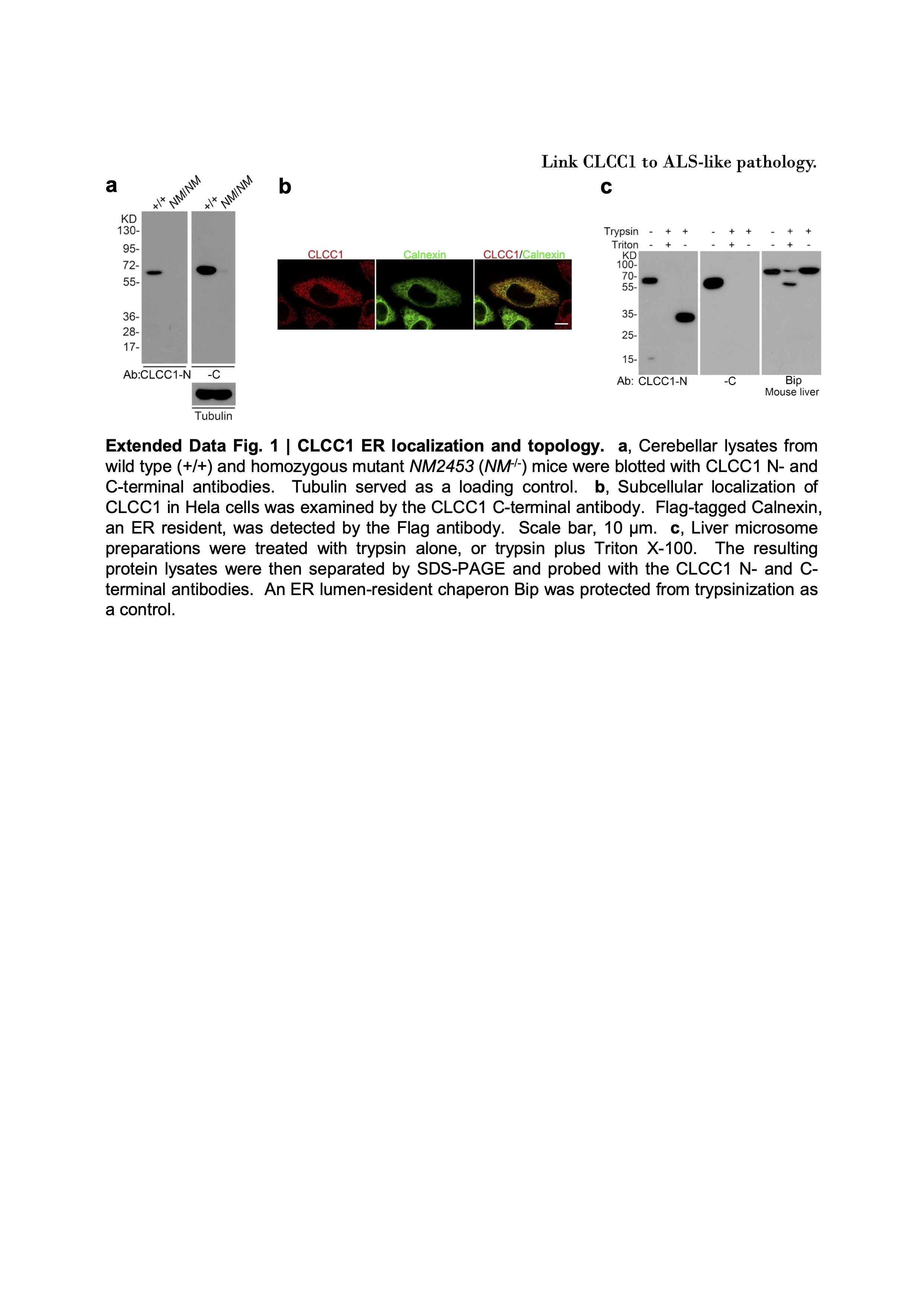
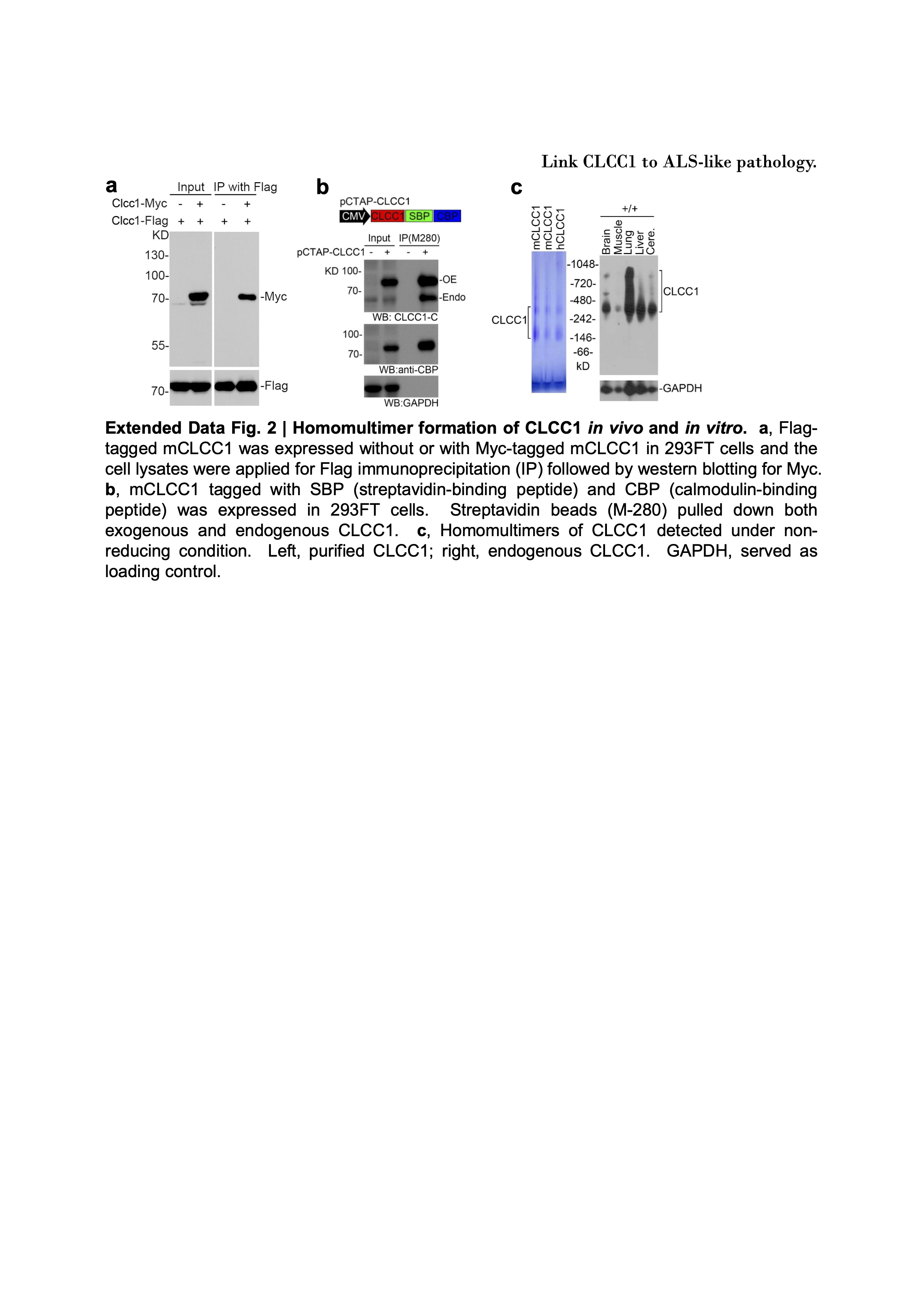
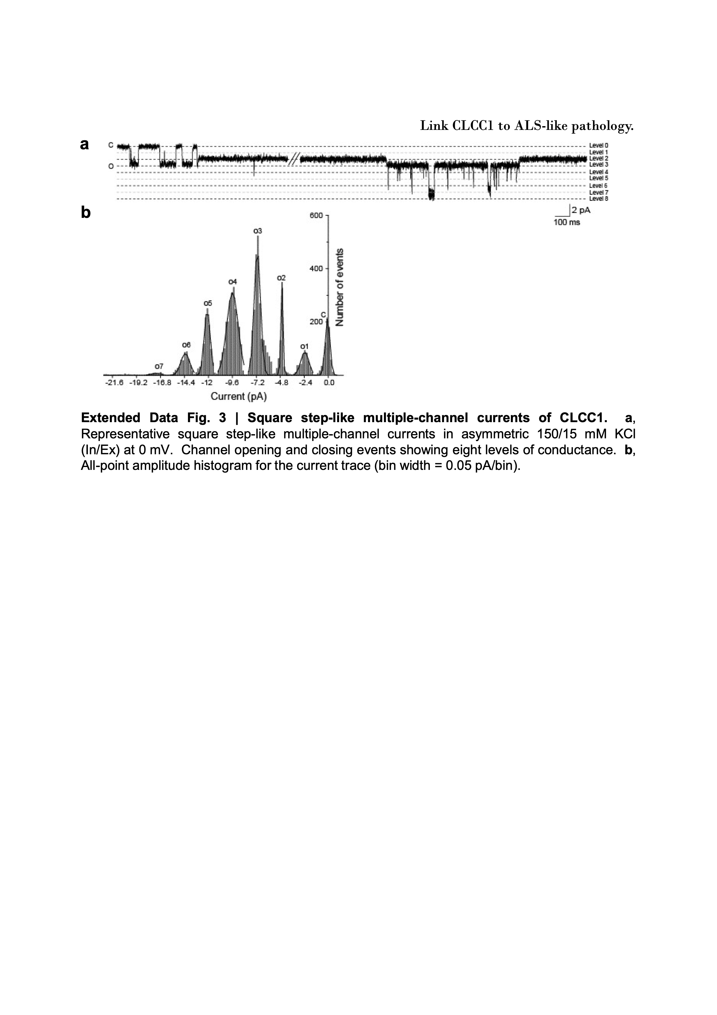
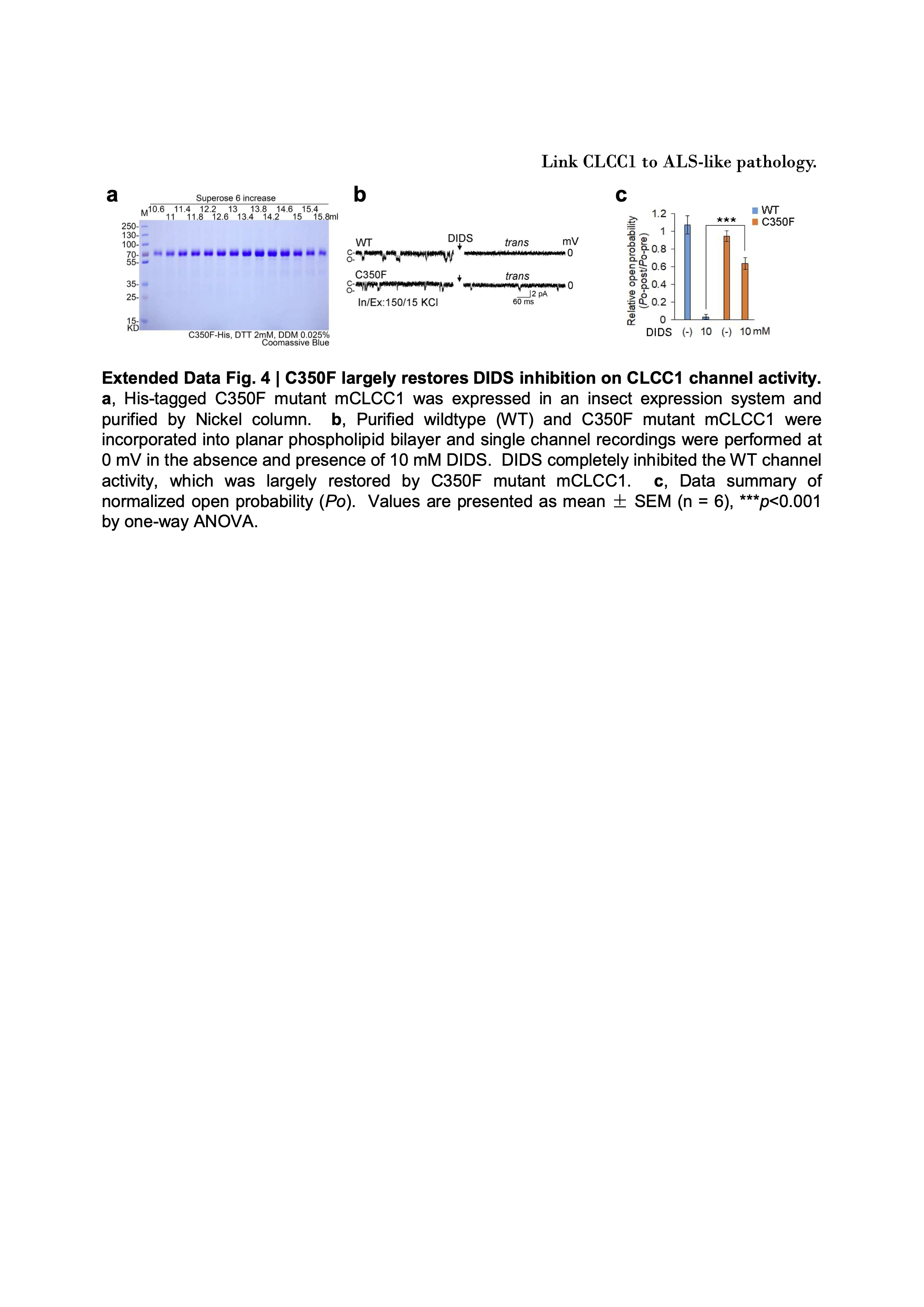
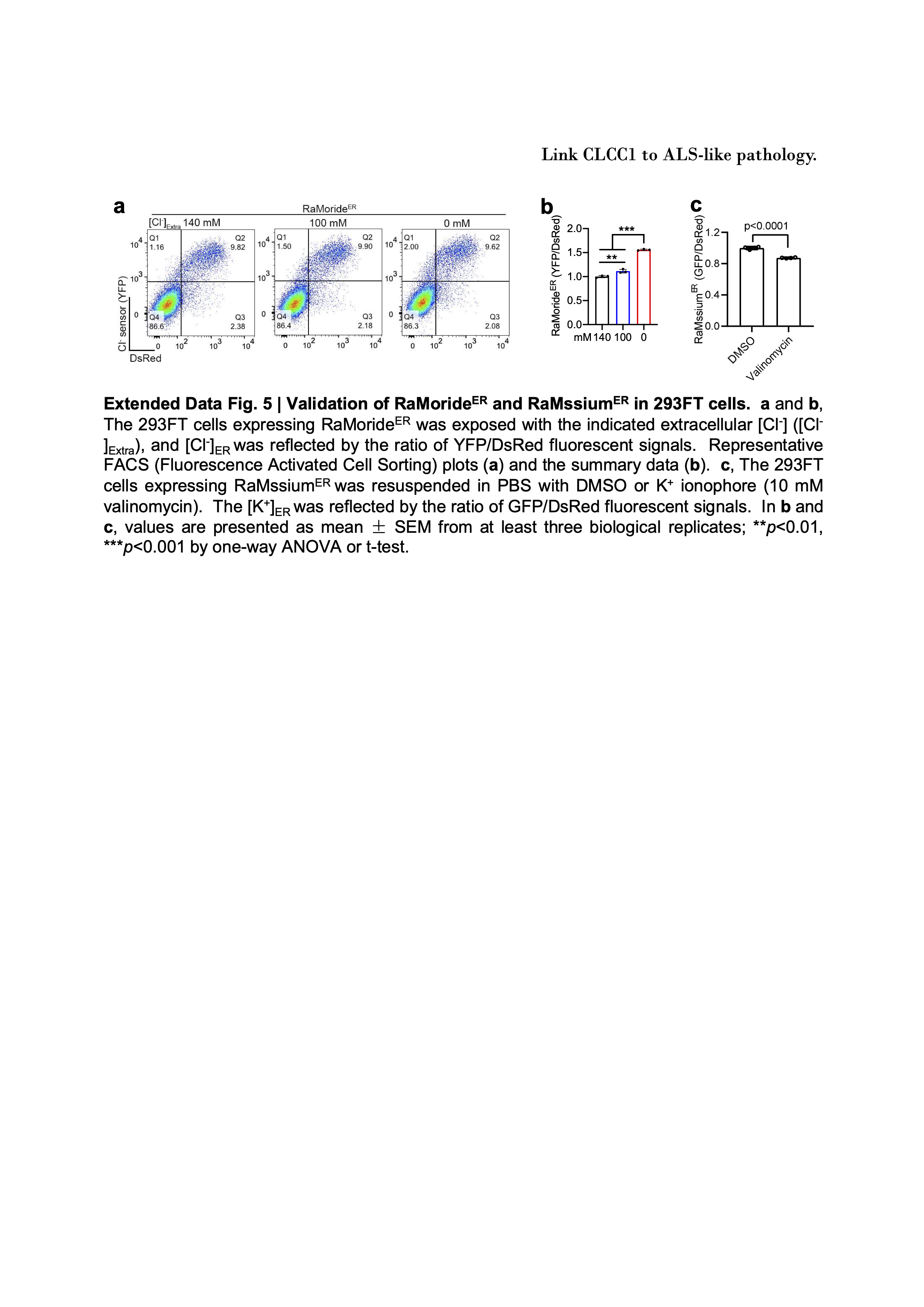
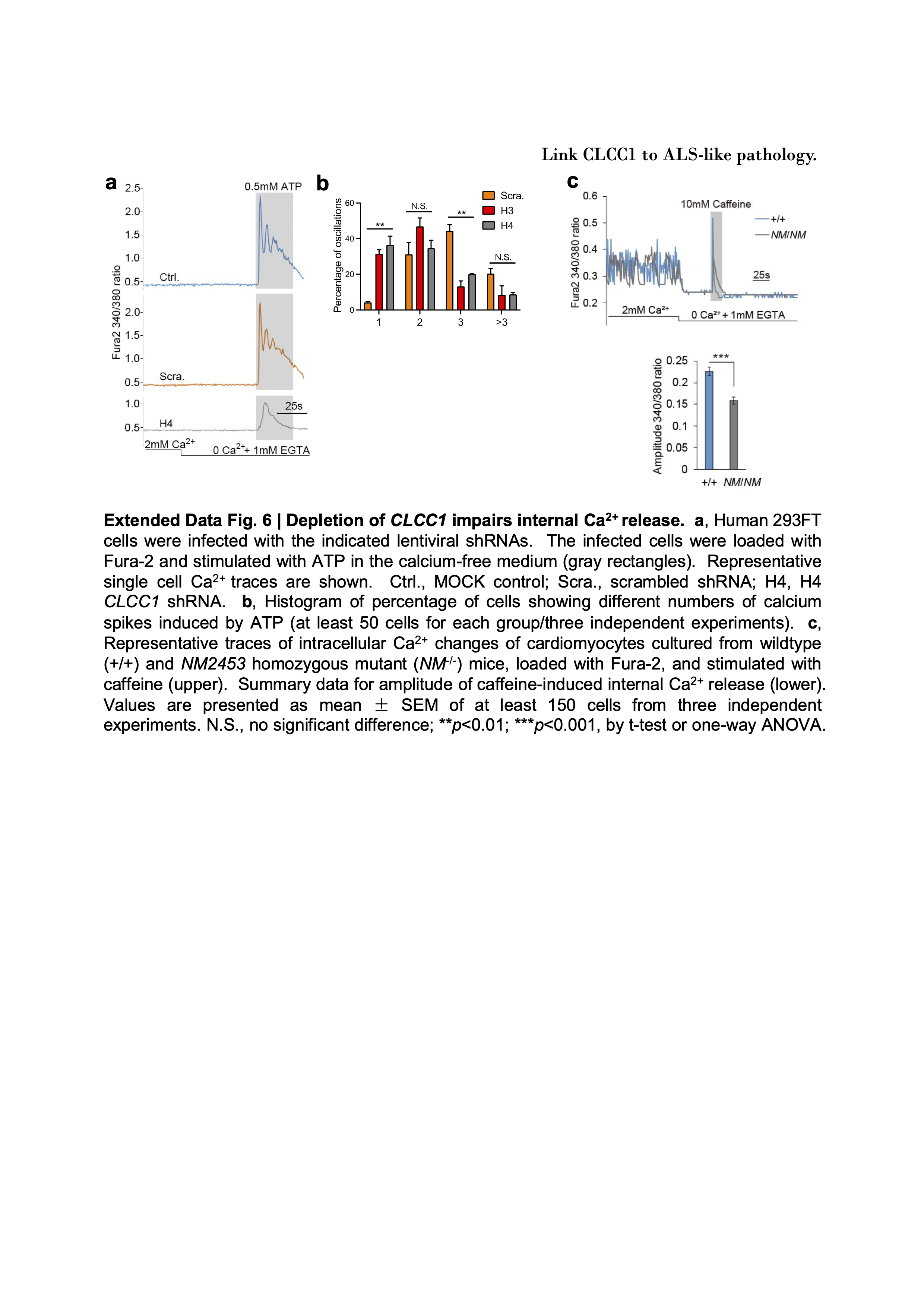
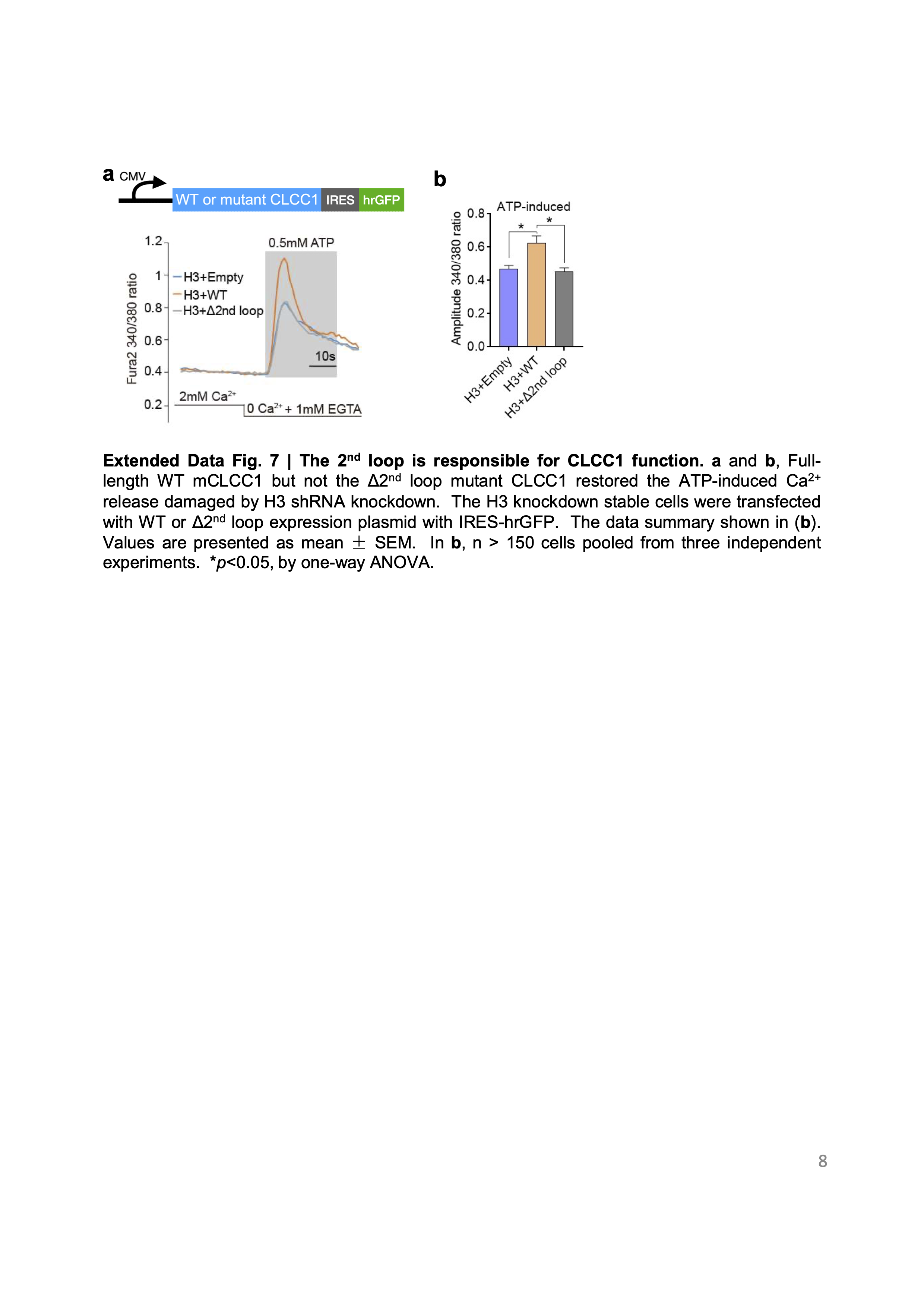
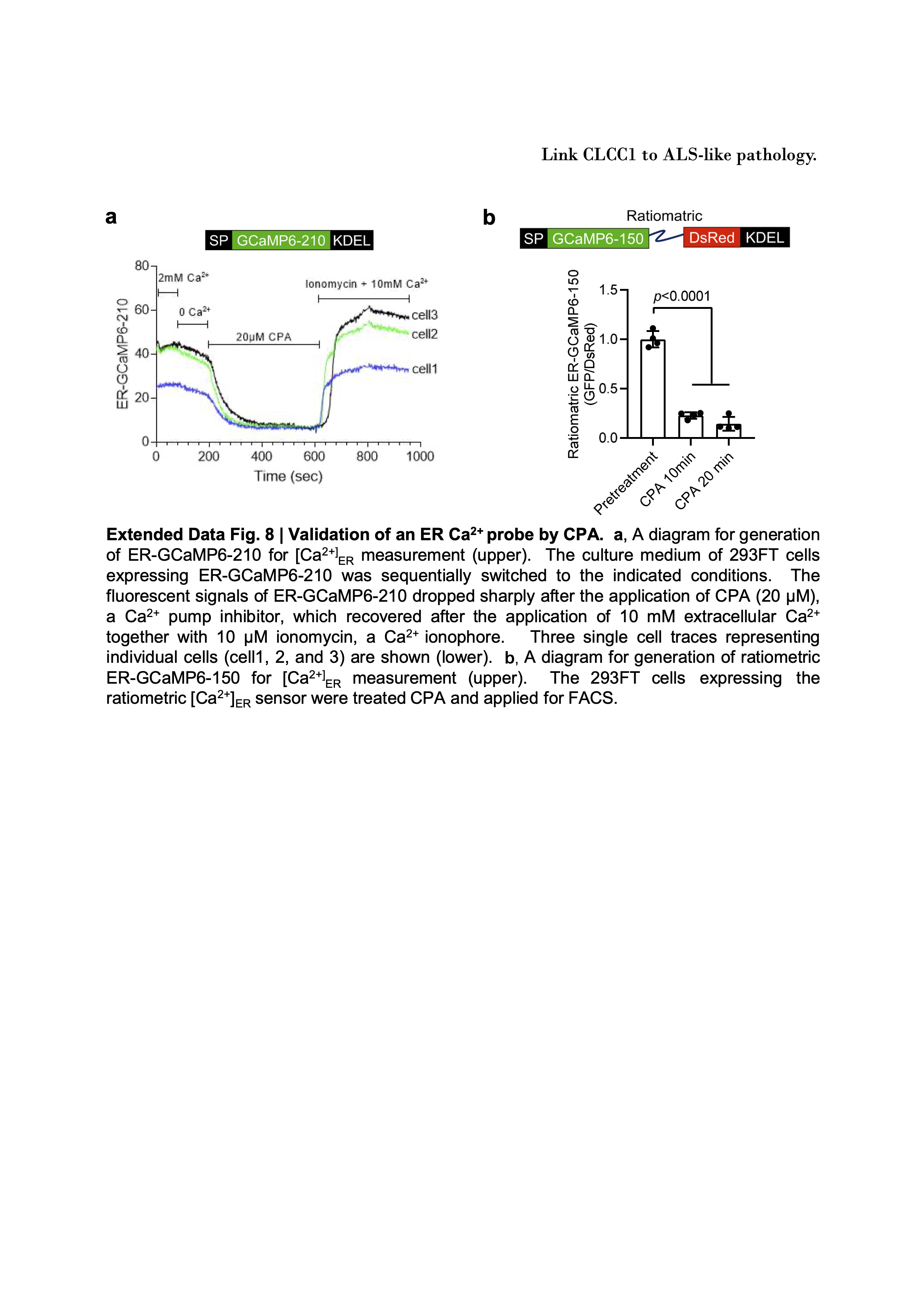
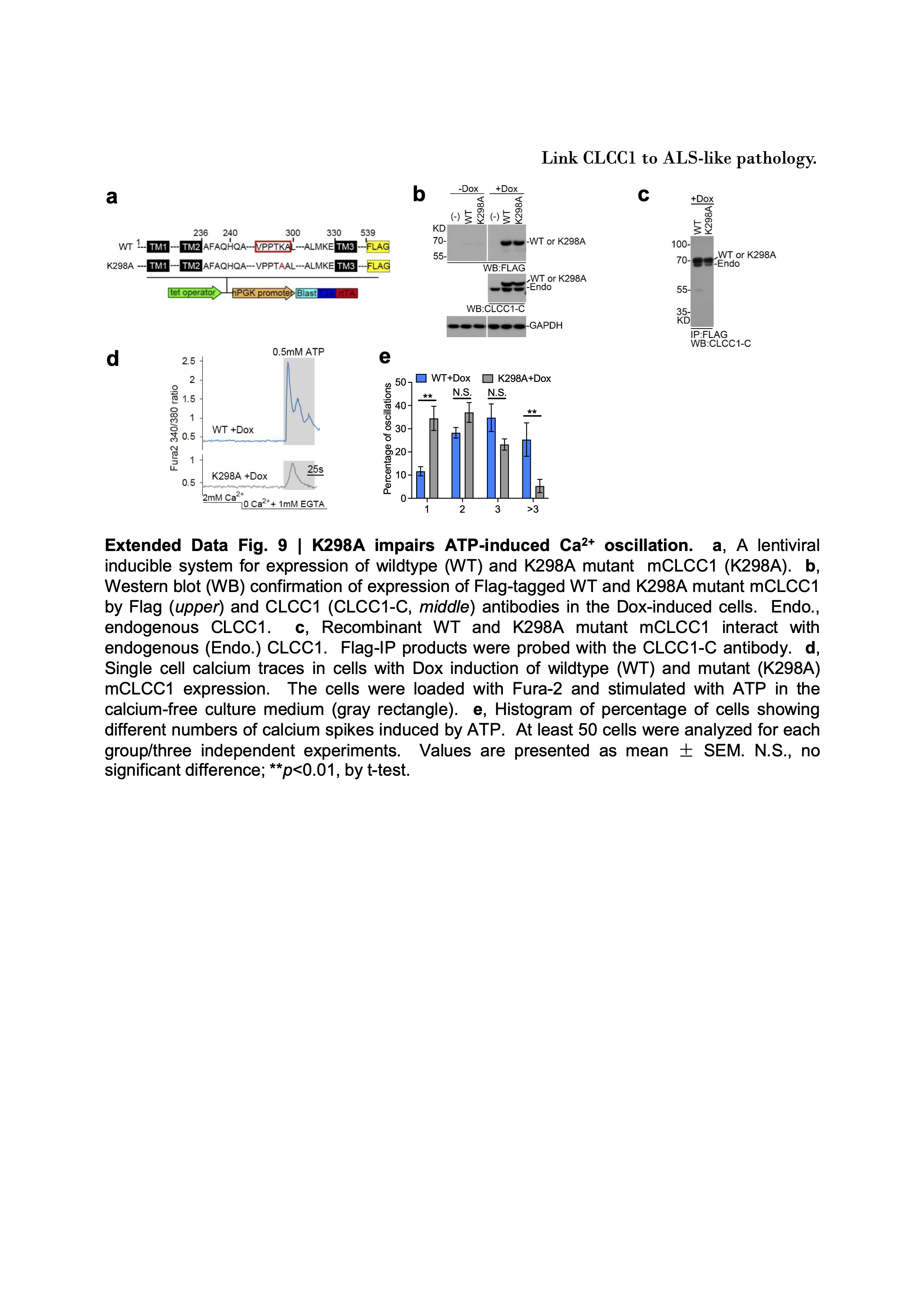
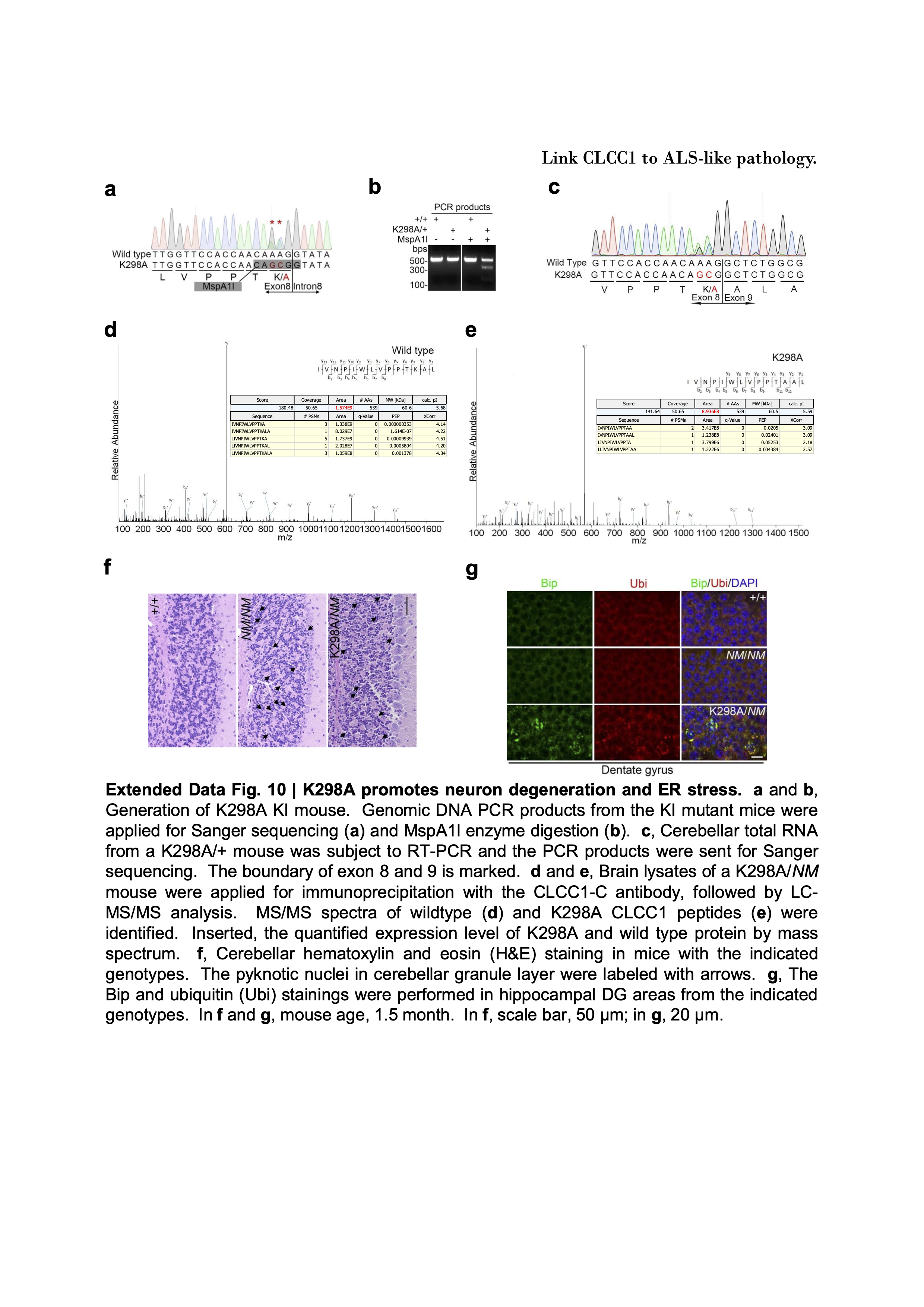
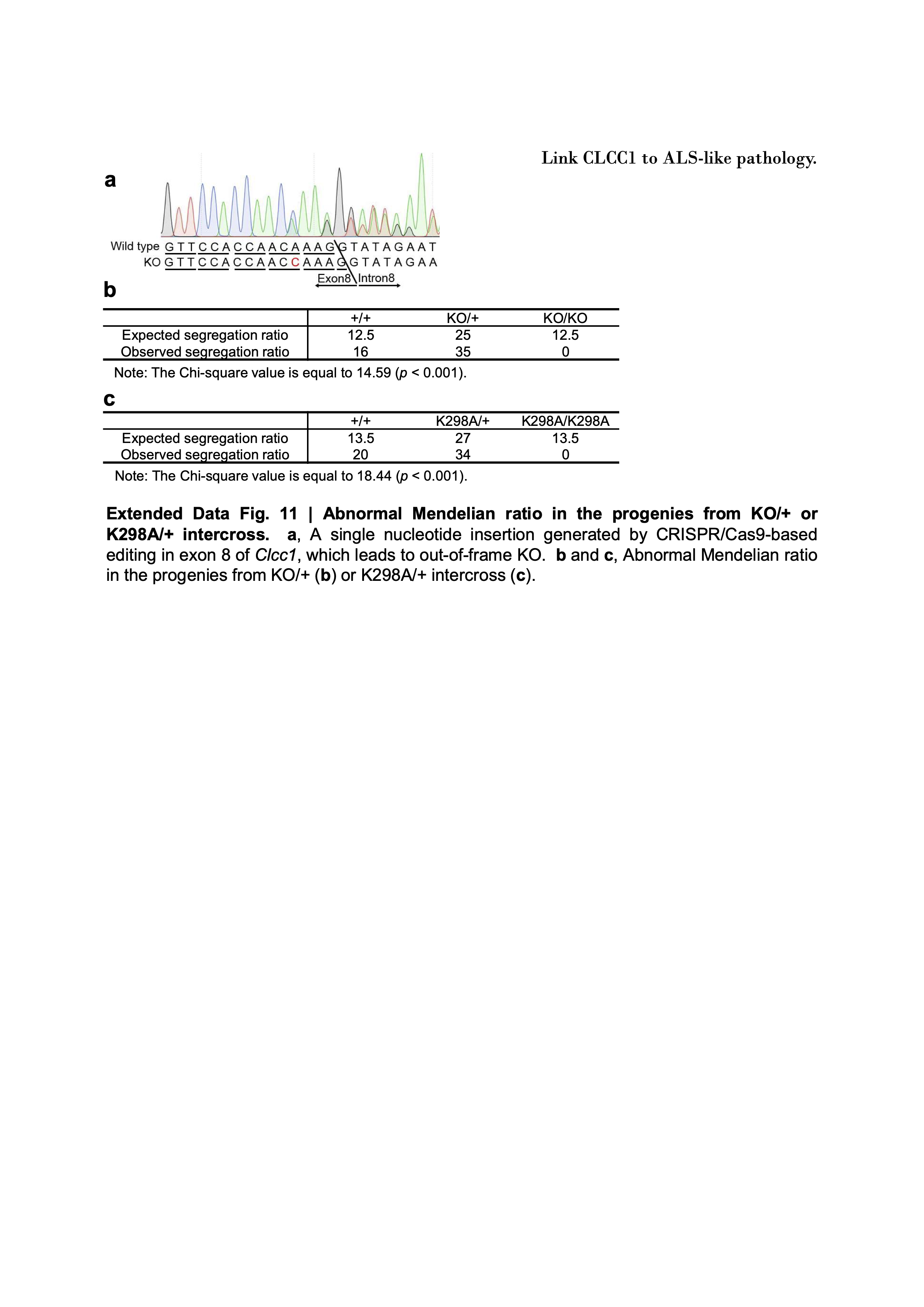
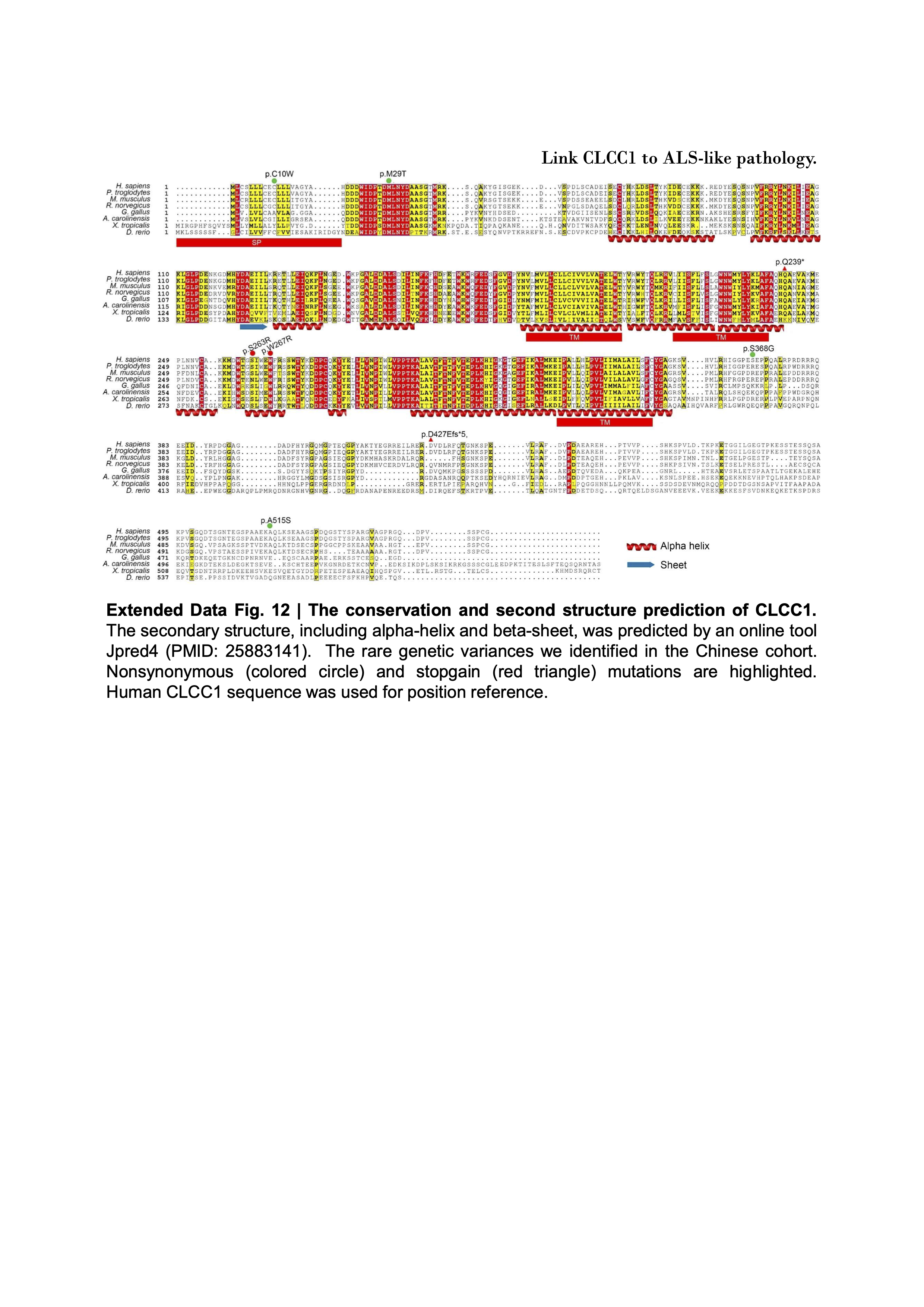
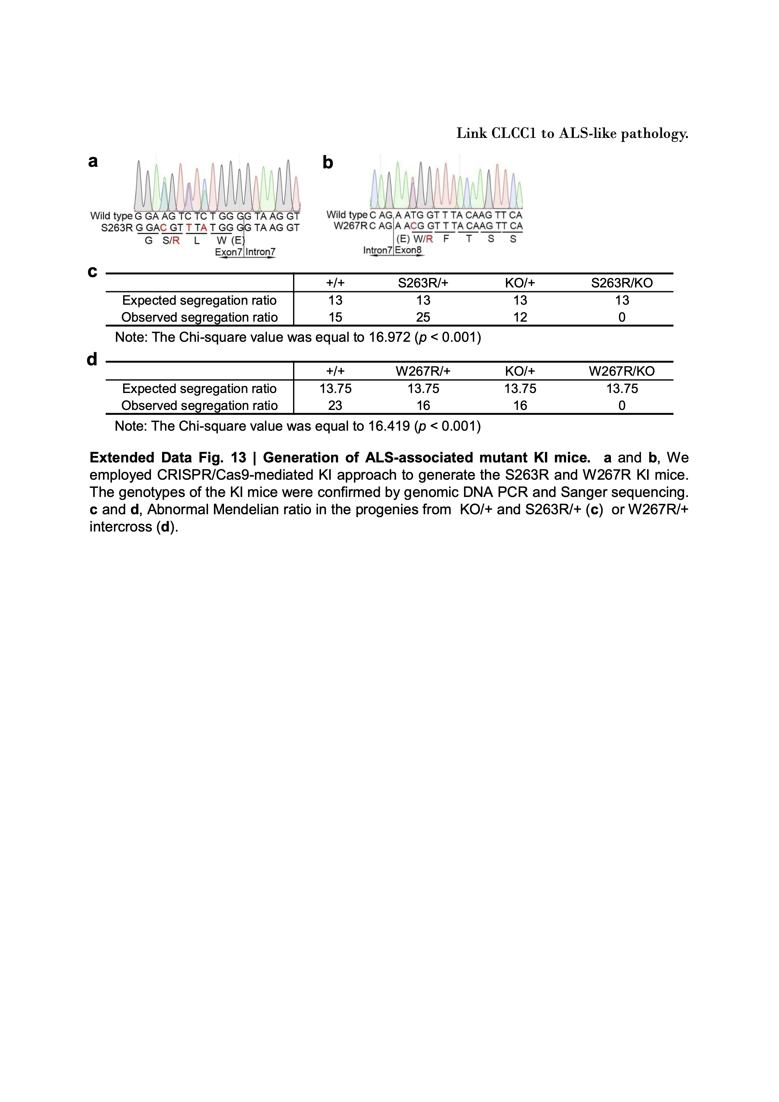
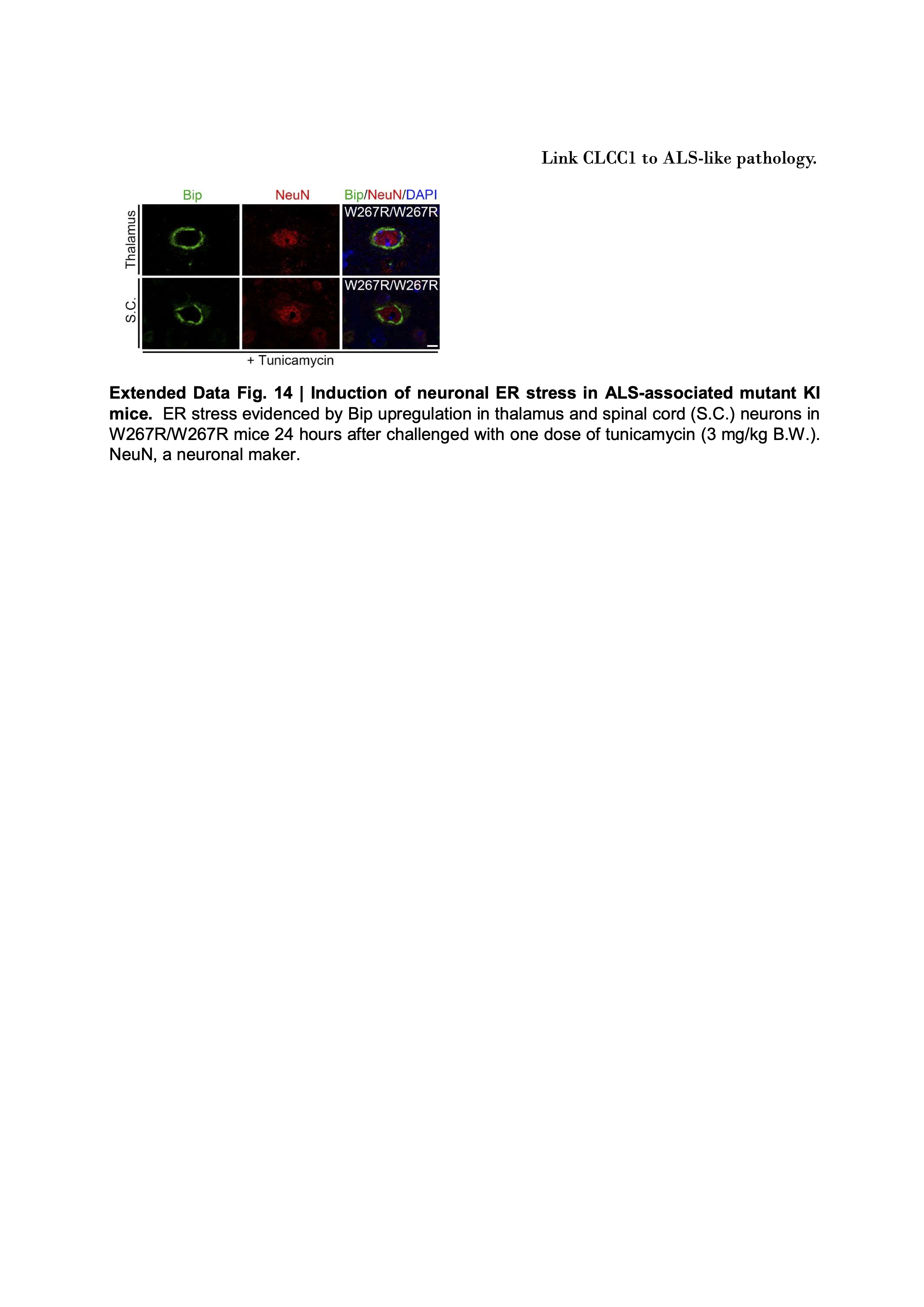
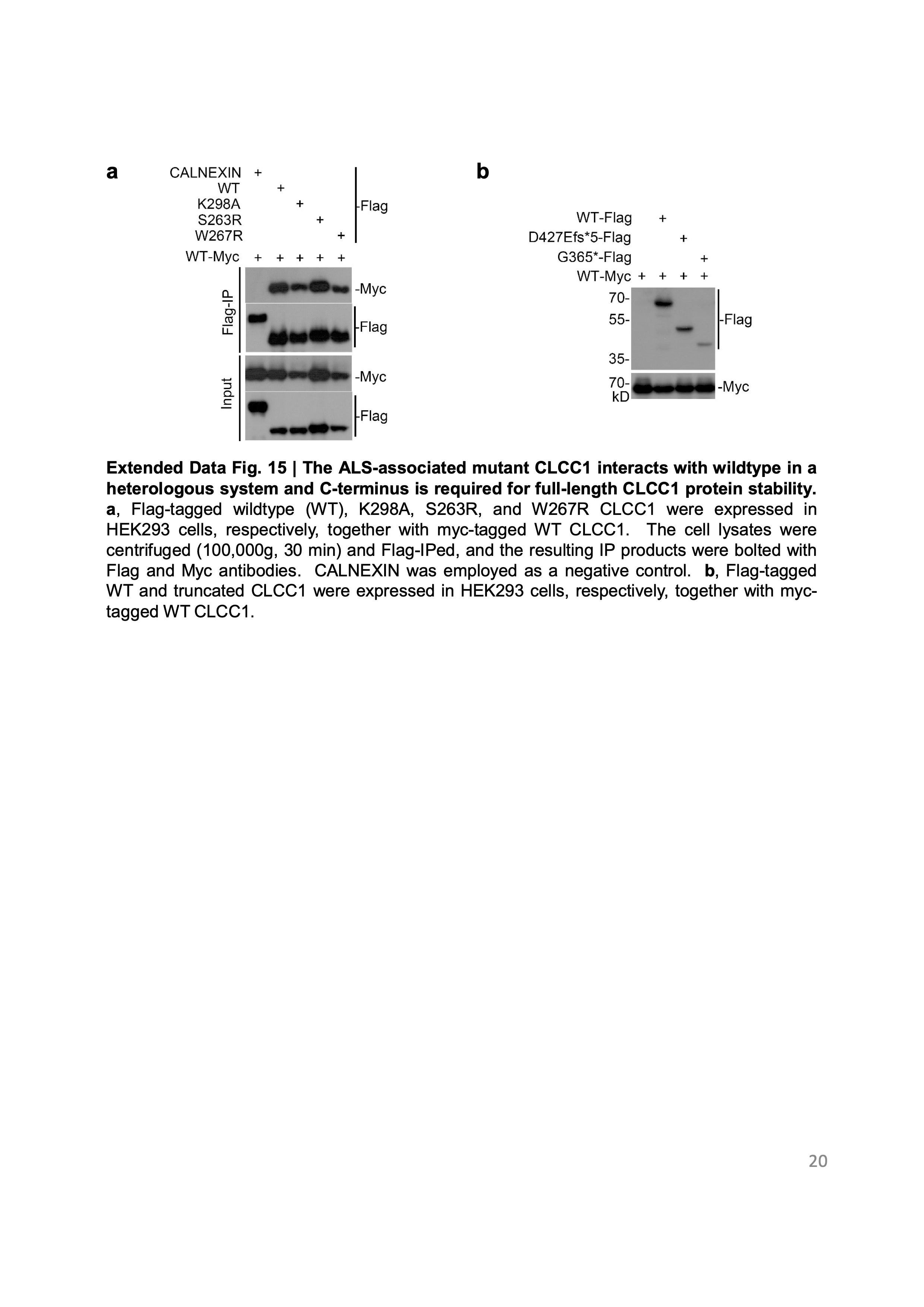
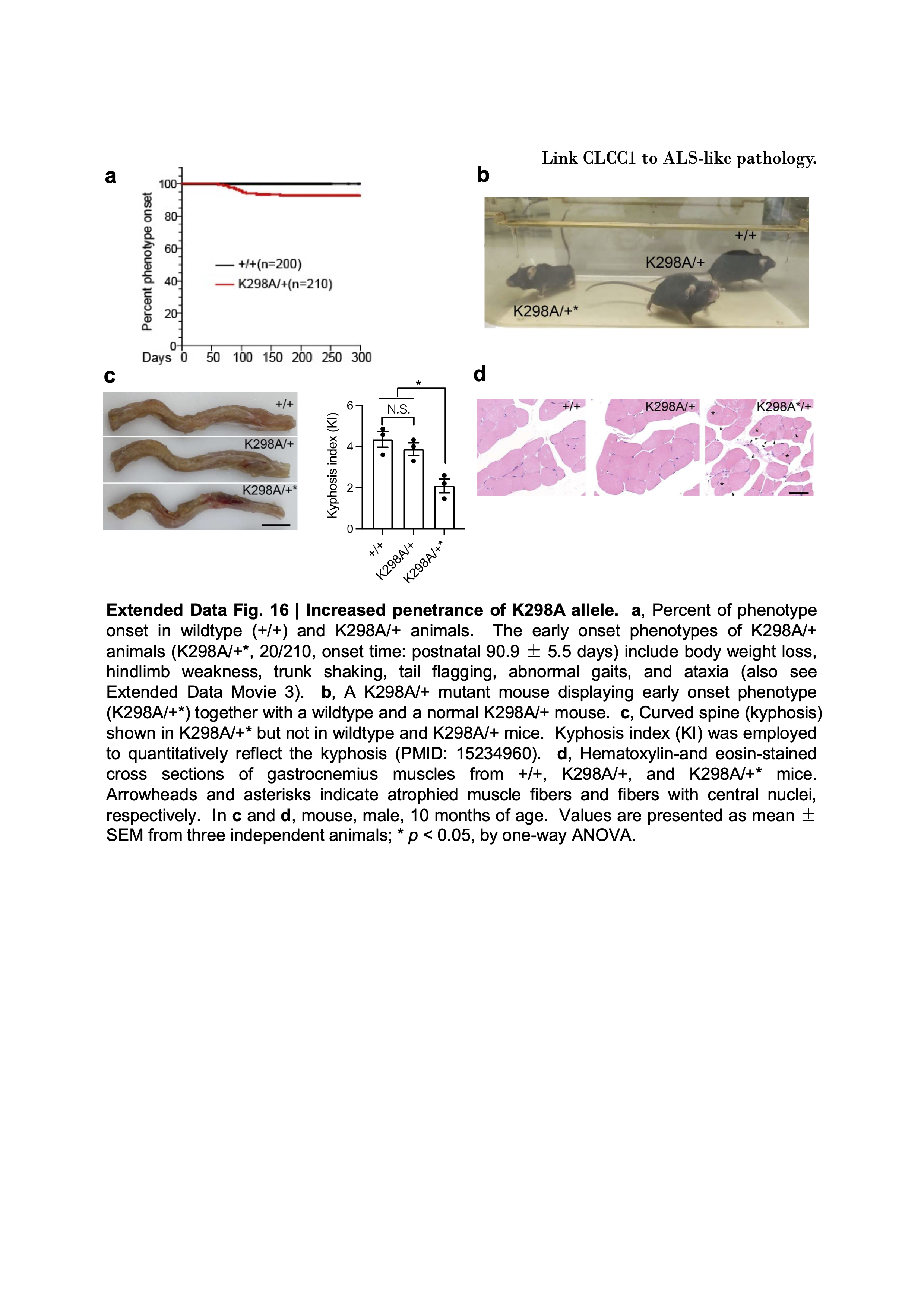
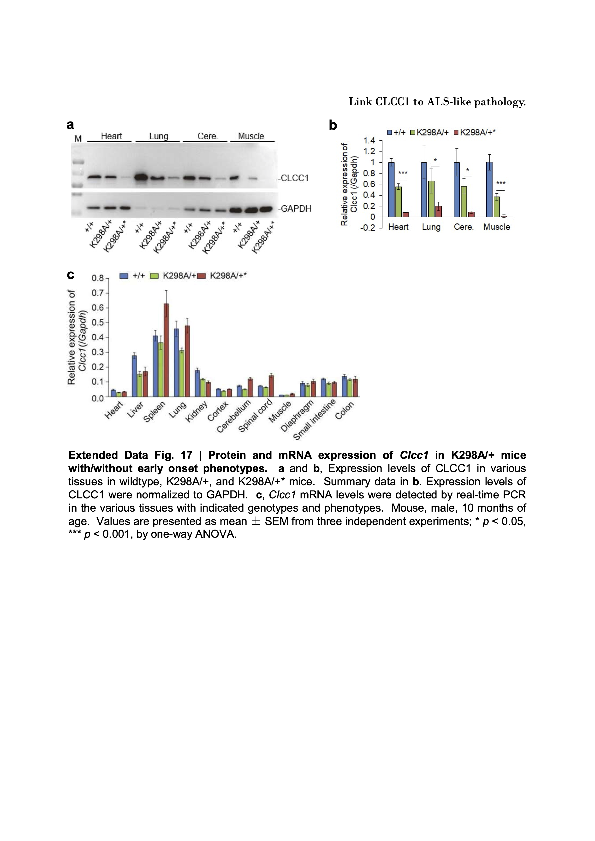
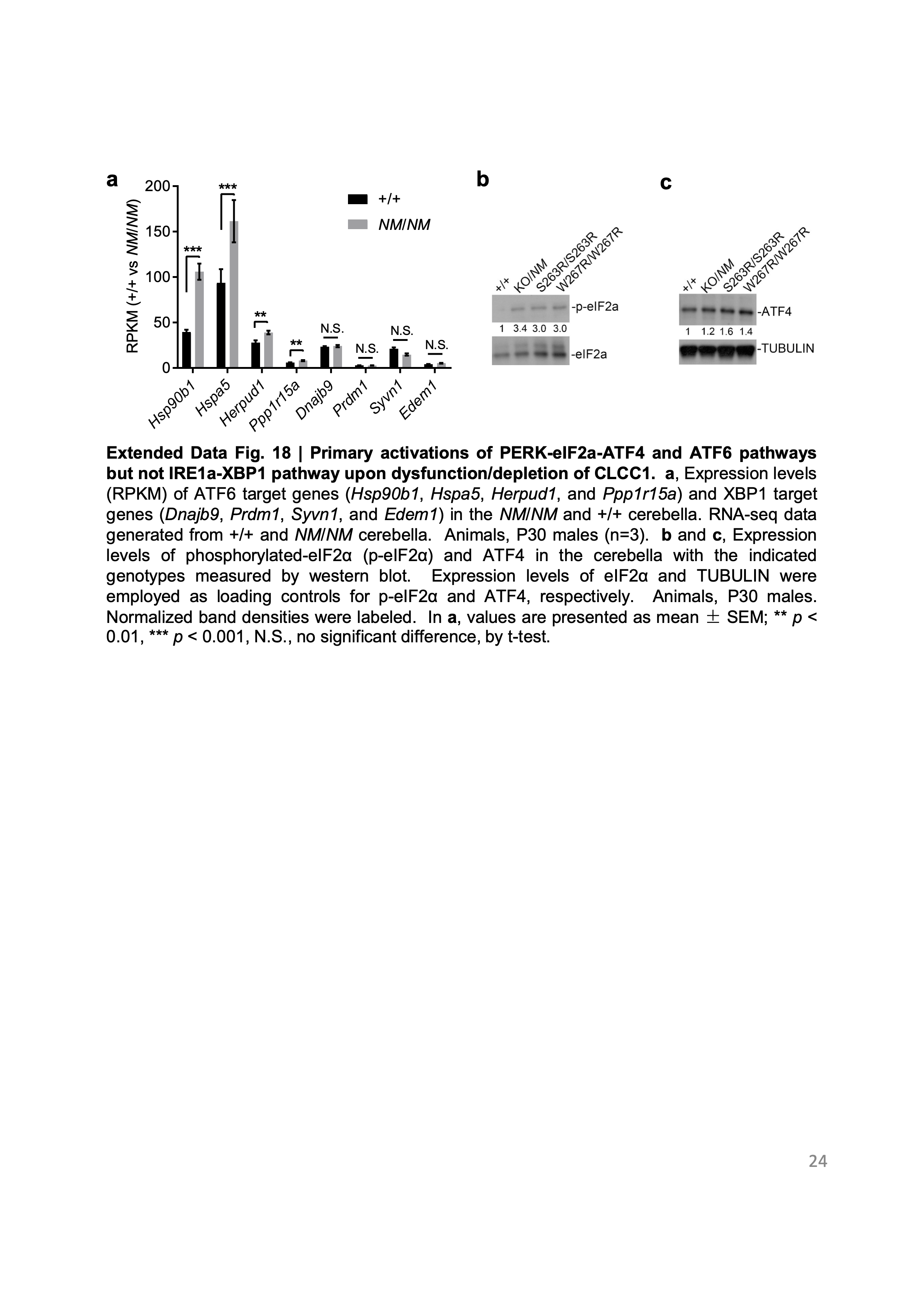
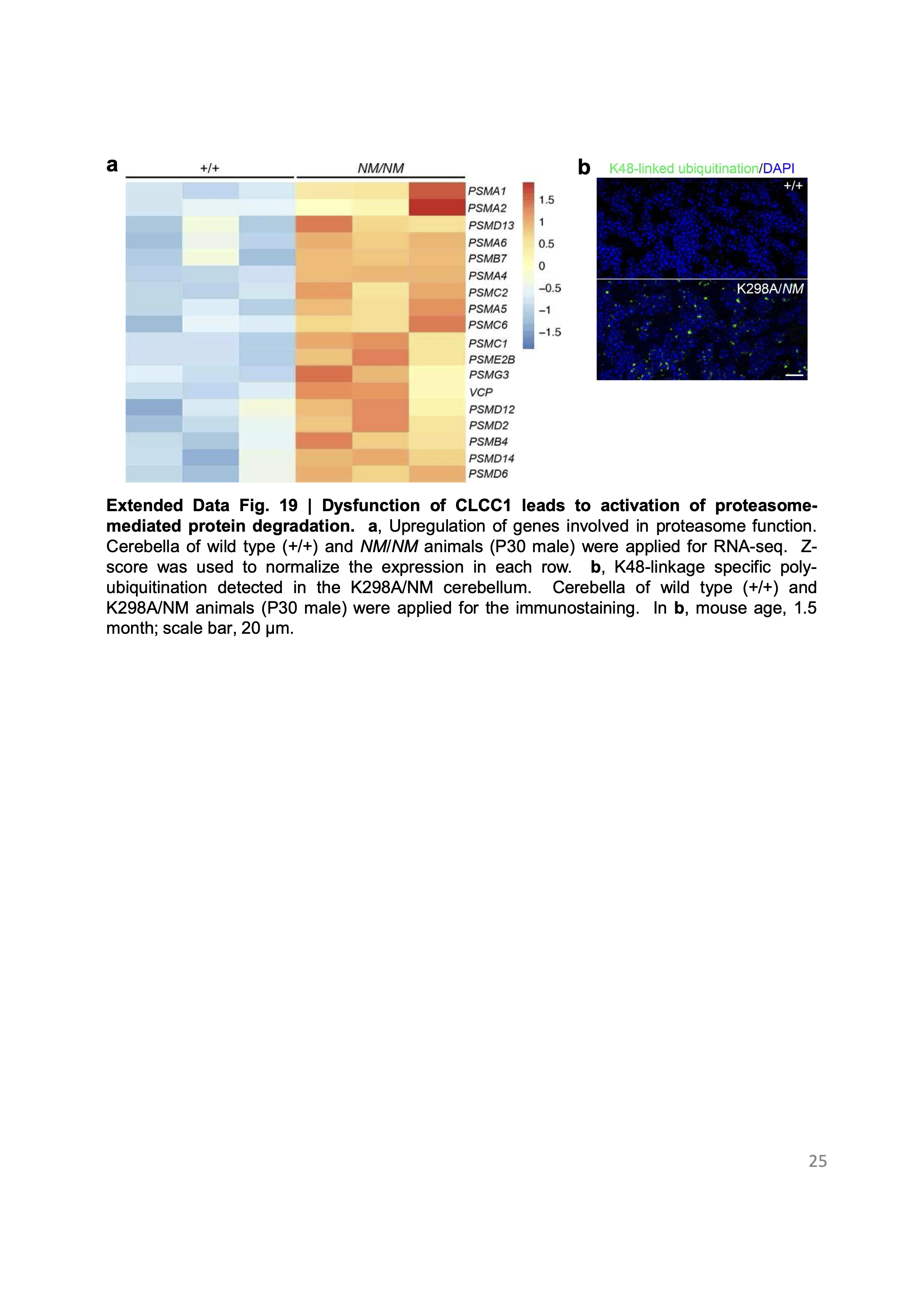
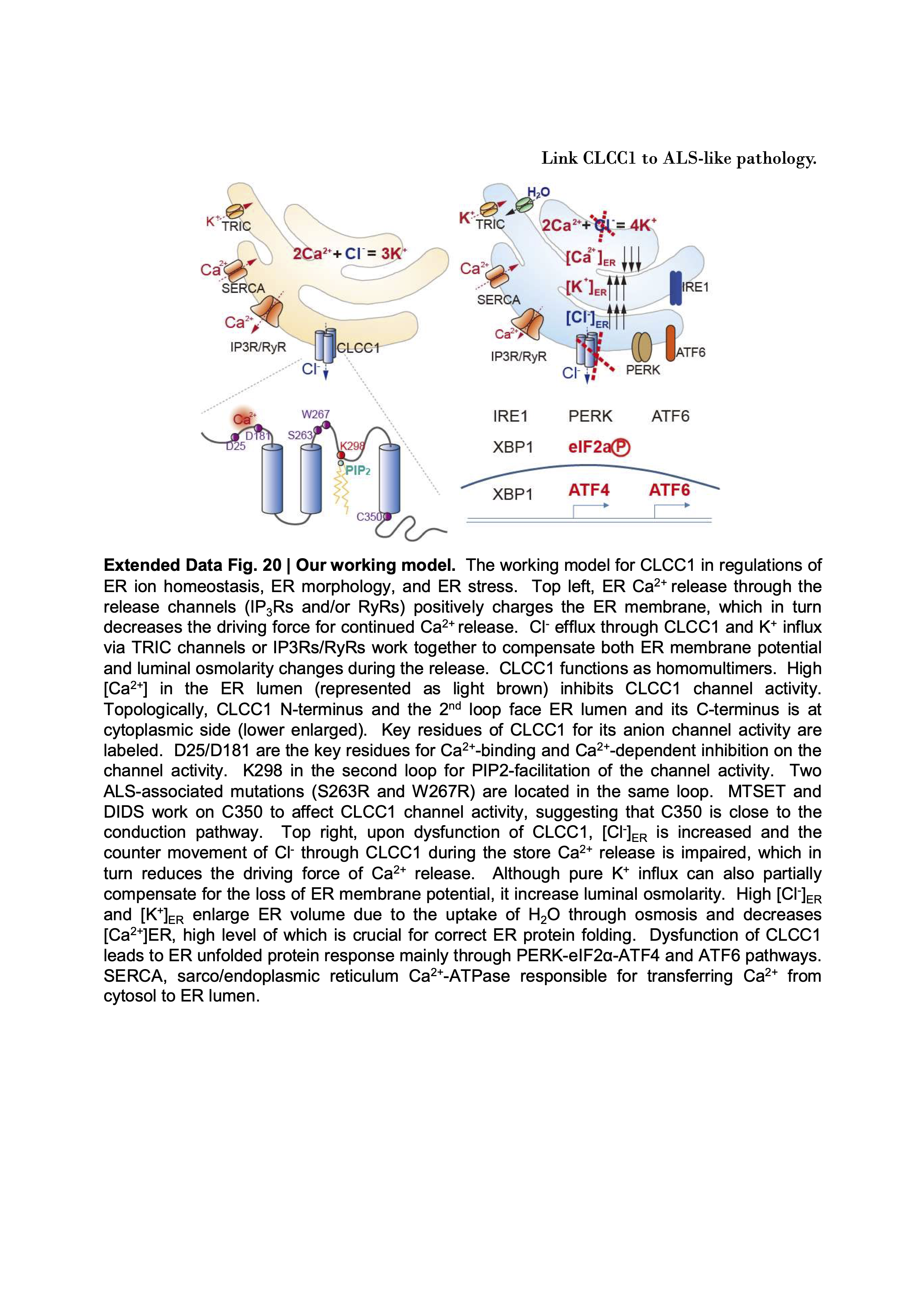
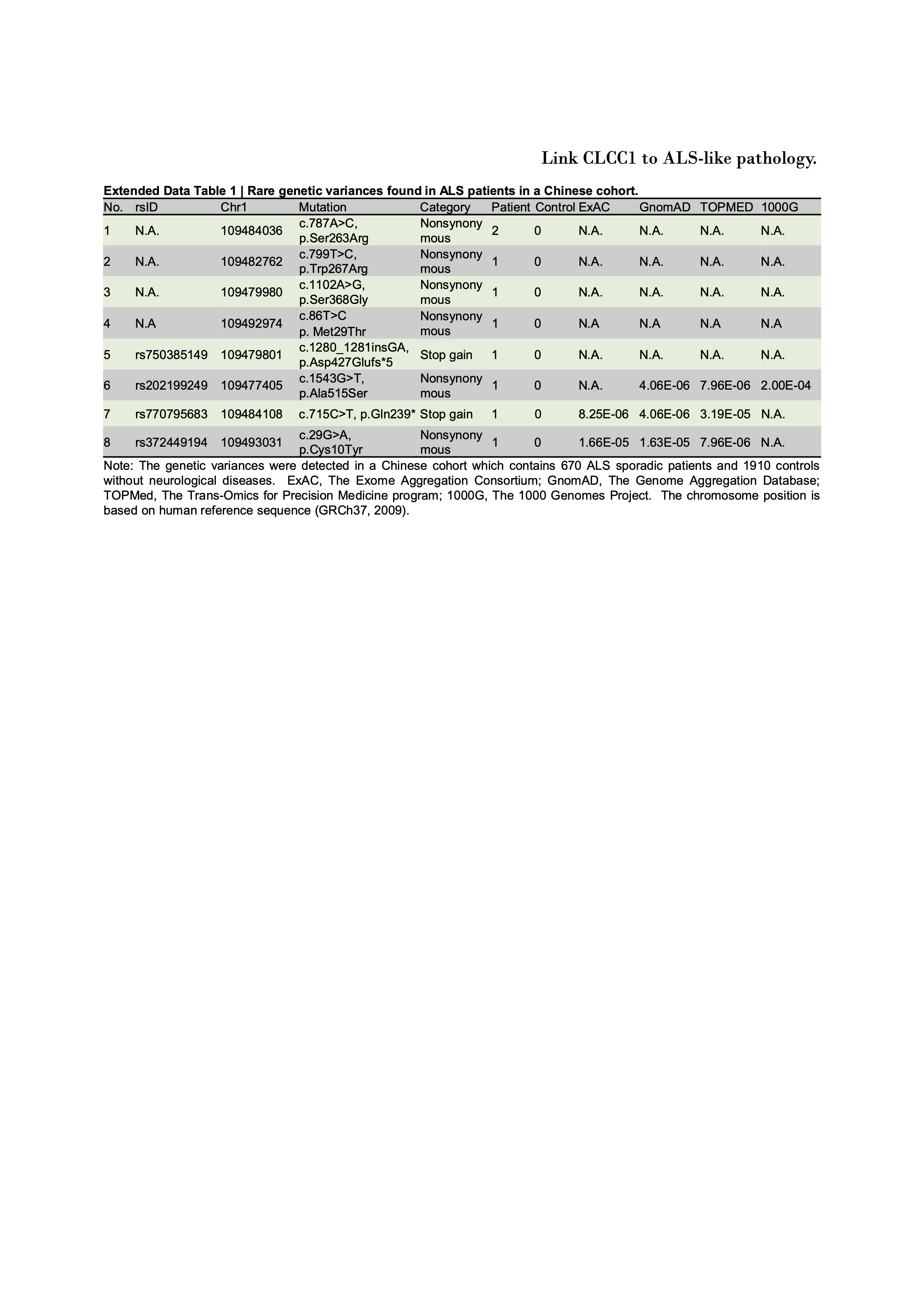
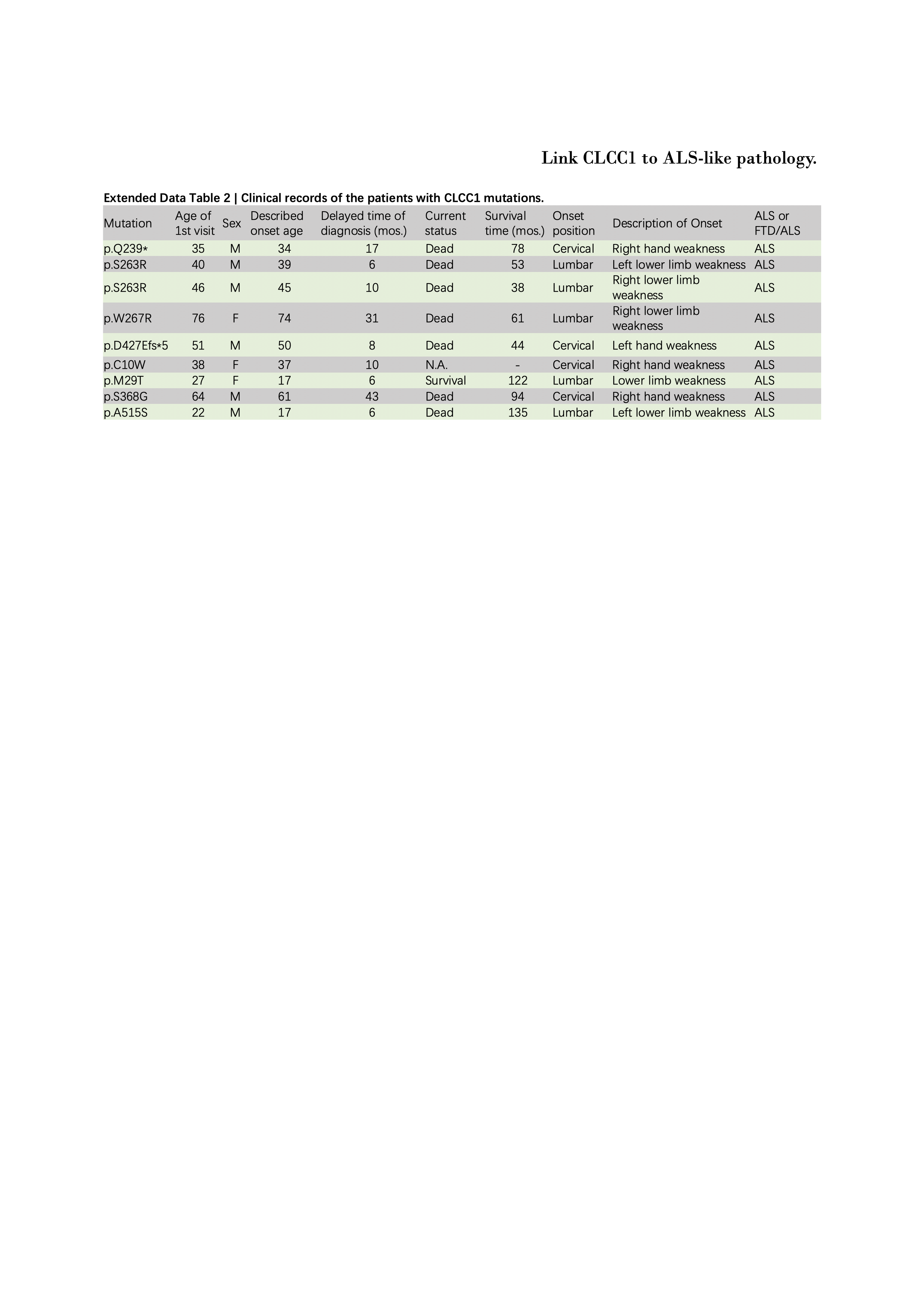