2.1 Experimental Animals
Fifty specific pathogen-free male Sprague-Dawley (SD) rats(250±30g,6-8weeks) were purchased from Experimental Animal Center of Liaoning University of Traditional Chinese Medicine(No. 210726211100566367,Liaoning, China), The rats were housed under 12h light-dark cycle in separate plastic cages under standard laboratory conditions, and had free access to food and water in Animal Laboratory Center of Liaoning University of Traditional Chinese Medicine. Adapting to the environment for 1 week.
2.2 Drugs
SJHG comprises 10 Chinese herbs: Reddle 6g, Paeoniae radix alba 10g, Radix bupleuri 12g, Paeoniae radix alba 12g, Ligusticum wallichii 12g, Fructus aurantii 1
2g, Tangerine peel 12g, Liquorice root 6g, Rhizoma cyperi 10g, inula japonica 12g. SJHG was prepared by Jiangyin Tianjiang Pharmaceutical Co., Ltd. (2109333). The inspection report certificates of all the herbs were provided by the company, and all the herbs met the requirements of the Chinese Pharmacopoeia (2020). Omeprazole Rat to human drug dosage conversion ratio is 6.3, So the granules needed were 11.07g/kg per day.The granules were mixed into the solution and stored at 4℃ for further use. Omeprazole Enteric Capsules, produced by Shandong Luoxin Pharmaceutical Group Stock Co., Ltd was 1.80mg/kg per day according to the same ratio of conversion.
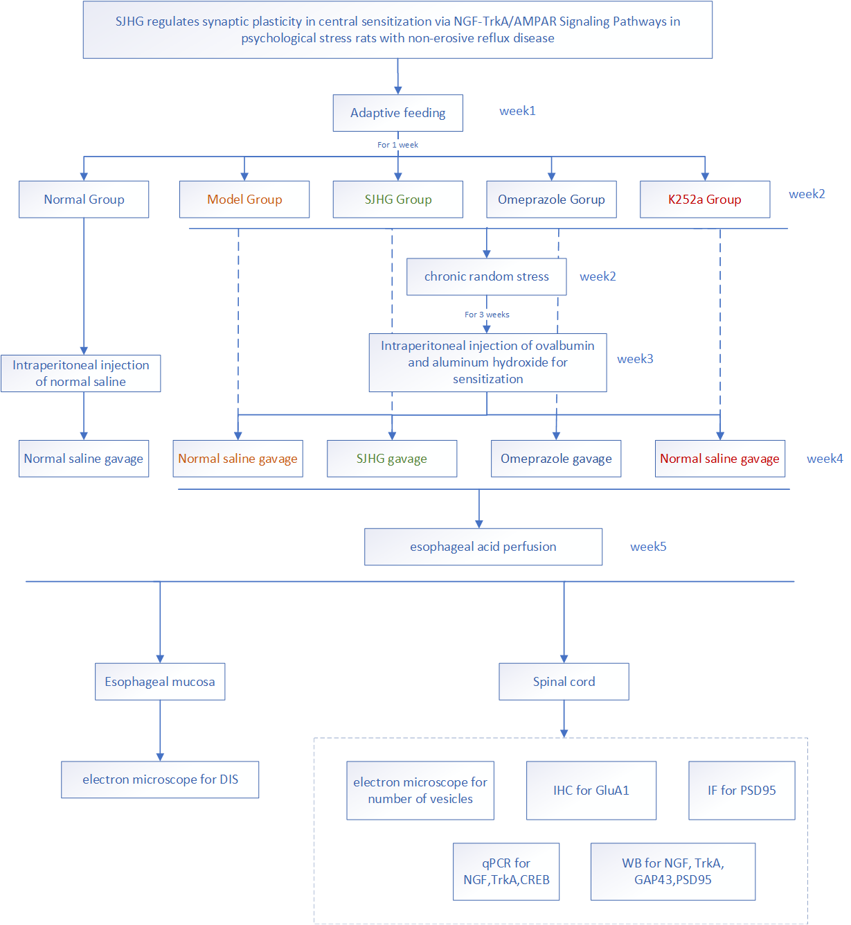
2.3 Animal Grouping
Rats were randomly divided into five groups by a random number table(10 rats in each group): including normal (healthy rats), model (model rats without any treatment), SJHG (model rats with SJHG treatment), Omeprazole(model rats with Omeprazole treatment), k252a (model rats treated with 100μmol/kg NGF/TrkA pathway inhibitor) groups, After the injection of mixture of ovalbumin and aluminum hydroxide. Rats in SJHG group and Omeprazole group were treated with drug gavage. The other groups were given 10 mL/kg of distilled water by gavaged at the same time once a day for two weeks, and K252a group were intraperitoneally injected with 100μmol/kg K252a once a day for 7 days.
2.4 Establishment of psychological stress with non-erosive reflux rats models
Other than the normal group, the other groups were subjected to chronic random stress for 21 days in the second week. One of the following eight stimuli was given randomly every day.The specific stress stimuli were as follows: ① Fasting for less than 20 hours; ②Water-deprived for 17hours; ③4℃swimming for 5minutes; ④45 degree angle cage tilt for 17hours; ⑤Trembling stress (high-speed shaking) for 10minutes; ⑥Restraint stress for 2hours; ⑦Wet litter (100g sawdust with 200ml water, 5h); ⑧Tail pinching for 2minutes. After each stressing, the rats were kept alone under normal conditions. The sequence and timing of stress should follow a random and unpredictable principle, and the same stress stimuli should not be repeated every week.
In the third week, the other groups were intraperitoneally injected with 1.5 mL of a mixture of ovalbumin and aluminum hydroxide (ovalbumin 100 mg/aluminum hydroxide 200 mg), and the rats in the normal group were intraperitoneally injected with the same volume of normal saline simultaneously. In the 29th day, except for the normal group, the other groups were treated with esophageal acid perfusion.Then the rats in the experiment were then sacrificed.ovalbumin(A8040) was supplied by Solarbio Technology Co., Ltd, Beijing, China.Aluminum hydroxide(77161) was supplied by Thermo Fisher Scientific Co., Ltd, Shanghai, China.
2.5 Esophageal Hydrochloric acid Perfusion Approach
After the rat was completely anesthetized, the abdominal and gastric walls were incised, the gastric cardia was inserted with a drainage cannula for collecting runoff solution from the esophagus. A single lumen clear vinyl tube was passed through the mouth into the esophagus. The tip of the tube was located 2cm-3cm above the esophagogastric junction. The anesthetized rats was positioned with its head elevated at a slight angle (20°-30°). Then, the tube was connected to a continuous perfusion pump. Hydrochloric acid (0.1 mol/L HCl, supplied by Yida Technology Co., Ltd., Quanzhou, China.) was perfused continuously at a rate of 10 mL/h for 50 minutes.
2.6 Sample Collection
After 10% chloral hydrate (0.3ml per100g in weight) for deep anaesthesia, the normal group rats were directly sacrificed, the other group rats were sacrificed after the acid perfusion. Then we took their spinal segments (L4-L6), and collected the lower third of esophagus (from 15mm above and 2mm below the esophageal sphincter). Some of the samples were put into fixative for TEM (Transmission EM), and some were taken into paraformaldehyde for immunohistochemistry (IHC), others were frozen for western blot (WB) and quantitative PCR (qPCR) and immunofluorescence (IF). The experiments were approved by Animal Experiment Ethics Committee of Liaoning University of Traditional Chinese Medicine (Ethical Approval Number:21000042021049) and flowchart of this experiment is presented in the supplemental file.
2.7 Testing Indexes and Methods
2.7.1 Observation of General Condition of rats.
Throughout the whole experiment, all rats' general condition was observed daily, including their mental state, activity, and fur appearance.
2.7.2 Transmission Electron Microscopy (TEM) staining of esophageal mucosa and synaptic vesicles in spinal cord.
The tissues were cut quickly within 1-3 minutes by a sharp blade.The size of tissue block should be no more than 1 mm3. Then the tissue blocks were transferred into an EP tube with fresh TEM fixative for further fixation, which was fixed at 4℃ for preservation and transportation. The embedding models with resin and samples were cut to 60nm-80nm thin on the ultra microtome, and the tissues were fished out onto the 150 meshes cuprum grids with formvar film. The esophagus and vesicles from spinal were observed under TEM.
2.7.3 Western Blot Analysis.
k252a(Lot.104529) were supplied by MedChemExpress Co.,Ltd.,Shanghai,China), Target protein was detected by antibodies: anti-NGF (Cat No.ab52918,1:1000, abeam), anti-TrkA (Cat No.Ab109010, 1:1000,abeam), anti-GAP43(Cat No.GB11095, 1:1000, Servicebio), anti-PSD95 (Cat No.GB11277,1:1000, Servicebio) subsequently incubated with secondary antibodies (Cat No. GB25301,1:5000,Servicebio). The values were corrected by reference to the value of β-actin (Cat No. GB15001, 1:2000, Servicebio) and the levels of target protein were analyzed by using Image J. The values were corrected by reference to the value of and the levels of target protein were analyzed by using AlphaEase FC.
2.7.4 Quantitative real-time PCR analysis.
RT-qPCR was conducted as previously described (10). Briefly, the RNA was reverse transcribed to cDNA using the Servicebio® RT First Strand cDNA Synthesis Kit according to the manufacturer's protocol. qPCR was performed using heat-activated SYBR Premix EX Taq DNA polymerase in a TaqMan ABI 5700 Sequence Detection system (Applied Biosystems; Thermo Fisher Scientific, Inc.). β-actin served as internal control. The sequences of the primers are listed in able1. PCR amplification was performed in three stages: Pre-denaturation at 95℃ for 10 min, denaturation at 95℃ for 15 sec and annealing, extension at 60℃ for 30 sec for 40 cycles. A melting curve analysis was performed between 65℃and 95℃, with a 0.5℃ raise in temperature every 5 s. The fluorescence signal was recorded every 0.5 ℃. Primer sequences were listed in Table 1.
Table 1
Primer sequences in q-PCR analysis
|
Primer name
|
Primer sequences(5'-3')
|
Fragment length
|
|
β-actin
|
Forward:TGCTATGTTGCCCTAGACTTCG
|
240
|
|
Reverse:GTTGGCATAGAGGTCTTTACGG
|
|
NGF
|
Forward:CATCACTGTGGACCCCAAACTGT
|
247
|
|
Reverse:GTCCGTGGCTGTGGTCTTATCTC
|
|
TrkA
|
Forward:AACAAGAAGAATGTGACGTGCTG
|
119
|
|
Reverse:TGATGCTGTTCCACGGCTT
|
|
CREB
|
Forward:CATTGCCCCTGGAGTTGTTAT
|
113
|
|
Reverse:CTCTTGCTGCTTCCCTGTTCTT
|
2.7.5 Immunohistochemical Analysis.
4 μm-sections were cut from the paraffin tissue block, and the GluA1 (Cat.No. 381355,1:200,Cellsignal). Antibody drips were prepared in a certain proportion with phosphate buffered solution (PBS) on the sections, incubated at 4°C overnight. HRP-labelled corresponding source of the secondary antibody (HRP-labeled Goat Anti-Rabbit, Cat No. G23303. 1:200, Servicebio) covered the tissue. The scanned files were collected by a tissue slice digital scanner on the immunohistochemical slices, then the image analysis system (Servicebio, Wuhan, China) automatically read the tissue measurement area, and first divide the positive grade (i): negative without staining, 0 points ; Weak positive light yellow, 1 point; medium positive tan, 2 points; strong positive tan, 3 points.
2.7.6 Immunofluorescence Analysis.
Frozen sections were used for immunofluorescence staining.The expressions of PSD95 were detected by immunofluorescence staining. Sections were incubated overnight with primary antibodies in PBS at 4℃. After washes with PBS, the sections were incubated with the fluorescent secondary conjugated Alexa Fluor-488 at room temperature for 2 hours. The cell nuclei were counterstained with DAPI (G1012,Servicebio). The slides were dried and sealed with anti-fluorescence quenching sealing tablets (G1401,Servicebio). Stained sections were examined and photographed with a fluorescence microscope (Nikon Fluorescence Microscope, Tokyo, Japan).
2.8.Statistic Analysis
The measurement data were presented as mean ± standard deviation (x¯±s) standard deviation and plotted using Graphpad Prism 8 software. The Shapiro-Wilk test was used to determine normal distribution and F test was used to evaluate the homogeneity of variance. Differences between two groups were compared using t tests. Differences between multiple groups were performed using analysis of one-way analysis of variance (ANOVA), or nonparametric test was used. All data were processed with SPSS 25.0 (provided by IBM, USA) software. P<0.05 was considered as statistically difference. P<0.01 was considered as significant difference.