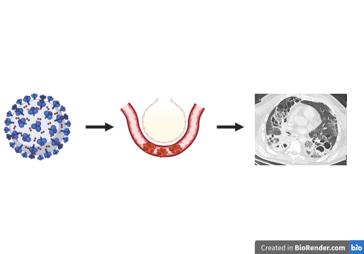
Our findings indicate that lung cavitations of variable size, at least in part a consequence of liquefying ischemic lung infarcts, contribute to lung pathology and loss of functional lung parenchyma in COVID-19 patients. These observations extend the reported spectrum of lung CT findings that is considered as typical for COVID-19, including ground glass opacities, consolidations and a “crazy paving pattern” of the lung parenchyma.15,16
In general, infectious causes and ventilator induced lung injury are recognized as the main etiologies of lung cavities in critically ill patients.17 Ischemia is less commonly considered as a cause, although cavitations are described in up to 32% of patients with pulmonary embolism and are a common finding in patients suffering from chronic thromboembolic pulmonary hypertension.18,19 While the precise pathogenesis is difficult to ascertain in individual cases in our study, we believe that several lines of evidence indicate that an ischemic pathogenesis rather than alternative causes play a major role.
First, several of our findings are not consistent with primarily ventilator induced lung injury. Ventilator settings were chosen to minimize lung trauma and did not differ significantly between the groups with and without lung cavities. The distribution of the cavities with a significant proportion of central lesions and involvement of the lower parts of the lung argues against ventilator induced lung injury, since from our experience one would expect mainly peripheral lesions in the upper lobes, if mechanical overdistension played the main role. The observation that a high percentage of lesions occurred in preexisting opacities also seems rather untypical for classical ventilator induced lung alterations. Due to higher compliance of the less affected regions, overdistension tends to occur preferentially in non-opaque regions of the lung. Most striking is the fact, that three of five patients, who did not need mechanical ventilation, also developed cavitations. Still mechanical ventilation might play an important role in the development of the cavitary lesions in that it aggravates the damage done by microvascular and macrovascular thrombosis. We therefore think that sticking to the principles of lung-protective ventilation is of great importance in this group of patients to minimize pulmonal destructions even if the lung compliance does not seem to be altered in every case.
Second, patients who developed cavitations did not exhibit positive cultures more often than the patients without, nor did we find typical abscess inducing species more frequently in the patients with lung cavities. However, a secondary bacterial infection of infarcted areas, transforming into cavitations cannot be ruled out.
Third, we found evidence for a prothrombotic state both in terms of coagulation parameters and thromboembolic complications. Although thromboembolic complications were evenly distributed between patients with and without lung cavities, notably pulmonary embolism was more frequent in patients with cavities (4 vs. 1). Patients who developed cavitations during the course of their disease were significantly older and had a higher BMI. Obesity has been associated with a prothrombotic state and hypofibrinolysis.20–24 The “mosaic pattern” of the lung-parenchyma that we observed in accordance with previous publications and the lung cavities strikingly resemble findings in patients with chronic thromboembolic pulmonary hypertension.15,18,19
During the time of this observational study critical care resources for COVID-19 patients were in no way compromised in our regional setting, enabling long-term ICU therapy. Together with a very large proportion of secondary referrals this might explain why others have to the best of our knowledge not yet reported similar observations. However, several other observations support the concept of impaired lung perfusion in COVID-19 patients. Ackermann et al. found a high incidence of microvascular thrombi and signs of endothelitis during autopsy of seven COVID-19 patients.6 Lang et al., using dual source computer tomography, discovered severe perfusion abnormalities in the lungs of three COVID-19 patients and postulated a significant contribution of altered perfusion to the etiology of respiratory failure in COVID-19.12
In terms of the mechanisms potentially causing pulmonary hypoperfusion, SARS-CoV-2 binds to the angiotensin converting enzyme 2 (ACE-2) receptor of alveolar epithelial cells.25 There is evidence for consecutive downregulation of ACE-2 leading to increased levels of angiotensin II.25 High levels of angiotensin II in the pulmonary circulation may lead to endothelial activation and vasoconstriction and promote a prothrombotic state.26 Hypoperfusion of pulmonary artery branches either due to vasoconstriction or due to thromboembolic occlusion will lead to increased dead space ventilation and impaired gas exchange, consistent with the ventilation pattern observed in COVID-19.5,27 Selective perfusion of the pulmonary circulation through anastomoses between the bronchial and the pulmonary circulation might further contribute to respiratory failure due to an increased right-to left shunt fraction.28
By implying a link between a prothrombotic stage, pulmonary hypoperfusion and structural lung changes, our data add to the considerations for therapeutic anticoagulation in COVID-19 patients. Interestingly in this regard Wang et al. recently reported a positive impact of tissue plasminogen activator treatment on oxygenation in a case series.29 An association of higher therapeutic targets of systemic anticoagulation and improved survival has also been reported.11,30
Our study has several limitations. Being retrospective and non-interventional it can only be hypothesis-generating. It is monocentric and focusses on severely ill patients, many of whom received ICU treatment for several weeks and our findings may not be generalizable to less severely ill patients. The etiology of lung cavitations is possibly heterogeneous and multifactorial. Finally, the number of autopsies supporting our interpretation is small and although all point to the exact same pathogenesis of the cavitations we do not know whether the same results would have been found in the lungs of the other patients of the cohort.
In conclusion, we found that cavitating lung lesions occur frequently in severely ill COVID-19 patients and provide evidence that pulmonary hypoperfusion and occlusion of pulmonary arteries plays an important role in the pathogenesis of these lesions.