Despite consistent progress in the diagnosis and management of PC, few breakthroughs for effective biomarkers and treatment strategies have emerged (19). Identification of the biological and molecular mechanisms as well as discovery of timely diagnostic, prognostic, and therapeutic biomarkers for PC is therefore necessary (20). Various studies have attempted to identify biomarkers and construct predictive models to diagnose or predict the survival of PC. Cheng et al. identified diagnostic and prognostic biomarkers for PC by a comprehensive analysis, which may promote proliferation and migration of PC cells (21). Wu et al. developed a nine-gene signature based on GEO and TCGA datasets and constructed a nomogram combining the gene signature and clinical prognostic features to predict OS in PC (22). Our present study provides a different approach to selecting candidate biomarkers and establishing a diagnosis and survival prediction model. To screen potential biomarkers that may enable diagnosis and prognostic prediction for PC, RankProd and GA-ANN were used to construct a model. Finally, four DEGs (LCN2, SLC6A14, SPOCK1, and VCAN) were identified, and a four-gene signature was developed by our data processing system. Both AUCs and Kaplan-Meier analyses of the gene signature for OS and DFS showed a stably high value for diagnosis and prognosis of PC.
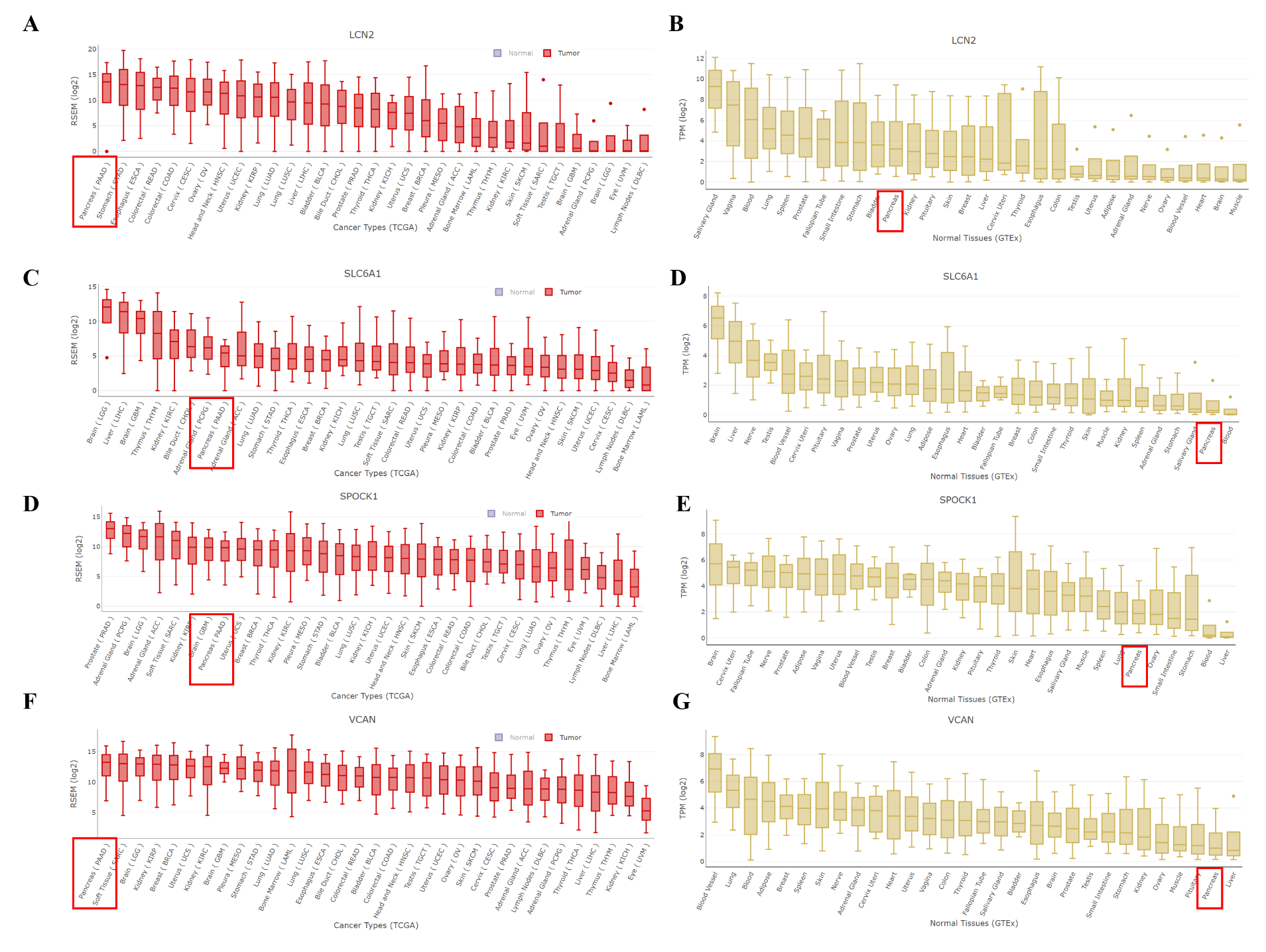
In the current study, the four candidate genes LCN2, SLC6A14, SPOCK1, and VCAN were selected for further study. Studies have shown that these four genes are vital in cancer diagnosis and progression, especially in PC. The role of LCN2 in PC was contradictory. Its expression is increased in pancreatic neoplasia, and this up-regulated level is correlated with malignant progression to PC (23–25); the increased expression of LCN2 has also been observed in various mouse models of PC (26). However, LCN2 depletion was also found in poorly differentiated PC (mesenchymal-like) and considered to be essential for invasion and metastasis (27). This down-regulation may be brought about by the activation of the EGFR signaling pathway, which inhibits E-cadherin and epithelial-to-mesenchymal transition (EMT) (28). LCN2 is also reported to inhibit angiogenesis and cause hypovascular conditions in tumor microenvironment (29). Thus, a therapeutic strategy involving the inhibition of LCN2-induced hypovascularity may potentially enhance the delivery of chemotherapeutic drugs and improve treatment effectiveness. As a member of the SLC6 family, SLC6A14 is a Na+- and Cl−-dependent solute transport molecule that activates the transport of neutral and basic amino acids (30). Previous studies have revealed that the expression of SLC6A14 is increased in cervical cancer, colorectal cancer, breast cancer, as well as PC (31). In our study, we also confirmed its increased expression in PC tissue and its correlation with poor survival of PC patients. It has been shown that blockade of SLC6A14 with either α-methyl-L-tryptophan (α-MT), a pharmacological inhibitor, or shRNA-mediated gene silencing causes amino acid starvation, inhibits the mTORC1 signaling pathway, and decreases PC cell growth and proliferation in vitro and in vivo (30). Thus, α-MT exhibited convincing specificity and potency as a pharmacological blocker of SLC6A14 and drug target for PC therapy. Gemcitabine-based chemotherapy is the main treatment for PC patients with or without surgery. Resistance to gemcitabine is a growing challenge to the effective treatment of PC because of the down-regulation of drug transporters SLC29A1 (ENT1) and SLC28A1 (CNT1) (32, 33). Because the expression of SLC6A14 is increased in PC, amino acid-based prodrug forms of gemcitabine could be used as substrates for SLC6A14 to enhance the chemotherapeutic sensitivity of gemcitabine in this form of cancer. SPARC (Osteonectin), Cwcv and Kazal like Domains Proteoglycan 1 (SPOCK1), one of the Ca2+-binding proteoglycan family members, was shown to be highly expressed in several cancer types (34). Studies have indicated that SPOCK1-mediated EMT regulates proliferation and invasion in various malignancies (34). A recent study has shown that SPOCK1 induces EMT to promote PC metastasis and inactivates the PI3K/Akt signaling pathway to attenuate PC cell apoptosis, in vitro and in vivo (35). Our work also showed that the expression level of SPOCK1 was up-regulated and associated with a shorter overall survival in PC. VCAN, an ECM macromolecule, induces several biological activities such as apoptosis and is known to accumulate in several types of cancers (36). It has been reported that VCAN interacts with numerous ECM components including hyaluronan, fibronectin, thrombospondin 1, and fibrillin to create an active biopolymer that affects cell morphosis, adhesion, proliferation, and migration (37, 38). However, studies of VCAN in PC are limited; therefore, further research is needed to investigate the roles of the post-translational modifications of VCAN.
Although the gene signature presented here possessed satisfactory predictive value, there are a few limitations to our study. First, PC is a heterogeneous tumor at the genetic and molecular level; therefore, the established model needs to be further validated in other clinical studies. To better understand the potential roles of LCN2, SLC6A14, SPOCK1, and VCAN in PC, further experiments and in vitro or in vivo studies are necessary to validate our results and explore the underlying molecular mechanisms.
In conclusion, the application of RankProd and GA-ANN enabled the identification of novel biomarkers for the diagnosis and prognostic prediction of PC in this study. Using this data processing approach, we developed and validated a prognostic gene signature that showed excellent predictive capacity for patient survival in PC.