The synthesis of CD-PEI
CD-PEI was fabricated by the microwave-assisted method with PEI-passivated at the surface. The glycerol and PBS solution (pH 7.4) have been mixed togather, and PEI (MWCO, 25 kDa) was added into the homogeneous solution. The mixed solution has been transform into a beaker, and placed into a microwave oven (80P) for heating 10 minutes. Then, the reaction product was diluted by 10 mL ultrapure water and dialysised for 2 days to remove the unreacted agents. The solution after dialysis was lyophilized and stored at 4 ℃.
The characterization of CD-PEI and a nanodrug system based on CD-PEI
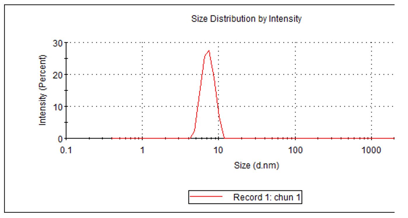
The TEM image of CD-PEI was obtained by high-resolution transmission electron microscopy (JEM-2100) and the X-ray photoelectron spectroscopy of CD-PEI and CD-PEI-DOX were measured by a Thermo 250Xi Thermo K-Alpha. The UV-Vis absorbance spectra of the CD-PEI, CD-PEI-DOX, CD-PEI-DOX-siNC, and CD-PEI-DOX-siMRP1 were measured by a Shimadzu UV3600. The PL spectra were characterized by a flourescence spectrofluorometer (Edinburgh, FLS 980-STM).
The loading and releasing of DOX
The DOX was loaded onto CD-PEI by electrostatic interactions, which has been studied in our previous work[10]. Brifely, the CD-PEI solution mixed with DOX was shaked for 24 h at 4 ℃ and then dialysised for 3 days to remove the excess DOX agent. The 40 nmol/L MRP1 siRNA was added into the CD-PEI-DOX solution and shaked for 24 h at 4 ℃ in order to combine them through electrostatic interactions.
The drug loading efficiency of DOX was calculated by the absobance at 480 nm. The DOX releasing by CD-PEI-DOX-siMRP1 was measured by the drug release experiment as following: 2 mL CD-PEI-DOX-siMRP1 solution was added into a dialysis bag (MWCO, 3 kDa), and soaked into PBS solution. After incubation for different interval, 1 mL PBS solution was gathered and fresh same volume PBS solution was added. The CD-PEI solution was placed under a 5 W UV flashlight with an excitation wavelength of 365 nm. The drug loading efficiency (DLE) was calculated as follows:
DLE% = (amount of DOX in CD-PEI-DOX/amount of DOX) ×100%.
Cell culture
The human lung cancer cells (A549) and doxorubicin-resistant lung cancer cells (A549/ADM) were cultured in Dulbecco’s Modified Eagle’s Medium (DMEM). The 10% fetal bovine serum (FBS), 1% penicillin(100 unit mL-1) and streptomycin (100 µg mL-1) (Gibco, Carlsbad, CA, USA) were added into the DMEM solution. The cells were cultured in a incubator with 5% CO2 at 37 ℃.
Cell cytotoxicity
The Cell Counting Kit-8 (CCK8) was used to measure the cell viability. 1×10⁴ of A549 and A549/ADM cells were seeded in 96-well plates for 24 hours. The PBS, free DOX, CD-PEI-DOX, and CD-PEI-DOX-siMRP1 were added into the DMEM with different concetrations and incubated for 48 hours. Then, the cells were rinsed and added medium with CCK8 for 1 hours. The absorbance at 450 nm was measured by a spectrophotometer.
Intracellular drug release
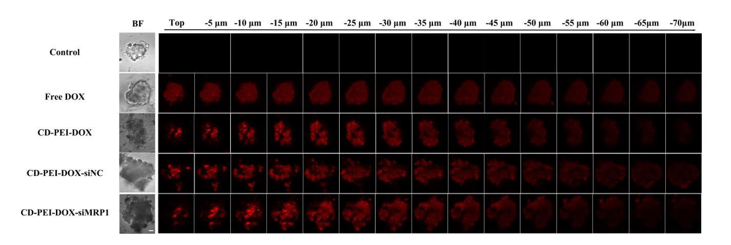
A549 and A549/ADM cells were seeded and cultured on the Ф15 mm glass battern cell culture dish, respectively. The PBS, free DOX (20μg/mL), CD-PEI-DOX (DOX concetration, 20μg/mL), CD-PEI-DOX-siNC (DOX concetration, 20μg/mL), and CD-PEI-DOX-siMRP1 (DOX concetration, 20μg/mL) have been added into the culture dish and incubated for 24 hours. After incubation, the cells were rinsed by PBS and observed by confocal laser scanning microscopy (Zeiss LSM 880, Jena, Germany). A549 and A549/ADM cells manmospheres were seeded in the cofocal dish and incubated with nanodrug for 24 houres.
Cellular uptake in vitro
6×10⁵ of A549 and A549/ADM cells have been seeded in the petri dish, respectively. And the cells were treated by different group, such as PBS, free DOX, CD-PEI-DOX, CD-PEI-DOX-siNC, and CD-PEI-DOX-siMRP1 for different inbucation time. And the cells were collected to measure the geometric mean fluorescence intensity by flow cytometry in Y610-mCHERRY channel (Beckman cytoflex LX).
The transwell assays
The migration and invasion assays of the A549/ADM and A549 evaluating metastasis ability were performed in 24-well transwell chambers with 8 µm size pores (Costar, Washington, D.C., USA). And the pores were covered with 100 µL of Matrigel (BD, USA) in the invasion assay. A549/ADM and A549 were added PBS, DOX, CD-PEI-DOX, CD-PEI-DOX-siNC and CD-PEI-DOX-siMRP1 and incubated for 24 hours. The DOX concetrations of A549/ADM and A549 are 60 μg/mL and 30 μg/mL, respectively. Then, 8×10⁴ cells in 200 μL serum-free DMEM were seeded in the upper chambers and 700 µL of medium supplemented with 10% fetal bovine serum was added in the lower chamber. After 24h of incubation in 37 °C, 5% CO2, the upper chambers were removed, and cells on the lower face of the membranes were fixed with 4% fixative solution (Solarbio, Beijing, China) and stained with Crystal violet. We counted the number of migrated or invaded cells under 5 randomly selected fields at ×100 magnification by inverted light microscope (Leica DMi1, Wetzlar, Germany) for three times independently.
RNA isolation and qPCR analysis
Total Cell RNA isolation and cDNA synthesis were finished with the PrimeScript™ RT reagent Kit (TaKara Bio, USA) following the protocol. The concentration and integrity of RNA was determined using the 260/280 ratios generated by a Nano-drop UV spectrophotometer. Gene-specific primers were designed and synthesized by IGE BIOTECHNOLOGY (Guangzhou, China). qPCR was performed using PrimeScript RT Enzyme Mix I according to the manufacturer’s instructions in CFX96™ Real-Time System (C1000 Thermal Cycler Class, Bio-Rad, California, USA). The two-step PCR conditions used are as follows: Pre-denaturation at 95 °C for 30 second; 40 cycles of (denaturation at 95 °C for 5 second, annealing and extension at 60 °C for 30 second), and denaturation for 10 second. And then melting curve stage was followed from 60 to 95 °C with increment 0.5 °C for 20 second while scanning for fluorescence. Relative quantitation was performed using the 2−∆∆Ct method and data were normalized against GAPDH. Primers sequences for MRP1 and GAPDH are as follows: MRP1: 5’-CCGTGTACTCCAACGCTGACAT-3’ and 5’-ATGCTGTGCGTGACCAAGATCC-3’; GAPDH: 5’-TGTGGGCATCAATGGATTTGG-3’ and 5’-ACACCATGTATTCCGGGTCAAT-3’.
In vitro transfection
A549/ADM cells (4×10⁵) were seeded in 6-well plates and incubated for 24h. The solutions of siMRP1-lipid complex complexes (50 nmol/L siMRP1) or CD-PEI-siMRP1 complexes were prepared before transfection. The medium was replaced by 1mL Opti-MEM per well and the two kind of siMRP1 mixture were separately added to each well. After 12 h transfection, the culture medium was replaced with the fresh complete medium and cells were incubated for an additional 36 h for flow cytometry (FCM). The siRNA are purchased from RiboBio Co., Ltd.(Guangzhou, China) and the number of NC is siN0000001-1-5 siR. The sequences for siMRP1 are as follows: stB0001371B genOFFTM st-h-MRP1_001: GACCTCCGCTTCAAGATCA; stB0001371B genOFFTM st-h-MRP1_002: CCGTCTACGTGACCATTGA; stB0001371B genOFFTM st-h-MRP1_003: CTGGGCTTATTTCGGATCA.
Western blot assays
Western blot assays were carried out as previously described[26]. Breiefly, total cell lysates were prepared in RIPA buffer (Beyotime, Shanghai, China). and proteins were separated by the sodium dodecyl sulfate-polyacrylamide gel electrophoresis (SDS-PAGE) and transferred to nitrocellulose membranes. After blocking with 5% skim milk for 2 h, the membranes were incubated with the primary antibodies against MRP1 (Cell Signaling Technology, Boston, MA, USA), ABCG2 (Cell Signaling Technology, Boston, MA, USA), P-gp (GeneTex, Irvine, CA, USA), and then incubated with the secondary HRP-conjugated antibody (1:5000, Zhongshan Goldenbridge, Beijing, China). Protein bands were visualized using ECL detection reagent and normalized by β-actin (Cell Signaling Technology, Cell Signaling Technology, Boston, MA, USA). Triplicate individual experiments were performed in this study.
Growth inhibition of sphere forming.
A549 and A549/ADM cells were treated by free DOX, CD-PEI-DOX, CD-PEI-DOX-siNC, CD-PEI-DOX-siMRP1 for 24h, respectively. The cells were digested and seeded onto 6-well plates and cultured in DMEM/F12 medium (GIBCO) contained with 4 μg/mL insulin,B27 (1:50), 20 ng/mL EGF and 10 ng/mL FGF. The morphology and diameters of spheres were measured after an additional 5 days.
Animal Study in vivo
All animal studies were approved by Animal ethics committee of the Fifth Affiliated Hospital Sun Yat-sen University. Briefly, A549 cells were subcutaneously inoculated into 4 weeks old male Balb/c nude mice to construct xenograft models. When tumors grew to palpable size, DOX and CD-PEI-DOX-siMRP1 were injected through tail vein. The bio-distribution of drugs were visualized by in vivo image system (IVIS) equipment. Hemolysis experiment were performed to assess the biocompatibility of drugs. The targeting of drugs on tumor were viusualized and photographed as the indicated time points. Penetration of the drugs into tumor were obtained using confocal microscopy LSM880 (Zeiss).
Immunohistochemical assays
Tumor and organs were obtained after mouse were scarificed humanely. Slices were prepared from frozen tissues. Immunohistochemical staining were conducted as prveiously described. Hematoxylin and eosin (H&E) staining (ZSBG-BIO, Beijing, China) were carried out to investigate the toxic effect of drugs. Representative pictures were shown and the distribution of different drugs were visulized undrer optical microscopes (BX53 System Microscope, Olympus, Japan).
Statistical analysis
The data between groups was statistical analyzed by Student’s t-test. P<0.05 was used as the criterion for statistical significance.