Separation of Milk-sEVs
sEVs were separated from fresh milk by ultra-high-speed centrifugation. Centrifugation was performed at 13,000 × g for 30 min, followed by 100,000 × g for 120 min, then the middle whey filter was collected, centrifuged at 135,000 × g for 90 min, then 100,000 × g for 120 min, and after filtration, centrifuged at 100,000 × g for 120 min, and the Milk-sEV suspension was collected.
Western blotting
The protein concentration of each sample was measured using a QuantiPro BCA Assay Kit (KeyGen Biotech, Shanghai, China). The membranes were then incubated overnight at 4ºC with specific anti-CD63 (diluted 1: 200; Abcam, Cambridge, MA, USA), anti-CD81 (diluted 1: 500; Abcam), anti-CD40 (diluted 1: 1000; Bioss Antibodies, Woburn, MA, USA), anti-ALIX (diluted 1: 1000; Abbexa Ltd., Cambridge, UK), anti-RUNX2 (diluted 1: 500, SAB, USA), anti-BMP-2 (diluted 1: 500, Bioworld Technology, St Louis Park, MN, USA USA), anti-ALP (diluted 1: 1000, Abcam), anti-GJA1 (diluted 1: 1000, Bioworld), anti-AP3B1 (diluted 1: 300, Proteintech, Rosemont, IL, USA) and anti-GAPDH (diluted 1: 5000, Bioworld). Incubation with the secondary antibody (diluted 1:500, ABclonal, Woburn, MA, USA) lasted 1 h. The ECL luminescent solution was configured to collect the blotting results with a Bio-Rad gel imaging system (Bio-Rad, Hercules, CA, USA), and the results were analyzed using Image Lab software.
Real-time PCR
Total cell RNA was separated using Trizol, and the corresponding cDNA template was generated according to the reaction conditions of the reverse transcription kit (Takara Bio Inc., Shiga, Japan). The primers were designed and synthesized by Sangon Biotech (Shanghai, China). The primer sequences were AP3B1: Forward: 5'-GCCTTCCAGCCAAGATAACGT-3', Reverse: 5'-CGCAGCAGAACAGAACCAATC-3'; USF2: Forward: 5'-CTGTCCAAGGCCTTGCGATTAC-3', Reverse: 5'-TCGAAGCAGGGCATTCTCAT-3'; GJA1: Forward: 5'-CAGCGCAGAGCAAAATCGA-3', Reverse: 5'-R-GGTCGCTGTCCACGATAGC-3'; ALP: Forward: 5'-TGAATCGGAACAACCTGACTGA-3', Reverse: 5'-R-GAGCCTGCTTGGCCTTACC-3' and GAPDH: Forward: 5'- GTATCGGACGCCTGGTTA-3', Reverse: 5'- CATTTGATGTTAGCGGGAT-3'.
Establishment of a mice skull defect model
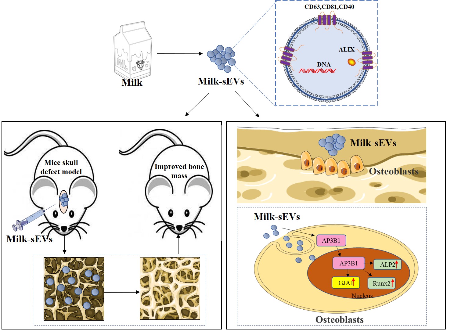
Twelve 8-week-old male Sprague-Dawley mice (180–220 g) were purchased from the Laboratory Animal Center, Dalian Medical University. The experimental protocol was approved by the Institutional Review Board of the School of Stomatology, Dalian Medical University (2021006). Milk-sEVs were added to GelMA (EngineeringForLife, China) to prepare a GelMA-Milk-sEVs composite material with a diameter of about 6.5 mm. After anesthesia (3% sodium pentobarbital, 0.8 mL/kg), the mice scalp was cut to strip away the periosteum, and the skull was drilled without pressure. A skull defect with a diameter of 1.5 mm was created and then the mice were randomly divided into three groups: a control group, a GelMA-PBS group and a GelMA-Milk-sEVs group. The control group was sutured directly without any treatment; in the GelMA-PBS group, GelMA-PBS hydrogel composite was placed in the defect and sutured; in the GelMA-Milk-sEVs group, GelMA-Milk-sEVs hydrogel composite was placed in the defect and sutured.
Transcriptome sequencing
A total of 1 μg RNA per sample was used as input material for the RNA sample preparations. Sequencing libraries were generated using a NEBNext® Ultra™ RNA Library Prep Kit for Illumina® (New England Biolabs (NEB), Ipswich, MA, USA) following the manufacturer’s recommendations and index codes were added to attribute sequences to each sample. The clustering of the index-coded samples was performed on a cBot Cluster Generation System using TruSeq PE Cluster Kit v3-cBot-HS (Illumina) according to the manufacturer’s instructions. After cluster generation, the library preparations were sequenced on an Illumina Novaseq platform and 150 bp paired-end reads were generated.
Luciferase reporter assay
Luciferase reporter assay was performed in 293T cells. DNA fragments encoding mice GJA1 promoters were ligated into pEZX-PG04.1 (GeneCopoeia Inc., Rockville, MD, USA) GJA1 promoter-luciferase reporter systems. Cells were then transfected in triplicate with one of the four vectors (GJA1-pomoter, Con-GJA1, Over-AP3B1, and Con-AP3B1). Gaussia luciferase (GLuc) activity and alkaline phosphatase activity were assayed after 48 h of transfection using a Secrete-Pair™ Dual Luminescence Assay Kit (GeneCopoeia) according to the manufacturer's instructions.
Statistical Analysis
Data are expressed as means ± SEM. Significant differences between test groups were analyzed by one-way analysis of variance. P < 0.05 was considered to be significant.