In the present study, the potential protective effects of CAR on testicular toxicity caused by arsenic, which influence millions of people around the world, were examined and it was seen that CAR could be a promising compound against SA-induced testicular toxicity.
Oxidative stress significantly affects fertility [26]. Previous studies have reported that arsenic impairs spermatogenesis by inducing oxidative stress, resulting in a decrease in sperm quality and quantity [6]. SOD, CAT and GPx enzymes are at the base of the cellular antioxidant defense line [30, 31]. SOD is responsible for scavenging superoxide radicals, and catalase is responsible for the decomposition of H2O2 into water and molecular oxygen [32–34]. While GSH helps maintain the redox state in cells, it also plays a role in removing metals from cells. Mice treated with arsenic have been reported to have a significant reduction in GSH levels [8]. In another study, it was reported that LPO, which is an important indicator of oxidative stress in the testicles, increased after SA administration, while a decrease in GSH, SOD, CAT and GPx levels occurred [6]. In this study, it was observed that GSH stores were significantly reduced in the testes of rats given SA, probably to compensate for excessive ROS production. it was thought that SA inhibited SOD, CAT and GPx enzymes and this occured either by directly acting on sulfhydryl groups or by excessive increase in ROS production, as mentioned before. These factors may have combined to cause a significant increase in MDA in testicular tissues after SA treatment. On the other hand, it was determined that CAR treatment increased GSH stores. This may be due to the fact that the hydroxyl group in its structure contributes to the clearance of ROS, restoring the activity of SOD, CAT and GPx enzymes, as well as reducing the load of GSH. All of them may have combined to cause a decrease in MDA levels.
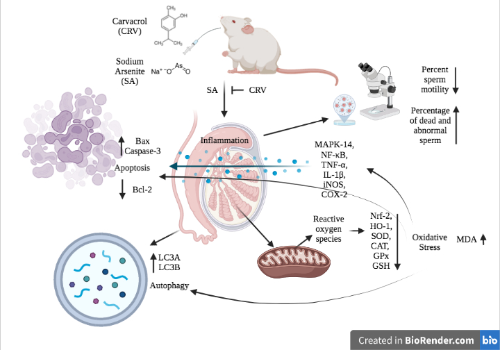
Nrf-2 is a redox-sensitive transcription factor that has an important role in protecting cells against oxidative stress [35, 36]. It is known that with the occurrence of oxidative stress, Nrf-2 and Keap-1 in complex in the cytoplasm are decomposed and Nrf-2 migrates to the nucleus, stimulating the transcription of phase II drug metabolizing enzymes such as HO-1, glutathione S-transferase and glutathione peroxidase [35, 37, 38]. In a previous study, it was reported that Nrf-2 was downregulated due to excessive oxidative stress in arsenic poisoning, and 3,5,7,3′,4′-Pentahydroxy flavone isolated from Madhuca indica triggered Nrf-2 expression [35]. In another study, it was reported that arsenite suppressed the expression of Nrf-2 and accordingly decreased HO-1 expression, and after naringin treatment, Nrf-2 and HO-1 expressions were triggered in rats [36]. Similarly, in the presented study, it was observed that the Nrf-2 and HO-1 mRNA transcript levels decreased in the testicular tissues of rats, possibly due to high oxidative stress, in the administration of SA, and that CAR upregulated Nrf-2 and HO-1 expressions by attenuating oxidative stress with its ROS scavenging feature.
Increasing evidence shows that in addition to oxidative stress, inflammation, which is triggered significantly by oxidative stress[39] and caused by the increase of pro-inflammatory cytokines has an important role in tissue damage induced by arsenic [4]. NF-κB is an important factor regulating the transcription of the pro-inflammatory cytokines IL-1β, TNF-α, iNOS and COX-2, and together they play a key role in spermatogenesis, testicular steroidogenesis and semen maturation [40]. MAPK14 is involved in the regulation of the inflammatory response by activating NF-κB in various cell types [11, 41, 42]. In a previous study, it was reported that administration of SA to mice increased IL-1β, IL-6 and TNF-α levels and Nos2 was up-regulated. In this report, it was also reported that reproductive dysfunction develops with testicular atrophy as a result of excessive NO production and increased pro-inflammatory cytokines [6]. Another study has similarly confirmed that arsenic exposure increases the production of pro-inflammatory cytokines and this plays a role in the pathogenesis of testicular damage [9]. Wang et al. (2013)[43] reported that there is a relationship between the MAPK pathway and COX-2, and that COX-2 is activated by the activation of the arsenite MAPK pathway in the bladder. In the present study, it was determined that MAPK14, NF-κB, TNF-α, IL-1β, COX-2 and iNOS expressions were up-regulated, possibly in a chain manner, in testicular tissues of rats given SA, in line with the literature. It is thought that the expression of pro-inflammatory cytokines increases due to the triggering of oxidative stress by SA and the subsequent activation of NF-κB. Thus, it is thought that SA may affect fertility by causing inflammation in the testicular tissue. Additionally, in the present study, it was thought that there was a suppression of MAPK14, NF-κB, TNF-α, IL-1β, COX-2 and iNOS expressions that were up-regulated by SA as a result of CAR treatment, and this was due to the ROS scavenging property of CAR.
Controlled by apoptotic proteins, apoptosis is a physiological event accompanied by oxidative stress in many cells, including spermatogenic cells [6]. Excessive apoptosis can cause infertility in men by affecting spermatogenesis [6, 44]. It has been reported that SA up-regulates Bax and Caspase-3 expressions, which have important roles in the regulation of apoptosis, and down-regulates Bcl-2 expression, thus stimulating apoptosis in testicular tissues by causing excessive ROS accumulation [6]. In another study, it was reported that arsenic increased the number of apoptotic germ cells, while the apoptotic index decreased significantly with the administration of melatonin [45]. In the present study, there was a remarkable increase in Bax and Caspase-3 expressions in testicular tissues of rats given SA, while Bcl-2 mRNA transcript levels were significantly decreased. On the other hand, it was observed that CAR treatment attenuated the apoptotic effect of SA by suppressing Bax and caspase-3 expressions and up-regulating Bcl-2 expression. Oxidative stress plays an important role in apoptosis [46]. In the study, it is thought that SA triggers caspase-3 expression by causing cytochrome c release from mitochondria. It is thought that this may protect against male infertility by reducing the testicular toxicity induced by SA.
Based on the knowledge that arsenite can induce oxidative stress and oxidative stress can induce autophagy [47], which is the cell's self-eating process, the effects of CAR against SA toxicity were also investigated with autophagic markers. The data obtained showed that the expressions of LC3A and LC3B [48], which are involved in autophagosome formation, increased significantly after SA administration, triggering autophagy in testicular tissue. Although autophagy is an essential event required by cells in normal physiological processes, it causes tissue damage when it occurs at a high level [49, 50]. This situation has also been reported in many previous studies, which is consistent with our results [47, 51]. In the study, it was also observed that CAR down-regulated LC3A and LC3B expressions and protected testicular tissue from SA-induced autophagy.
It is known that there is a close relationship between arsenic exposure and male reproductive dysfunction [3]. There was no significant difference between testicular weights and sperm densities in the study. However, it was determined that sperm motility decreased significantly after SA administration, and the percentages of dead and abnormal sperm were significantly increased compared to other groups. In addition, it was observed that CAR administration brought these values closers to the control group levels. In a previous study, it was reported that a significant decrease in sperm counts occurred in rats given arsenite [2]. In another study, it was reported that arsenic administration caused a decrease in testicular weights and sperm motility, and this occurred as a result of increased lipid peroxidation [4]. Baltaci et al. (2016)[18] reported that SA and quercetin administrations did not cause any change in testicular weights in accordance with the present study.