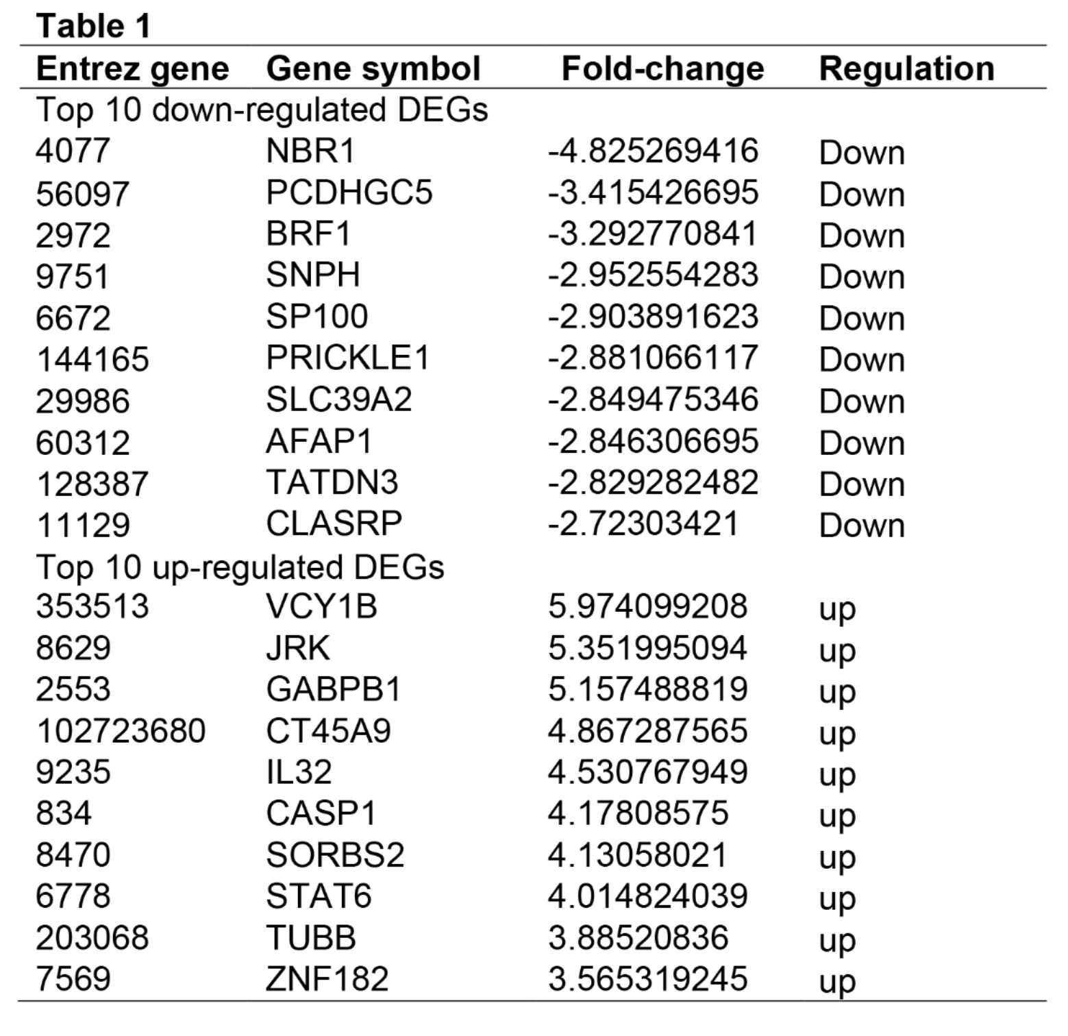
Identification of DEGs in fibrosarcoma cells with FV infection
To determine the effects of FV on fibrosarcoma cells, we analyzed the RNA-seq data of PFV-infected HT1080 cells. A total of 1210 genes were identified with a threshold of P < 0.05. The top up- and down-regulated genes were indicated by the heatmap and volcano plot (Fig. 1). The top ten DEGs were listed in Table 1.
KEGG and GO analyses in fibrosarcoma cells with FV infection
To determine the potential mechanism of FV infected fibrosarcoma cells, we performed the KEGG and GO analyses by using the RNA-seq data (Figure 2). We identified the top ten KEGG signaling pathways, including “Sphingolipid signaling pathway”, “Carbon metabolism”, “Small cell lung cancer”, “Glycerophospholipid metabolism”, “Pancreatic cancer”, “Colorectal cancer”, “Mitophagy – animal”, “Endometrial cancer”, “Thyroid cancer”, and “2−Oxocarboxylic acid metabolism”. We identified the top ten biological processes (BP) of GO, including “Regulation of membrane potential”, “Regulation of response to DNA damage stimulus”, “Gonad development”, “Development of primary sexual characteristics”, “Regulation of DNA repair”, “Regulation of double−strand break repair”, “Release of cytochrome c from mitochondria”, “Positive regulation of blood pressure”, “Positive regulation of double−strand break repair”, and “Calcium−dependent cell−cell adhesion via plasma membrane cell adhesion molecules”. We identified the top ten cellular components (CC) of GO, including “Cell−substrate junction”, “Focal adhesion”, “Postsynaptic density”, “Extrinsic component of plasma membrane”, “Peroxisomal membrane”, “Microbody membrane”, “Cell division site”, “Sarcoplasm”, “Magnesium−dependent protein serine/threonine phosphatase complex”, and “Extrinsic component of synaptic membrane”. We then identified the top ten molecular functions (MF) of GO, including “Phospholipid binding”, “Phosphatidylinositol binding”, “Phosphatidylinositol phosphate binding”, “Structural constituent of muscle”, “Polyubiquitin modification−dependent protein binding”, “Peptidase activator activity involved in apoptotic process”, “Steroid hormone receptor activity”, “Acylglycerol lipase activity”, “CARD domain binding”, and “Cysteine−type endopeptidase activator activity involved in apoptotic process”.
PPI network and Reactome analyses
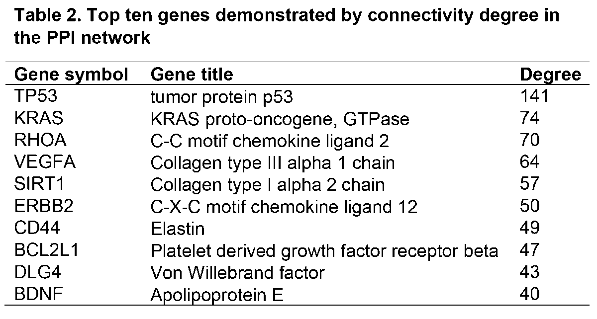
To explore the potential relationship among the DEGs, we constructed the PPI network by using 1118 nodes and 3661 edges. The combined score > 0.2 was set as a cutoff by using the Cytoscape software. Table 2 showed the top ten genes with the highest scores. The top two significant modules were presented in Figure 3. We further analyzed the PPI and DEGs with a Reactome map (Figure 4) and identified the top ten biological processes including “Transcriptional activation of cell cycle inhibitor p21”, “Transcriptional activation of p53 responsive genes”, “Regulation of TP53 Expression”, “Insulin-like Growth Factor-2 mRNA Binding Proteins (IGF2BPs/IMPs/VICKZs) bind RNA”, “BH3-only proteins associate with and inactivate anti-apoptotic BCL-2 members”, “TP53 Regulates Transcription of Genes Involved in G1 Cell Cycle Arrest”, “Defective Intrinsic Pathway for Apoptosis”, “The NLRP1 inflammasome”, “FOXO-mediated transcription of cell cycle genes”, “Intrinsic Pathway for Apoptosis” (Supplemental Table S1).