Dental caries can be defined as an infectious, multifactorial, transmissible process that leads to the progressive destruction of dental hard tissues (enamel and dentine). As one of the most prevalent and consequential oral diseases globally, it causes serious health and economic burdens throughout the world [1, 2]. Therefore, it is very important to detect carious lesions sensitively and accurately, which can protect dental health and reduce substantial costs [3].
Visual tactile method is a traditional and preferred route in everyday clinical practice, which is widely used for screening and diagnosis of dental caries [4, 5]. Dentists inspect dental caries based on changes of the colour, texture, and structure of teeth according to their experiences. Hence, it is highly subjective, and the sensitivity and specificity of visual tactile diagnosis can not meet the accuracy requirements [6, 7].
X-ray radiography is an irreplaceable aid for the diagnosis of dental caries, especially hidden or inaccessible dental caries. There are three common types of radiographs widely used in clinical practice, which are periapical, bitewing, and panoramic X-rays [8]. Periapical and bitewing X-rays concentrate on only one or a few teeth and can not detect the full mouth in one attempt. Panoramic X-rays can capture the entire mouth in a single image, but it is a two-dimensional X-ray examination.
Cone-beam computerized tomography (CBCT) is a new radiographic technique which can generate three dimensional (3-D) images of dental structures, soft tissues, and bone in the craniofacial region in a single scan. The focused x-ray beam reduces scatter radiation, resulting in better image quality. Meanwhile, it produces a wide variety of views and angles which can provide more complete evaluations. Thus, CBCT has advantages such as high accuracy and low artifact, allowing for more precise treatment planning [9, 10]. Walsh et al searched several databases and studied related articles about the detection and diagnosis of early caries. They found that CBCT showed superior sensitivity and had the potential to be used as a reference standard in diagnostic studies of dental caries [11]. However, there were also controversial results in this respect. Felemban et al evaluated the diagnostic accuracy of CBCT, extraoral bitewings, and intraoral bitewings in the detection of interproximal caries. Results showed that no significant differences were found in sensitivity, specificity, and area under the curve values among these methods [12]. By far, the detection of dental caries with radiography was still complicated due to the complexity of the tooth anatomy, the artifacts from restorations, etc.
Nowadays, there are several other methods being developed to detect dental caries, such as fluorescence, transillumination, optical coherence tomography, and electrical conductance or impedance technologies [13]. Salama et al investigated occlusal surface of 45 molar and 49 premolar teeth using three diagnostic methods, which were visual examination using the International Caries Detection and Assessment System (ICDAS-II), light fluorescence-based devices (Vista Proof iX HD smart), and fissurotomy. Among them, Vista Proof showed the highest level of agreement in dentine carious lesion detection with the highest sensitivity value (95%). However, the level of agreement in enamel carious lesion detection was very low with 48% sensitivity value [14]. The near-infrared light transillumination (NILT) method provides noninvasive, nonionizing radiation choices. This technique has been proven to be successful as a screening tool for the early detection of demineralized dental caries lesions and can be considered as an adjunct to bitewing radiographs [15–17]. It can be advantageous in screening pregnant, growing adolescent patients and in cases where multiple follow-ups are needed and ionizing radiation must be avoided. Vanella et al summarized that NILT using light ranging from 700 to 1700nm had better optical properties compared to conventional optical systems using light in the visible spectra. It exhibited increased sensitivity compared to radiographs and was more suitable to identify approximal enamel lesions [18]. However, there were also controversial reports. Alrayyes et al pointed out that the use of NILT as a stand-alone caries detection method for interproximal primary molar surfaces was limited [19]. Stratigaki et al warned that treatment decisions should not be based on NILT alone [20]. Other methods such as bioluminescence measurements (Calcivis imaging system, Cis) and enhanced truncated-correlation photothermal coherence tomography (eTC-PCT) using long-wave infrared (LWIR) cameras were also investigated for the visualization of early caries [21, 22]. However, further studies still needed to be explored considering about penetration depth or background noise.
Recently, the intraoral scanner (IOS) has been raised as a new caries diagnostic tool. Schlenz et al compared three intraoral scanners to established diagnostic methods. They set µ-CT as reference. Results showed that caries diagnostics with IOS seemed to be interesting strategies that should be further investigated in clinical studies [23]. There were many studies investigated IOS in implant placements. The improved accuracy of scan templates fabricated using the IOS can eliminate the possible laboratory errors associated with the conventional technical procedures as well as reduce the inaccuracies resulting from the image processing and segmentation of CBCT data [24]. Meanwhile, IOS were combined with CBCT in measuring dimensional changes of bone and soft tissue surface [25]. Gómez-Polo et al merged IOS with CBCT for implant-supported complete-arch fixed dental prostheses. They confirmed that such methodology could improve the dimensional accuracy of IOS in edentulous arches [26].
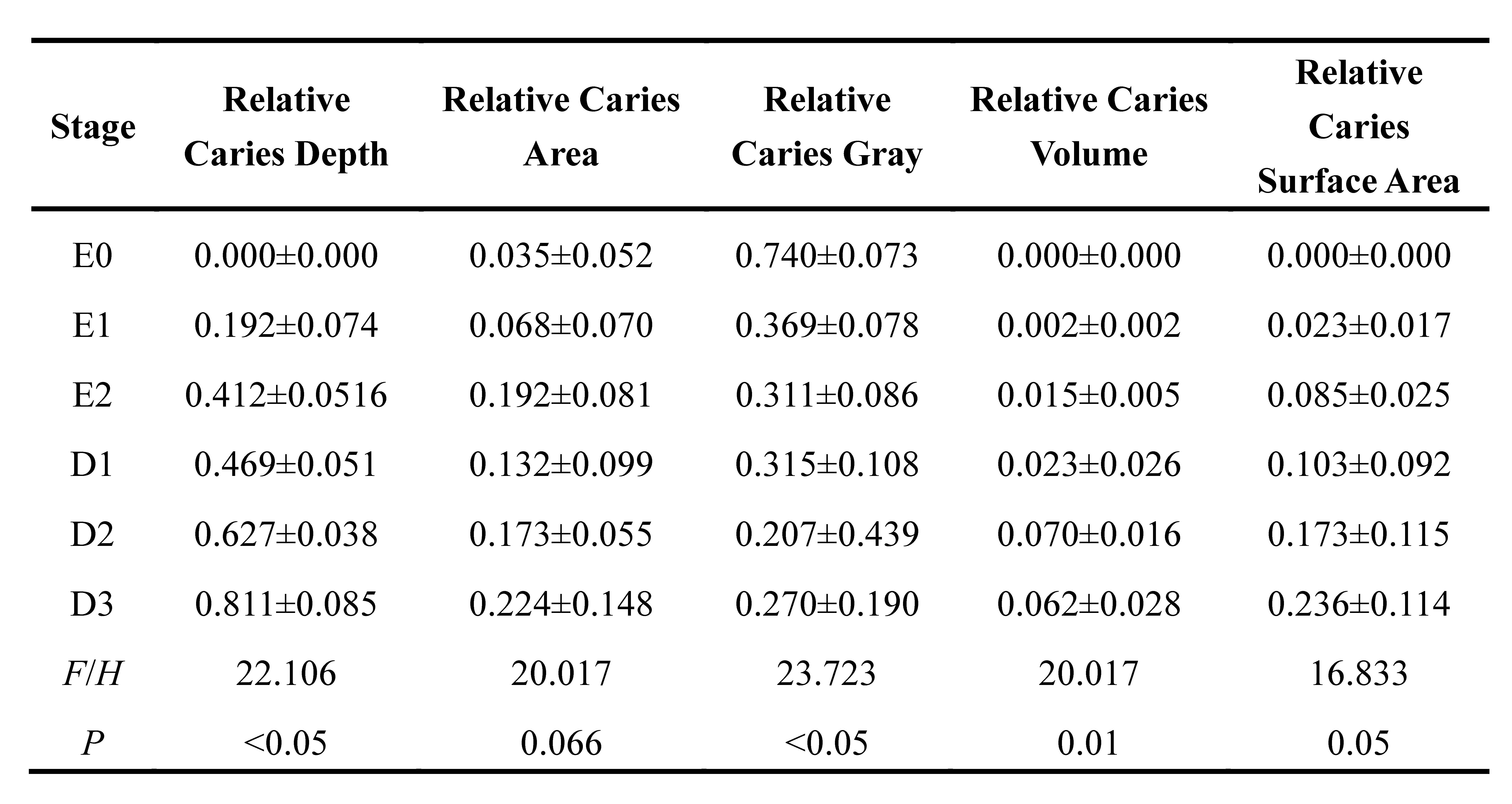
Our team developed a multimodal fusion method based on IOS and CBCT in dental caries detection. We collected the IOS and CBCT images of mandibular molar teeth with various degrees of dental caries. The 3D model data format was raised by data fusion and precise registration of these images, which provided both the surface texture features and internal voxel structure features. Quantitative measurements were made according to the color and structure characteristics of dental caries. Such results were compared with results from visual-tactile and histological assessments. This study hypothesized that the combined usage of IOS and CBCT will lead to the creation of a new digital methodology for the precise diagnosis of dental caries. The novelty in this paper lied in the development of the quantitative geometric measurement and potential usage in diagnosis based on fusion modeling of external surface texture obtained from IOS and internal structure information from CBCT images. The usage of such method can be helpful in the identification and diagnosis of dental caries. Schematics of the whole study were shown in Fig. 1.