1.2.1 Materials
5-bromoindole-2-carboxylic acid (B2761), MTT powder (M2128), α-MEM Powder, Alginate sodium (Alg, A2033), Chondroitin sulfate (CS, C6737), Calcium chloride (CaCl2, 55670), Phosphate buffer saline (PBS, P4417), Calcein AM (17783), Ethidium homodimer-1 (46043) and Agarose (A0169) were obtained from Sigma.
1.2.2 Selection of the best CS and PL concentration
For choosing the best concentration of CS, we prepared the hydrogel by blending a 1.5% (w/v) Alg with three different concentrations of CS 0.5, 1, and 2.5% (w/v) and then the hydrogels were incubated in agarose mould wells (6 mm Ø x 3 mm H) at RT. The agarose molds were prepared from 3% w/v agarose in CaCl2 150mM for in-situ gelling of the Alg solution. The obtained gels were kept overnight in a solution of 150mM CaCl2 before using for evaluation of mechanical properties by compression testing. The hydrogels were also prepared in a larger agarose mold of 25mm Ø x 2 mm H, for rheology tests.
We also prepared the Alg hydrogel with different concentrations of fresh and freeze-dried PL. Briefly, three different concentrations of fresh PL (10, 20, and 50% (v/v)) and also three different concentrations of freeze-dried PL (0.5, 1, and 2 % (w/v)) were mixed with 1.5% (w/v) sodium alginate, then cast into the agarose mold, covered with 150mM CaCl2 and incubated at RT.
Preparation of the PL was performed as follows:
Blood samples (300ml) were collected after obtaining written consent from healthy volunteers according to the ethical approval from Ethics Committee of Royan institute (IR.ACECR.ROYAN.REC.1395.174). The samples were centrifuged at 200g for 30min at RT. Then, the resulting plasma supernatants were pooled, transferred into a 15mL Falcon tube, and centrifuged at 2000g for 5min at RT to produce a platelet pellet. The platelet pellet was resuspended in PBS (1/10th of the initial blood volume), sonicated for 15 min at RT and then stored at -20°C to use later as PL.
1.2.2.1 Compression Mechanical test
Unconfined hydrogels were compressed using an Instron 5866 electromechanical test device equipped with a static load cell of 100N, at a displacement of 1mm/min and stopped at 50% sample’s height (n=6). Young’s modulus was calculated according to the stress-strain-curve.
1.2.2.2 Rheological analysis
The viscoelastic properties of the various hydrogel formulations were investigated using an Anton Paar MCR-302 rheometer equipped with a Peltier temperature control unit and cone-plate geometry (Disposable Measuring Plate PP25 with a 25 mm diameter). A fixed gap of 0.8 mm was selected, and a time sweep was conducted at 100% strain at 25°C. The storage modulus (G') and loss modulus (G'') values were calculated at 0.1% strain (n=6).
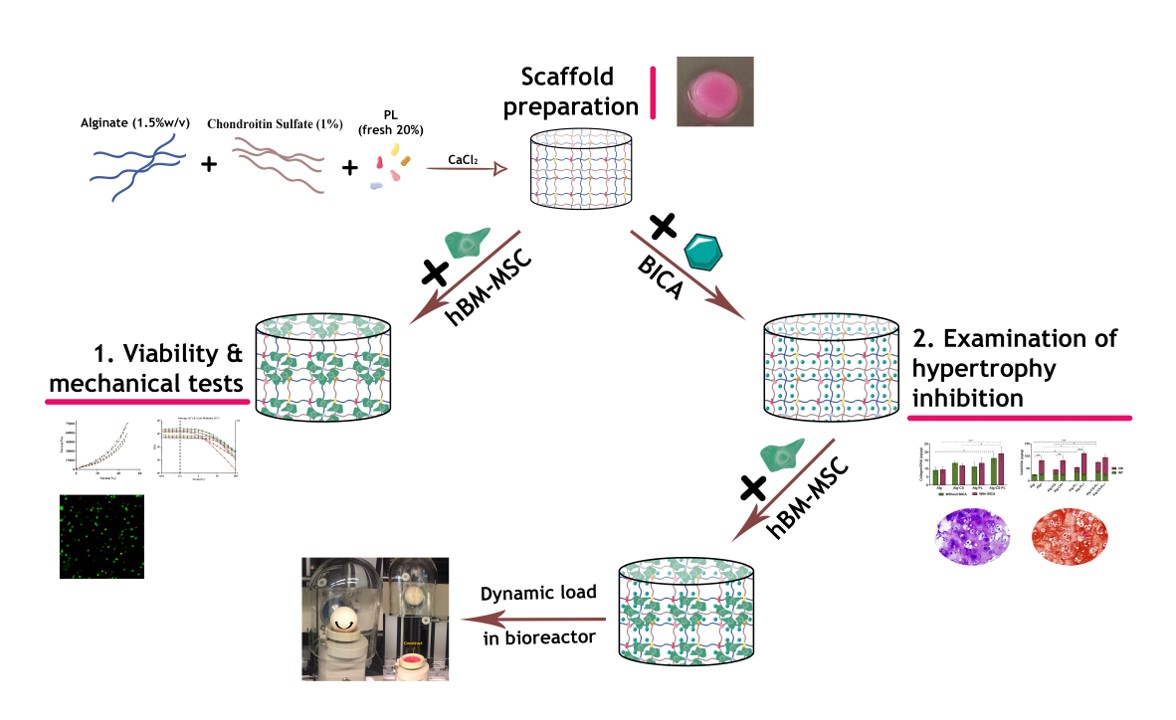
1.2.3 Hydrogel Preparation and Characterization
Hydrogel was prepared by mixing alginate sodium solution, CS and PL to a final concentration of 1.5% (w/v) Alg, 1%(w/v) CS and 20%(v/v) PL. The aqueous hydrogel mixture was cast into the agarose mould, covered by 150mM calcium chloride (CaCl2) and incubated at room temperature (RT) overnight before characterization. The detailed hydrogel formulations are shown in table 1.
The mechanical properties of cell-free and BM-MSC embedded hydrogels were assessed. hBM-MSCs were embedded in hydrogels (5×10⁶/ml) and the mixture was incubated in a solution of 150mM CaCl2 for 1h prior to mechanical analysis (n=3).
Table 1: List of hydrogel formulations
|
Hydrogel
Description
|
Alginate
(%w/v)
|
Chondroitin Sulfate (%w/v)
|
PL
|
|
Alg
|
1.5
|
0
|
0
|
|
Alg-CS
|
1.5
|
0.5, 1, 2.5
|
0
|
|
Alg-PL
|
1.5
|
0
|
Freeze-dried: 0.5, 1, 2 (%w/v)
|
|
Fresh: 10, 20, 50 (%V/V)
|
|
Alg-CS-PL
|
1.5
|
1
|
Fresh: 20(%V/V)
|
1.2.4 Determination of ideal concentration of BICA
1.2.4.1 Cell viability evaluation
Cell toxicity of BICA was evaluated in duplicate using MTT assay at two time points (day 3 and 7). hBM-MSCs were isolated from bone marrow samples (2 donors aged 56 and 60 years old) according to the ethical approval from Ethics Committee of Royan institute (IR.ACECR.ROYAN.REC.1395.174). Briefly bone marrow aspirates were harvested during routine procedures from each patient’s iliac crest, after isolation, hMSCs were subcultured an initial cell density of 1 × 10⁴ cells/cm2 in α-MEM supplemented with 15% fetal bovine serum (FBS) and 100 mg/mL penicillin–streptomycin. The cells were expanded through subcultures until passage 3 to use for other procedures. The cells were also characterized in terms of trilineage differentiation and expression of some surface markers (CD44, CD73, CD105, CD 90, CD11b, CD45, and CD34). hMSCs were cultured in a 24-well plate at a density of 1×10⁴ cell/cm2 in α-MEM supplemented with 10% FBS and six different concentrations of BICA (1, 10, 20, 31, 39, and 50 µM). Cells were washed with PBS followed by removing the culture medium, then incubated in 0.5% of 3–(4,5-dimethylthiazol-2-yl)-2,5-diphenyltetrazolium bromide (MTT) for 2h at 37 ℃ in a 5% CO2 atmosphere incubator. MTT was replaced with an equal volume of DMSO to dissolve the formazan crystals. Absorbance was measured at 540nm by a Multiskan Spectrum microplate reader (ThermoScientific, USA).
1.2.4.2 Effect of BICA on Chondrogenesis of hBM-MSC
In order to investigate the effect of BICA on chondrogenic differentiation, standard pellet culture method was used [61]. Briefly, hBM-MSC pellets were formed by centrifuging 2.5×10⁵ hBM-MSCs at 300 g for 5 minutes in chondrogenic culture medium; high glucose DMEM supplemented with ITS (1%), ascorbic acid (50 µg/ml), non-essential amino acid (1%), Dexamethasone (10-7M), L-Glutamine (2 mM), and TGF-β1(10 ng/ml). Six different concentrations of BICA, as above, were added to the chondrogenic medium. A control group without BICA was also prepared. The medium was changed every 3 days until the pellets were harvested at day 28 for further analysis. The collected media were also pooled and kept at 4℃ until further investigation.
1.2.4.2.1 Biochemical analysis for glycosaminoglycan, Collagen, and DNA Content assay
Glycosaminoglycan (GAG) assay was performed on intact pellets (INT) and the conditioned media (CM). Pellets were rinsed with PBS and digested overnight in proteinase-K (0.5 mg/ml) at 56℃. DMMB (1,9-Dimethyl-methylene blue) was used in order to quantify the sulfated glycosaminoglycan. Briefly, 200µl of DMMB color reagent was added to 20µl of sample or chondroitin sulfate standard. The absorbance was measured immediately at 535nm. The results were normalized to the DNA content quantified by PicoGreen assay on the same proteinase-K digest as above. For this purpose, PicoGreen was added to each sample or DNA standard and incubated at RT for 5 min. The fluorescence was measured at excitation 485nm and emission 535nm. On the same digested samples, the collagen content was evaluated with Sircol™ Insoluble Collagen Assay kit, according to the manufacturer’s instructions. Briefly, 1ml Sircol dye reagent was added to 50µl of sample or standard and gently mixed on a mechanical shaker for 30 min. The tubes were drained followed centrifugation at 12000 rpm for 10 min. The pellet was dissolved in Alkali reagent after removing the unbound dye with an ice-cold Acid-Salt Wash reagent. The absorbance was recorded at 550 nm and the results were normalized to the DNA content. All absorbance/fluorescence measurements were performed by a Victor3 micro plate reader. GAG, Collagen, and DNA assays were done in triplicate.
1.2.4.2.2 Real-time PCR analysis
The pellets were washed with PBS and snap frozen in liquid nitrogen. Total RNA was extracted using TRI Reagent (Molecular Research Center TR-118, Cincinnati, USA). Reverse transcription was performed with random hexamer primers and TaqMan reverse transcription reagents. All real-time quantitative polymerase chain reactions (PCR) for the pellet culture studies were performed with a SYBR Premix Ex TaqTM II (TaKaRa RR820L, Kusatsu, Japan) on an ABI StepOnePlusTM Q-PCR system (applied Biosystems Life Technologies) for Coll X and MMP-13 genes. The expression level of target genes was normalized to β-actin as the reference gene. The analysis was performed by the comparative ∆∆CT method. Primers are listed in Table S1(supplementary material).
1.2.4.2.3 Histological evaluation
hBM-MSC pellets were fixed in 4% paraformaldehyde and incubated in 4℃ for 3 days. After processing in a tissue preparation machine (Did Sabz co, Iran), the samples were embedded in paraffin. Six µm sections were stained with safranin O & fast green, and toluidine blue.
1.2.5 Release kinetics of BICA in tri-part hydrogels
In order to examine the kinetic release of BICA, alginate-based hydrogels were used. The best concentration of BICA was added into four hydrogels with different compositions (Table 1). The hydrogels were prepared in the cap of a 1.5 mL Eppendorf tube and cross-linked with a 150mM CaCl2 solution at RT for 1h. The remaining solution was stored to calculate the amount of non-encapsulated drug. The BICA-embedded hydrogels were incubated in 1ml PBS at 37℃. Aliquots from the 1ml PBS solution were collected and replaced with 1 ml fresh PBS at specific time points and the BICA concentration was determined by a Thermo Scientific™ Multiskan™ GO Microplate Spectrophotometer (Thermo Scientific™, USA) at a wavelength of 290 nm.
1.2.6 Embedded hydrogels with mesenchymal stem cells
Human BM-MSCs were isolated from bone marrow samples (3 donors aged 15, 18, and 37 years) after ethical approval from the cantonal ethical commission of Bern (KEK: Req-2016-00141) using previously described protocols [62]. Bone marrow samples were aspirated of vertebral bodies from each donor, after isolation, at 70-80% confluence, hBM-MSCs were harvested by trypsinization and subcultured at a density of 3 × 10³ cells/cm2 in Minimum Essential Medium supplemented with 10% Sera Plus bovine serum, 100-U/mL penicillin, and 100-mg/mL streptomycin, and 5ng/ml FGF and grown until passage 3. Cultures were maintained at 37°C/5% CO2, and the medium was refreshed every second day. hBM-MSCs were also seeded into the hydrogels at a density of 5×10⁶ cell/ml at passage 3. The cell-laden hydrogels were cast into a mold (8 mm Ø x 5 mm H) and incubated in 150mM CaCl2 for 1h at RT. Then, washed with PBS and incubated in chondrogenic culture medium for more analysis.
1.2.6.1 Live& Dead assay
After embedding hBM- MSCs into the Alg-based hydrogels, the cell-laden constructs were cultured in αMEM supplemented with 10% v/v SeraPlus, 1% v/v of Pen/Strep and 5 ng/ml recombinant human fibroblast growth factor-2 (FGF-2). Then, cell viability was assessed at 2-time points (day 3 and 7) on the bulk hydrogel, using live/dead assay, where living cells were stained with Ca-AM and dead cells with EthD-1. At each time point, the hydrogels were removed from the culture medium and incubated in a staining solution containing 5µM Ca-AM and 1µM EthD-1 prepared in serum-free DMEM LG for 1h at 37°C, within a humidified atmosphere of 5% CO2. After incubation, cells were imaged with confocal laser scanning microscopy (LSM810, Zeiss). For each sample, single plane and Z stack (1300 μm) were acquired and tile scans were generated to image a larger sample area. Four different fields of view per sample were used to quantify cell viability by counting the red (dead) and green (viable) cells in Image J software.
1.2.6.2 chondrogenesis assessment
Chondrogenic differentiation of cell-laden hydrogels (with and without BICA) was investigated by qRT- PCR, biochemical assessment and histological staining after 28 days. Total RNA was isolated using TRI Reagent® Solution (Molecular Research Centre Inc., Cincinnati, OH, USA) according to the manufacturer’s protocol. Reverse transcription of 1 µg total RNA was performed by TaqMan Reverse Transcription Kit (Applied Biosystems, Foster City, USA). Relative gene expression (quantitative polymerase chain reaction (qPCR)) reactions were set up in 10µL reaction mixtures containing TaqMan Universal Master Mix (Thermo Fisher, Zürich, Switzerland), the appropriate set of primers and probes, DEPC-H2O and cDNA template. For gene expression, Coll I, Coll II, ACAN, ALP, Coll X, and MMP-13 were evaluated, and 18S was used as a housekeeping gene. The expression of genes was normalized to a control of encapsulated hBM-MSCs into Alg- hydrogel, incubated for 24h. Technical triplicates were used for each target gene and for the different donors (n=3). Primer and probe sequences are shown in supplemental Table S2 (supplementary material), while catalogue numbers of Assays-on-Demand (Applied Biosystems, Foster City, USA) are listed in the supplemental Table S3 (supplementary material). GAG and collagen content in the samples were normalized to DNA content. In addition, GAG in the conditioned medium was also measured. Histological staining analysis was performed.
1.2.7 The effect of mechanical loading on the chondrogenesis of hBM-MSCs in hydrogels
Chondrogenesis of hBM-MSCs in the best hydrogel (Alg-CS-PL with and without BICA) was evaluated in the presence or absence of mechanical loading. The mechanical loading was produced by multiaxial loading bioreactor [63]. Briefly, a ceramic ball (32mm in diameter) was pressed onto the scaffold. An interfacial shear load was generated by ±25° oscillatory rotation of the ball about the axis perpendicular to the scaffold’s axis (at a fixed indentation of 0.2mm with a frequency of 0.1Hz). After an initial 2-week culture without loading, mechanical stimulation was applied 1h per day for five consecutive days per week over a period of 2 further weeks. After a total of four weeks culture, samples were harvested for qRT-PCR, histology, and biochemical analyses. (2 different donors with 2 replicates.)
1.2.8 Statistical analysis
Data were obtained from the samples and represented as the mean ± standard deviation (SD). Statistical assessment was carried out by analysis of two-way ANOVA and post-hoc Tukey’s tests. Statistical analysis was performed by means of Prism software (GraphPad Software, La Jolla, CA, USA).